Histology Prelab - Respiratory System
 Call Kai
 Learn
 Practice Test
 Spaced Repetition
 Match
 Flashcards
 Knowt Play
1/161

There's no tags or description
Looks like no tags are added yet.
162 Terms
Upper and lower respiratory tracts
Anatomically, what are the divisions of the respiratory system?
Nasal cavity, nasopharynx, larynx
What is included in the upper respiratory tract?
Extrapulmonary and intrapulmonary
What are the divisions of the lower respiratory tract?
Trachea and primary bronchi (extrapulmonary bronchi)
What is included in the extrapulmonary lower respiratory tract?
Intrapulmonary (secondary/tertiary) bronchi
Primary bronchioles
Terminal bronchioles
Respiratory bronchioles
Alveolar ducts
Alveolar sacs (clusters of alveoli)
What is included in the intrapulmonary lower respiratory tract?
Conducting portion and respiratory portion
Functionally, what are the divisions of the respiratory system?
Trachea, bronchi, conducting bronchioles, and terminal bronchioles
What is included in the conducting portion of the lower respiratory system?
Respiratory bronchioles, alveolar ducts, and alveoli
What is included in the respiratory portion of the lower respiratory system?
Condition the air and conduct/carry the air to the respiratory portion
What is the function of the conducting portion of the respiratory system?
Site of gaseous exchange between oxygen and carbon dioxide
Exchange occurs through the alveolar wall/septum between pulmonary capillaries and alveoli
What is the function of the respiratory portion of the respiratory system?
Higher or greater
The smaller the diameter, the _________ the resistance of the individual structure
Higher
The smaller the diameter, the _________ the pressure of the individual structure
Less (due to extensive branching)
The overall resistance of the respiratory portion will be _________ than the trachea
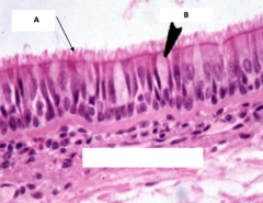
Pseudostratified ciliated columnar
Identify the type of epithelium labeled A
Goblet cell
Identify the cell labeled B
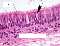
Upper part of trachea
Identify the part of the respiratory system this slide is from

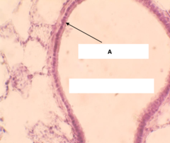
Simple cuboidal
Identify the type of epithelium labeled A

Terminal bronchiole
Identify the part of the respiratory system that this slide was taken from
Pseudostratified ciliated columnar
Identify the type of epithelium labeled A

LCT
Identify the type of tissue labeled B

Smooth muscle
Identify the type of tissue labeled C

Tertiary bronchi or primary bronchiole
What part of the respiratory system is this slide taken from?

Simple squamous
Identify the type of epithelium that the arrow is pointing to in the image shown

Alveolar duct/alveoli
What part of the respiratory system is this slide taken from?

Lamina propria
What is the LCT just under the PSCC in the trachea called?
BALT tissue (Bronchiole Associated Lymphatic Tissue)
What is the tissue that is under the PSCC in the trachea that contains lamina propria with high numbers of WBCs called?
Submucosa
What is the layer of CT denser than lamina propria that contains seromucous glands called in the trachea?
Towards the bottom
In the trachea, where would you find more glands in the submucosa, towards the top, or towards the bottom?
Trachealis m.
What is the smooth muscle associated with the trachea called?
Hyaline cartilage
What type of cartilage forms the C-shaped rings in the trachea?
Adventitia
What is the areolar CT that contains adipose and large blood vessels on the outside of the trachea called?
Layer of DICT and numerous lymphatic nodules
What tissues are associated with lymph nodes?
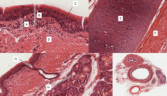
Pseudostratified Ciliated Columnar
Identify the type of epithelium labeled A
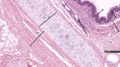
Mucosa
Identify the tissue layer labeled B

Submucosa
Identify the tissue layer labeled C
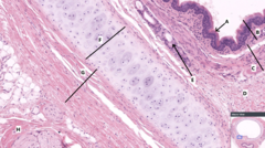
DICT
Identify the type of tissue labeled D

Seromucous glands
Identify the tissue labeled E

Hyaline cartilage
Identify the type of the tissue labeled F

Perichondrium
Identify the name of tissue labeled G

Epineurium
Identify the name of tissue labeled H

Pseudostratified ciliated columnar
Identify the tissue labeled A

Mucosa
Identify the layer of tissue labeled B

Lamina Propria
Identify the layer of tissue labeled C
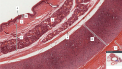
Submucosa
Identify the layer of tissue labeled D

Perichondrium
Identify the name of the tissue labeled F

Hyaline cartilage
Identify the type of tissue labeled G
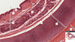
Adventitia
Identify the layer of tissue labeled H

Lamina Propria
Identify the layer of tissue labeled A
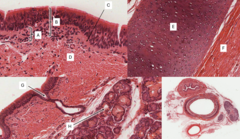
Mucosa
Identify the layer of tissue labeled B
Basement membrane
Identify C
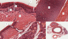
Submucosa
Identify the layer of tissue labeled D

Hyaline cartilage
Identify the type of tissue labeled E

Perichondrium
Identify the name of the tissue labeled F

Duct of gland
Identify the structure labeled G

Seromucous glands
Identify the structure labeled H

Arrangement of cartilage (c-shaped = trachea, plates = PB)
How do you tell the difference between primary bronchi and the trachea, histologically?
Presence of lung tissue surrounding the structure
How do you tell whether you are extrapulmonary or intrapulmonary?
Lymph node
Identify the tissue labeled A

Lung tissue - alveoli
Identify the tissue labeled B
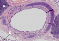
Primary bronchus
Identify the part of the respiratory system shown in the image

Thyroid gland
Identify the tissue labeled A

Skeletal muscle
Identify the tissue labeled B
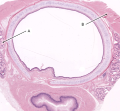
Trachea
Identify the part of the respiratory system shown in the image?

PSCC lining lumen
Underlying LCT in lamina propria
Muscularis mucosa (smooth muscle) increases in wall
Seromucous glands seen in submucosa
Hyaline cartilage forms incomplete plates
Adventitia comprised of LCT visible
What is the histological appearance of primary bronchi?
Primary bronchi have adventitia and large lymph nodes and large blood vessels
Intrapulmonary bronchi will have alveoli and BVs surrounding bronchi interspersed b/w lung tissue
How do you tell the difference between primary bronchi and intrapulmonary bronchi?
Secondary bronchi
Tertiary bronchi
Conducting/primary bronchiole
Terminal bronchiole
What are the intrapulmonary parts of the conducting portion of the respiratory system?
Bronchi have PSCC, cartilage, glands, only some with smooth muscle
Bronchioles have no cartilage and no glands and smooth muscle is present
What is the difference between bronchi and bronchioles?
Primary/conducting bronchiole
Identify the part of the respiratory system shown in the image

Primary leading to terminal bronchii
Identify the part of the respiratory system shown in the image

Intrapulmonary bronchus
Identify the part of the respiratory system indicated A
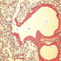
Primary bronchiole
Identify the part of the respiratory system indicated B

Terminal bronchiole
Identify the part of the respiratory system indicated C

Respiratory bronchiole
Identify the part of the respiratory system indicated D

Alveolar duct
Identify the part of the respiratory system indicated E

Pulmonary v
If a vessel is large and thin-walled travelling alone in the lung, it's a ________________________
pulmonary a.
If a vessel is large and thin-walled travelling along the respiratory tree in the lung, it's a ________________________
Bronchial aa.
What vessels are found in the CT of the walls of bronchi and bronchioles that are small and thick-walled?
Size and amount of cartilage (we don't need to ID for lab, just label intrapulmonary bronchi)
What is the histological difference between a secondary and tertiary bronchus?
Alveoli
Identify A

Alveolar duct
Identify B

Arteriole
Identify C

Smooth muscle
Identify D

Bronchial a.
Identify A

BALT
Identify B

Pulmonary a.
Identify D

BALT
Identify E
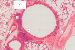
Conducting Bronchiole (no cartilage)
What part of the respiratory tract is this slide taken from?

Arteriole (branch of bronchial a.)
Identify A

Diffuse lymphatic tissue of BALT
Identify B

Smooth muscle
Identify C

No cartilage or glands
Smooth muscle present as complete rings
Wavy epithelium
Epithelium ranges from PSCC to ciliated columnar to ciliated cuboidal
Goblet cells scattered and disappear as diameter decreases
What is the histological appearance of primary/conducting bronchioles?
Alveoli
Identify A

Terminal bronchiole
Identify B

Arteriole (from pulmonary a.)
Identify C

Smooth muscle
Identify D

Primary/conducting bronchiole
Identify E

Primary/conducting bronchiole
Identify F

Terminal bronchiole
Identify G

Simple ciliated cuboidal to simple cuboidal non-ciliated interspersed by club cells
Goblet cells absent
Appears as smooth lined due to limited smooth muscle
What is the histological appearance of terminal bronchioles?
Terminal bronchiole
What is this?
