Health Assessment Test 2
 Call Kai
 Learn
Learn Practice Test
 Spaced Repetition
 Match
Match Flashcards
 Knowt Play
1/99

There's no tags or description
Looks like no tags are added yet.
Name | Mastery | Learn | Test | Matching | Spaced | Call with Kai | Chat |
|---|
No analytics yet
Send a link to your students to track their progress
100 Terms
Primary Lesion
Arise from previously healthy or unaltered skin as a result of a specific triggering agent or disease process.
Macule
Flat, circumscribed area of color change <1 cm (e.g., freckle).
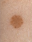
Papule
Elevated, firm, circumscribed area <1 cm (e.g., mole, wart).

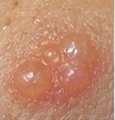
Vesicle
Elevated, circumscribed, fluid-filled <1 cm (e.g., chickenpox, shingles).
Patch
Flat, non-palpable, irregular-shaped macule greater than 1 cm in diameter (e.g., mongolian spots, vitiligo).

Plaque
Elevated, firm, and rough lesion with a flat top, with a surface area greater than 1 cm in diameter (e.g., psoriasis, eczema).

Wheal (Urticaria)
Elevated, irregular-shaped area of cutaneous edema; solid, transient, with a variable diameter (e.g., allergic reaction, insect bite).

Nodule
Elevated, firm, circumscribed lesion that extends deeper into the dermis than a papule; 1 to 2 cm in diameter (e.g., melanoma).

Tumor
Elevated and solid lesion; deeper in the dermis; greater than 2 cm in diameter (e.g., lipoma).

Bulla
Larger than 1 cm, superficial, thin-walled (e.g., drug reactions, blisters from severe burns).

Pustule
Elevated, superficial lesion similar to a vesicle, but filled with purulent fluid (pus) (e.g. acne, impetigo).

Cyst
Elevated, circumscribed, encapsulated lesion located in the dermis or subcutaneous layer; filled with liquid or semisolid material (e.g., cystic acne).

Secondary Lesion
Result from changes over time, evolution of a primary lesion, trauma, or healing
Scale
Heaped-up, keratinized cells; flaky skin; irregular; thick or thin; dry or oily; variable size (e.g., psoriasis, eczema).

Lichenification
Rough, thickened epidermis secondary to persistent rubbing, itching, or skin irritation (e.g., chronic dermatitis, psoriasis).

Keloid
Irregular-shaped, elevated, progressively enlarging scar that grows beyond the boundaries of the original wound caused by excessive collagen formation during healing (e.g., following surgical incisions or piercings).

Scar
Thin-to-thick fibrous tissue that replaces normal skin following an injury or laceration to the dermis (e.g., healed surgical wound or traumatic laceration).
Excoriation
Loss of the epidermis; linear, hollowed-out, crusted area (e.g., abrasions, scratches)

Fissure
Linear crack or break extending from the epidermis into the dermis; may be moist or dry (e.g., athlete’s foot, chapped hands).

Erosion
Loss of part of the epidermis; depressed, moist, glistened area; follows the rupture of a vesicle or bulla (e.g., candidiasis).
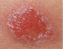
Ulcer
Loss of epidermis and dermis; concave shape; varies in size (e.g., pressure injuries).

Crust
Dried exudate, blood, or pus on the skin surface; slightly elevated; variable size; red-black, brown, tan, or yellow-mixed color (e.g., scabs on abrasions, impetigo).

Striae
Silver/pink "stretch marks" from rapid skin stretching.
Vascular Lesions
Result from blood leaking from blood vessels into the dermis/subcutaneous tissues or from dilated blood vessels
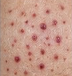
Petechiae
Tiny, flat, reddish-purple, non-blanchable spots in the dermal layer; 1 to 3 mm (or <0.5 cm) in size (e.g., bleeding disorders, tiny capillary hemorrhages due to infection)
Purpura
Flat, reddish-purple, non-blanchable skin discoloration greater than 0.5 cm (or >3 mm) in diameter (e.g., infection, bleeding disorders).

Ecchymosis (Bruise)
Reddish-purple, non-blanchable patch of variable size caused by trauma or bleeding under the skin; changes color (blue, green, yellow, brown) as it heals (e.g., trauma, physical injury).

Cherry Angioma (Angioma)
Small, smooth, slightly raised, bright red benign vascular lesion; ranges from tiny pinpoint up to 1–3 cm (e.g., benign tumor of small blood vessels)

Telangiectasia
Permanent dilation of preexisting small blood vessels (capillaries, arterioles, or venules) producing fine, irregular red line (e.g., rosacea, sun damage)
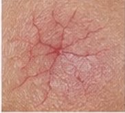
Spider Angioma (Vascular Spider)
Small, central red spot with radiating spider-like capillary legs; characteristically blanches when pressure is applied to the central core (e.g., pregnancy, liver disease)
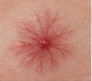
Capillary Hemangioma (Port-Wine Stain)
Vascular lesion involving capillaries that produces an irregular, flat or raised patch ranging from light red to dark purple (e.g., congenital vascular malformation of capillaries).
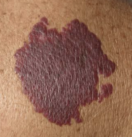
Venous Star
Non-blanching, bluish vascular star-shaped lesion that can be linear or irregularly shaped (e.g., increased pressure in superficial veins).

Stage 1 Pressure Injury
Intact skin with a localized area of non-blanchable erythema (redness that does not turn white when gentle pressure is applied).

Stage 2 Pressure Injury
Partial-thickness loss of skin with exposed dermis.
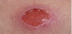
Stage 3 Pressure Injury
Full-thickness loss of skin; adipose (subcutaneous fat) tissue is visible in the ulcer, with granulation tissue and rolled wound edges (epibole) frequently present. Slough and/or eschar may be visible.

Stage 4 Pressure Injury
Full-thickness skin and tissue loss with directly visible or palpable bone, tendon, cartilage, ligament, or muscle.

Unstageable Pressure Injury
Full-thickness skin and tissue loss in which the extent of tissue damage cannot be confirmed because it is obscured by slough or eschar.
Deep Tissue Pressure Injury (DTPI)
Intact or non-intact skin with localized area of persistent, non-blanchable, deep red, maroon, or purple discoloration, or epidermal separation revealing a dark wound bed or blood-filled blister.
Braden Scale
Analyzes:
Sensory Perception 2. Moisture 3. Activity 4. Mobility 5. Nutrition 6. Friction and Shear.
Significance: Total score ranges from 6 to 23. A lower score indicates a higher risk for pressure ulcer development.
Melanoma Screening
A - Asymmetry: One half does not match the other half.
B - Border: Ragged, notched, or blurred edges.
C - Color: Variation in shades (brown, black, tan, red, or blue).
D - Diameter: Greater than 6 mm (size of a pencil eraser).
E - Evolving: Changing in size, shape, or color over time.
Color Variations Across Skin Tones
Pallor, Cyanosis, Erythema, Jaundice
Pallor
Light Skin Tone: ghostly white/pale
Dark Skin Tone: ashen/grey; check mucous membranes or conjunctiva
Cyanosis
Light Skin Tone: blueish tinge (lips, nail beds)
Dark Skin Tone: grayish/dull; check palms, soles, and oral mucosa
Erythema
Light Skin Tone: red/bright pink
Dark Skin Tone: purplish tinge; palpate for increased warmth/edema
Jandice
Light Skin Tone: yellow skin and sclera
Dark Skin Tone: yellowing most visible in hard palate and sclera
Lesion Shapes and Patterns
Annular: Circular (e.g., ringworm).
Zosteriform: Linear arrangement following a nerve dermatome (e.g., Herpes Zoster).
Polycyclic: Annular lesions that grow together (e.g., psoriasis).
Physical Exam Checklist of Skin
Inspection: Observe for color (congruency with genetic background), texture, moisture, and integrity (lesions/breakdown).
Palpation:
Temperature: Use the dorsal aspect of the hands.
Moisture: Diaphoresis vs. dehydration.
Texture: Smooth and firm.
Mobility/Turgor: Tested over the clavicle; "tenting" indicates dehydration.
Normal v. Abnormal Findings Nails
Normal Findings: Capillary refill <3 seconds; 160-degree nail base angle.
Abnormal Findings:
Clubbing: Angle >180 degrees. Indicates chronic hypoxia (COPD, congenital heart disease).
Beau's Lines: Transverse depressions (systemic illness/trauma).
Konylochia: "Spoon nails" (iron deficiency).
Hair Physical Exam
Normal Findings: Vellus (fine, faint body hair) vs. Terminal (darker, thicker scalp/axillary hair).
Abnormal Findings:
Alopecia: Significant hair loss.
Hirsutism: Excessive hair growth in women, often following male-pattern distribution.
Head Terminology
Normocephalic: Round, symmetric skull proportionate to body.
Hydrocephalus: Fluid accumulation causing head enlargement.
Microcephaly: Abnormally small head
Macrocephaly: Abnormally large head.
Facial Muscle Abnormalities
Bell’s Palsy (CN VII): Lower motor neuron lesion; total paralysis of half the face (cannot wrinkle forehead on affected side).
Stroke (CVA): Upper motor neuron lesion; paralysis of lower face only (can still wrinkle forehead due to bilateral innervation).
Red Flags: Lumps (potential malignancy), "Worst headache of life" (aneurysm/hemorrhage), and nuchal rigidity (meningitis).
Normal v. Abnormal Findings Lymph Nodes
Normal Lymph Nodes | Abnormal (Lymphadenopathy) |
|---|---|
Nonpalpable (often) | Large (>1 cm) |
Soft, mobile | Hard, fixed (suggests malignancy) |
Nontender | Tender (suggests infection) |
Thyroid & Neck Assessment
Technique: Use posterior or anterior approach. Ask patient to take a sip of water; the thyroid should move up with swallowing.
Hyperthyroidism: Symptoms include tachycardia, weight loss, heat intolerance, exophthalmos (bulging eyes).
Hypothyroidism: Symptoms include bradycardia, weight gain, cold intolerance, periorbital edema.
Visual Clinical Tests
Snellen Chart: Standardized at 20 feet. "20/40" means the patient sees at 20 feet what a normal eye sees at 40 feet. Used for far vision.
Rosenbaum: Used for near vision (14 inches).
Hirschberg (Corneal Light Reflex): Light should reflect in the same spot on both corneas. Asymmetry indicates muscle weakness.
6 Cardinal Fields of Gaze: Tests CN III, IV, and VI for conjugate movement and nystagmus.
Cover/Uncover Test: Detects small degrees of deviated alignment. If the uncovered eye jumps to fixate, it was previously out of alignment (tropia/phoria).
PERRLA Guide
P - Pupils: Observe shape and size (mm).
E - Equal: Compare left and right pupils.
R - Round: Ensure circularity.
RL - Reactive to Light:
Direct: Pupil constricts in eye with light.
Consensual: Other pupil constricts simultaneously.
A - Accommodation: Ask patient to focus on a distant object, then shift to a near object. Pupils should constrict and eyes should converge.
Normal v. Abnormal Findings Eyes
Structure | Normal Findings | Abnormal Findings |
|---|---|---|
Sclera | White | Yellowing (Icterus) |
Cornea | Clear, smooth | Cloudy, abraded (steamy) |
Pupils | 3-5mm, symmetric | Anisocoria (unequal size) |
Eyelids | Symmetrical | Ptosis (drooping), Lid lag |
Nystagmus | None | Oscillating movement (Red Flag if not lateral) |
Eye Abnormalities
Miosis: Constricted/fixed pupils.
Mydriasis: Dilated/fixed pupils.
Anisocoria: Naturally occurring unequal pupil size (found in ~5% of population).
Conditions: Cataracts (lens opacity), Glaucoma (increased intraocular pressure), Conjunctivitis (pink eye/erythema).
Tympanic Membrane Normal Findings
Tympanic Membrane (TM): Should be Pearly Gray. Landmarks include the Umbo, Malleus, and the Cone of Light (5 o'clock in right ear, 7 o'clock in left)
Hearing Tests
Whisper Test: High-frequency sound check.
Weber: Tuning fork on midline skull; sound should be equal bilaterally.
Rinne: AC (Air Conduction) should be > BC (Bone Conduction) 2:1.
Ear Conditions
Otitis Media: Middle ear infection (red, bulging TM).
Otitis Externa: "Swimmer's ear" (pain with movement of pinna/tragus).
Equilibrium Test
Romberg Test: Stand with feet together, eyes closed. Minimal swaying is normal. Significant loss of balance is a positive Romberg (indicates vestibular/proprioception issues).
Tonsil Grading Scale
1+: Visible.
2+: Halfway between tonsillar pillars and uvula.
3+: Touching the uvula.
4+: Touching each other ("kissing" tonsils)
Rhinologic Assessment
Nose: Check patency of nares.
Allergic Rhinitis: Pale, boggy, gray turbinates.
Common Cold: Bright red, swollen turbinates.
Sinuses: Palpate Frontal (above eyebrows) and Maxillary (cheekbones).
Palpation & Auscultation of Respiratory System
Tactile Fremitus: Palpate "99"; vibrations should be symmetric.
Ladder Pattern: Systematically move from side-to-side to compare lobes.
10 posterior sites, 8 anterior sites, 7 lateral sites.
Adventitious Sounds & Disease Exemplars
Crackles (Rales): Discontinuous popping (Pneumonia, Heart Failure).
Wheezes: High-pitched musical squeaking (Asthma, Emphysema).
Rhonchi: Low-pitched "snoring" sound (Bronchitis; may clear with cough).
Stridor: High-pitched crowing (Upper airway obstruction - Emergency).
Pleural Friction Rub: low-pitches, grating sound (Pericarditis)
Diminished: quieter (COPD)
CN I
Name: Olfactory
Type: Sensory
Assessment: Smell identification
CN 2
Name: Optic
Type: Sensory
Assessment: Snellen Chart
CN 3
Name: Oculomotor
Type: Mixed
Assessment: PERRLA; EOM (up, middle, down)
CN 4
Name: Trochlear
Type: Motor
Assessment: EOM (down and inward)
CN 5
Name: Trigeminal
Type: Mixed
Assessment: Clench teeth; light touch to face
CN 6
Name: Abducens
Type: Motor
Assessment: EOM (lateral movement)
CN 7
Name: Facial
Type: Mixed
Assessment: Smile, frown, puff cheeks
CN 8
Name: Vestibulocochlear
Type: Sensory
Assessment: Whisper test; Weber/Rinne
CN 9
Name: Glossopharyngeal
Type: Mixed
Assessment: Gag reflex; “Ahhh” (uvula rise)
CN 10
Name: Vagus
Type: Mixed
Assessment: Swallowing and speech quality
CN 11
Name: Accessory
Type: Motor
Assessment: Shrug shoulders; turn head against resistance
CN 12
Name: Hypoglossal
Type: Motor
Assessment: Stick out tongue
Bradypnea
Rate & Depth Characteristics: Regular rhythm with a rate slower than 12 breaths per minute
Clinical Significance: Can be caused by neurological injury, medication/sedation, or respiratory depression
Tachypnea
Rate & Depth Characteristics: Rapid, shallow breathing at a rate greater than 20 breaths per minute
Clinical Significance: Occurs with fear, fever, exercise, acute respiratory distress, or hypoxemia
Hyperventilation
Rate & Depth Characteristics: Rapid, deep breathing at a rate greater than 20–24 breaths per minute
Clinical Significance: Expels excessive CO2 leading to hypocarbia and respiratory alkalosis; caused by exertion, anxiety, pain, or metabolic disorders
Hypoventilation
Rate & Depth Characteristics: Irregular, shallow respirations at a rate below expected limits
Clinical Significance: Leads to carbon dioxide retention (hypercarbia); caused by overdose, airway obstruction, or sedation
Sighing
Rate & Depth Characteristics: Frequently interspersed deeper breaths within a normal pattern
Clinical Significance: Occasional sighing expands alveoli; frequent sighing indicates hyperventilation or altered emotional/respiratory status
Cheyne-Stokes
Rate & Depth Characteristics: Cyclic pattern of gradually increasing depth, followed by decreasing depth, and periods of apnea
Clinical Significance: Indicates severe CNS hypoxia, brain damage, or end-of-life respiratory failure
Kussmaul
Rate & Depth Characteristics: Rapid, deep, labored breathing without expiratory pauses
Clinical Significance: Compensatory mechanism for severe metabolic acidosis, most commonly diabetic ketoacidosis
Biot (Ataxic)
Rate & Depth Characteristics: Disorganized pattern with irregular depths and unpredictable periods of apnea
Clinical Significance: Caused by severe brain injury, meningitis, or profound CNS depression
Air Trapping
Rate & Depth Characteristics: Pattern of increasing difficulty in exhaling air
Clinical Significance: Caused by chronic airway resistance in conditions like COPD and emphysema
Normal Adult Thorax
Anteroposterior to Transverse Ratio 1:2
Barrel Chest
1:1 Ratio; caused by hyperinflation of lungs
Funnel Chest
Sunken or depressed sternum; congenital structure
Pigeon Chest
Forward protrusion of the sternum; congenital structure
Scoliosis
Lateral S-shaped curvature of the thoracic and lumbar spine; restricts chest expansion
Kyphosis
“hunchback” ; common in older adults
Infant Thorax
1:1
Acute Bronchitis
Etiology: Inflammation of bronchial mucous membranes
Clinical Signs: Coughing progressing to green/yellow sputum
Assessment Findings: Rhonchi or wheezes on auscultation, which often clear or shift after coughing
Emphysema/COPD
Etiology: Permanent destruction of alveolar walls leading to decreased gas exchange
Clinical Signs: underweight, barrel chest, SOB, pursed-lip breathing, tripod position
Assessment Findings: Diminished breath sounds, wheezes, prolonged expiration, hyperresonance on percussion
Asthma
Etiology: Hyperreactive airway disease causing bronchoconstriction, airway inflammation, and edema
Clinical Signs: dyspnea, tachycardia, tachypnea with prolonged expiration, wheezes, accessory muscle use, cough, diminished breath sounds
Assessment Findings: High-pitched musical wheezes
Pneumonia
Etiology: Infection of terminal bronchioles and alveoli causing exudative consolidation
Clinical Signs: Fever, chills, dyspnea, tachypnea, tachycardia, pleuritic chest pain, cough producing yellow/green or rust-colored sputum
Assessment Findings: Fine/coarse crackles, increased tactile fremitus, dullness to percussion
Pneumothorax
Etiology: Air accumulation in the pleural space causing partial or complete lung collapse
Clinical Signs: Absent breath sounds, sudden sharp chest pain, tachycardia, tracheal displacement
Pleural Effusion
Etiology: Accumulation of serous fluid in the pleural space
Clinical Signs: Dyspnea, sharp pleuritic chest pain (worse with deep inspiration/cough), tachypnea, tachycardia
Clinical Sings: Absent or decreased breath sounds over fluid level, dullness to percussion, decreased tactile fremitus, pleural friction rub
Lung Cancer
Etiology: Malignant tumor growth within lung tissue
Clinical Signs: Persistent cough, hemoptysis, dyspnea, chest pain, unintended weight loss, fatigue, hoarseness
Assessment Findings: Localized wheezing, diminished or absent breath sounds distal to tumor obstruction