Lecture 3 - Bacterial Cell Structure (PART 1/SLIDES 0-69))
 Call Kai
 Learn
Learn Practice Test
 Spaced Repetition
 Match
Match Flashcards
 Knowt Play
1/39

There's no tags or description
Looks like no tags are added yet.
Name | Mastery | Learn | Test | Matching | Spaced | Call with Kai | Chat |
|---|
No analytics yet
Send a link to your students to track their progress
40 Terms
Microbe of the day: Agrobacterium tumefaciens
Agrobacterium tumefaciens
Type: alpha-proteobacterium (related to Rhizobia)
Disease: Causes crown gall disease (tumor-like growths in plants)
Key Factor: Uses the Ti (tumor-inducing) plasmid to modify plant behavior to support bacterial growth
Biotech Use: Modified by scientists as the main tool for plant genetic engineering

What criteria determines if a species is a Prokaryote?
Lack of membrane-bond nucleus (still true).
What outdated and partially correct criteria determines if a species is a Prokaryote?
Lack of cytoskeleton (false; bacteria and archaea have cytoskeletons).
Lack of membrane-bound organelles (false; some have internal membrane structures).
Lack of internal membranous structures (mostly true, but not always).
Why did Norman Pace believe that the domain “Prokaryotes” should be abandoned in 2006 in favor of the specific terms "Bacteria" and "Archaea."?
Bacteria and Archaea have significant differences.
Archaea share a more recent common ancestor to Eukaryotes than Bacteria do.
Definition is vague; criticizes the absence of organelles, instead of distinct descriptions.
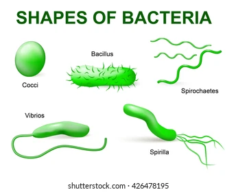
Bacteria Cell Morphology: What 5 shapes can bacteria cells be?
Cocci (spherical)
Bacilli / Rods (oblong)
Spirillium (Spiral)
Spirochete (spiral/corkscrew)
Vibrio (comma-shaped)
Bacteria Morphology: What are the 5 ways cocci bacteria cells can be arranged / filaments (cells remain attached after division)?
Diplococci: Pairs.
Streptococci: Chains (division in one plane).
Staphylococci: Grape-like clusters (division in random planes).
Tetrads: Groups of four (division in two planes).
Sarcinae: Cubic packets of eight (division in three planes).

Bacteria Morphology: What are the 3 unique bacteria cells shapes?
Spirochetes (flexible spiral)
Mycelium (network of filaments)
Pleomorphic (variable shapes).
What are the typical sizes of bacteria and examples?
Small = 0.3 μm
• Mycoplasma
Average = 1.1 to 1.5 μm wide by 2 to 6 μm long
• E. coli, Bacillus
Very large bacteria
• Epulopiscium fishelsoni (600 by 80 μm)
• Thiomargarita namibiensis (up to 1 cm long)
Thiomargarita magnifica(up to 2 cm long).
Ultramicrobacteria: Candidatus Saccharibacteria (<0.1 μμm).

What relationship was seen between size and shape in cells?
Cells tend to be small to maintain a high surface area-to-volume ratio (S/V ratio), which increases the efficiency of nutrient uptake and diffusion. Larger sizes or odd shapes may offer protection.
Bacterial Structures: What makes up the cellular envelope (layers surrounding the cell)?
Plasma membrane.
Cell wall (peptidoglycan, glycocalyx).
Outer membrane (in Gram-negatives) or mycomembrane (in mycobacteria).
Some have external layers (capsule, slime layer, S layer)

Bacterial Structures: What makes up the cytoplasm (internal contents)?
Nucleoid (contains chromosome).
Ribosomes.
Microcompartments (e.g., magnetosomes, carboxysomes).
Bacterial Structures: What are external structures of bacterial cells?
Pilli.
Flagella.
Secretion systems.
What is the Function of the plasma/cytoplasmic membrane?
Selectively permeable barrier for nutrient uptake and waste removal.
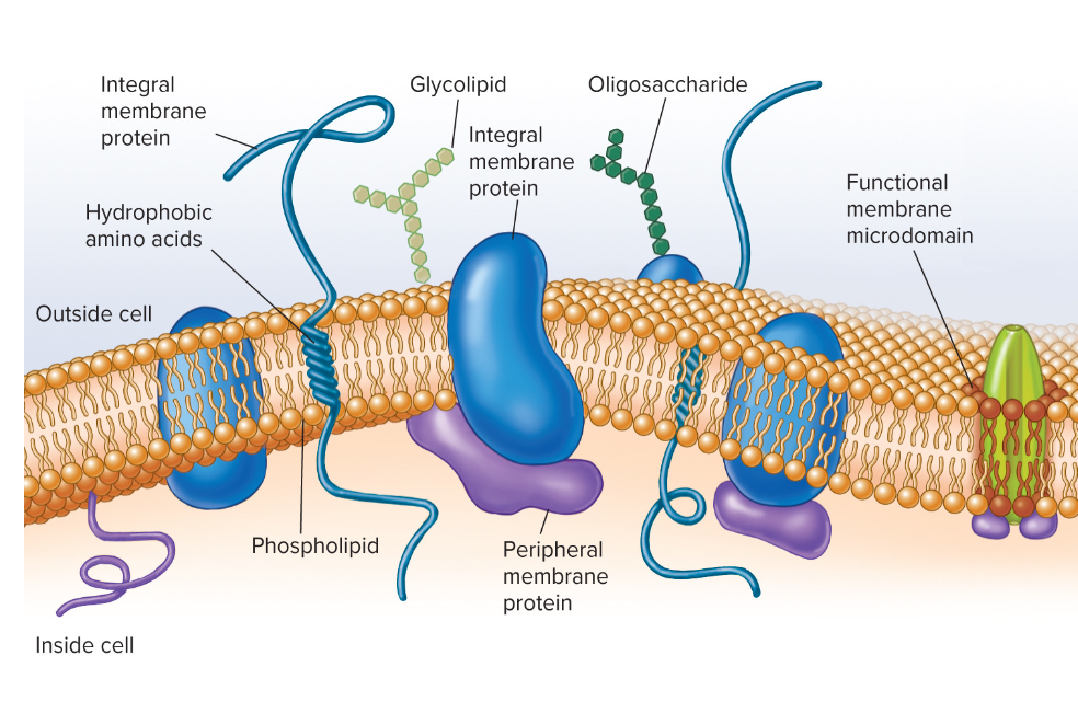
What is the structure of the plasma membrane? Does it form spontaneously (Dynamic)?
A thin (7-8 nm) lipid bilayer composed of amphipathic phospholipids.
Hydrophilic polar heads (interact with water) and hydrophobic non-polar fatty acid tails/R1 and R2 (insoluble in water, interact with each other) that forms spontaneously.

How can fatty acid tails on the phospholipid bilayer effect the plasma membranes permeability?
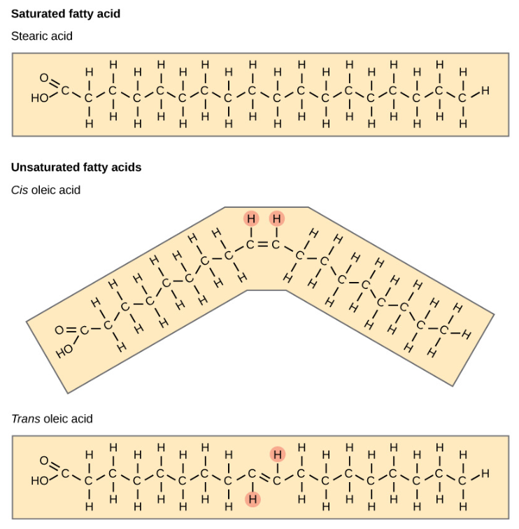
Saturation: No double bonds, tightly packed, less fluid.
Unsaturated: Double bonds, linked, more fluid.
What are Haponoids?
Hydrophobic molecules similar to cholesterol (found in eukaryotic membranes) found in 10% of bacterial membranes, affecting fluidity and shape.
They can form functional membrane microdomains, useful for protien assembly.

What are the 2 types of proteins that span across the membrane?
Integral proteins: Embedded; hydrophobic regions interact with lipids and hydrophilic regions exposed, involved in signaling and transport.
Peripheral Proteins: Loosely attached and easily removed.

What are the 2 types of nutrients microbes intake? What is the role of their nutrients?
Macronutrients: Required in large amounts.
Found in organic molecules (proteins, lipids, nucleic acids, carbohydrates).
Usually contain cations (e.g., K++, Ca2+2+, Mg2+2+, Fe2+2+) that are important as enzyme cofactors, for protein folding, and cell structure.
Micronutrients: Required in small amounts.
Often act as enzyme cofactors or assist in protein structure (e.g., Mn, Zn, Co, Mo, Cu).
What is the difference between essential and non-essential nutrients?
Non-essential: Microbes can synthesize these from simpler precursors, typically from macromolecules, don’t need to specifically eat them.
Essential: Microbes cannot synthesize these and must obtain them from the environment (e.g. humans needs vitamin C)
What is biosynthesis?
The biological process where living organisms convert simple precursor molecules into complex biological compounds (such as proteins, fats, nucleic acids, and hormones) using cellular energy and enzymes.
If your body lacks the genes or enzymes required for that specific biosynthetic pathway, it cannot manufacture the molecule itself—making that compound an essential nutrient that must be supplied by what you eat.
What are the 5 mechanisms of molecule transport across the plasma membrane?
Passive diffusion
Facilitated diffusion
Primary active transport
Secondary active transport
Group translocation
What is passive diffusion?
Spontaneous movement of molecules from high to low concentration, requiring a significant concentration gradient. The rate decreases as intracellular concentration increases.

What is facilitated diffusion?
Movement from high to low concentration, aided by transport proteins (channel proteins or carrier proteins). This process is not energy-dependent, rate increases with the concentration gradient (initially faster than passive), but the rate can plateau due to carrier saturation.

What is active transport?
Movement of molecules against the concentration gradient, requiring energy (ATP hydrolysis or proton motive force).
What role do carrier proteins play in active transport?
They facilitate the movement of molecules and control the overall rate of transport across the membrane.
What is primary active transport?
A process that directly uses energy from ATP hydrolysis to move substances against their concentration gradient without modifying them.
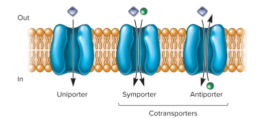
What is a Uniporter?
A carrier protein involved in primary active transport that moves a single type of molecule across the membrane.
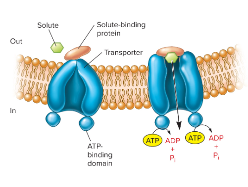
What are ATP-binding cassette (ABC) transporters?
Common primary active transport uniporters containing:
• two hydrophobic membrane spanning
domains.
• two cytoplasmic ATP-binding domains.
What is Secondary Active Transport?
A transport process that utilizes the potential energy stored in ion gradients to co-transport an ion and solute across the membrane without modifying them.
What is Symport?
A mechanism of secondary active transport where the ion and solute are moved across the membrane in the same direction.

What is Antiport?
A mechanism of secondary active transport where the ion and solute are moved across the membrane in opposite directions.

What is Group Translocation? What is a common example?
Energy dependent transport that chemically modifies (only this) the molecule as it is brought into cell.
Ex. Phosphoenolpyruvate: sugar phosphotransferase system (P T S): Imports sugars while phosphorylating them.
How does Bacteria Uptake Iron?
Ferric Iron (Fe^3+) is essential but insoluble, so they secrete siderophores that chelate and transport it into the cell through high-affinity transport systems.

What are the 3 functions of the bacterial cell wall?
Maintains shape.
Protects from lysis and toxic materials.
Contributes to pathogeny.
What is the bacterial cell wall made out of?
Peptidoglycan (murein): Rigid structure lying outside the plasma membrane.
What is the structure of peptidoglycan?
Two alternating sugars:
• N-acetylglucosamine (NAG).
• N- acetylmuramic acid (NAM).
Each disaccharide also contains a short
peptide attached to the NAM.

What is the structure of peptidoglycan 2D? What are the 2 ways it is formed?
Adjacent peptidoglycan chains are directly cross-linked or indirectly cross-linked by a peptide interbridge forming a strong helical 3D mesh.

Why is important for the peptidoglycan layer to be 3D?
Structural strength.
Creates varying sized pores.
Makes it flexible and permeable.

What is the 5-step process of peptidoglycan biosynthesis? (not sure if you really need to know this)
Cytoplasmic Precursor Synthesis
UDP-GlcNAc is converted into UDP-MurNAc by MurA and MurB.
Mur enzymes (MurC through MurF) sequentially attach amino acids to form a pentapeptide tail ending in D-Ala-D-Ala.
Membrane Anchor Assembly
MraY links the UDP-MurNAc-pentapeptide to the lipid carrier undecaprenyl pyrophosphate in the inner membrane, creating Lipid I.
MurG attaches GlcNAc to Lipid I, creating the full disaccharide precursor, Lipid II.
Translocation
Lipid II is translocated (flipped) across the plasma membrane from the cytoplasmic side to the outer cell wall side.
Transglycosylation
Transglycosylase enzymes extend the sugar backbone by connecting the new GlcNAc-MurNAc disaccharide units into long glycan chains.
Transpeptidation
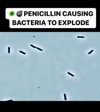
Penicillin-Binding Proteins (PBPs) create cross-links between peptide chains of adjacent glycan strands, releasing the terminal D-Alanine to give the cell wall its final structural strength.

Why is peptidoglycan synthesis a common antibiotic target?
Many PG biosynthesis enzymes are essential. Inhibiting these enzymes can lead to bacterial cell death.