Integumentary System
 Call Kai
 Learn
Learn Practice Test
 Spaced Repetition
 Match
Match Flashcards
 Knowt Play
1/28

There's no tags or description
Looks like no tags are added yet.
Name | Mastery | Learn | Test | Matching | Spaced | Call with Kai | Chat |
|---|
No analytics yet
Send a link to your students to track their progress
29 Terms
Epidermis
Composed of epithelial tissue
Completely replaced every 4-6 weeks

Dermis
Composed of connective tissue

Hypodermis
AKA: Subcutaneous layer
Also connective tissue, but different composition than dermis
Connects skin to the underlying fascia
Fascia: fibrous covering of muscles and organs
“Tied” to other layers by the interweaving of collagen fibers between hypodermis and fascia
Well-vascularized, loose areolar connective tissue and adipose tissue
Functions include fat storage, insulation and cushioning

Order of stratum top →bottom
Come → Stratum corneum
Lets → Stratum lucidum
Get → Stratum granulosum
Sun → Stratum spinosum
Burned → Stratum basale

Stratum corneum
The outermost layer of the skin
Exposed to the environment
15-30 layers of dead skin cells
Prevents penetration of microbes
Prevents dehydration of underlying tissues
Shed periodically, replaced every 4 weeks
Stratum lucidum
Reduces friction between stratum corneum and stratum granulosum
Found in palms of hands and soles of feet
Referred to as “thick” skin
Stratum granulosum
As the keratinocytes are pushed from the stratum spinosum, the cells become flatter, cells membranes thicken, and they accumulate large amounts of keratin
Characterized by the presence of granules, which are intracellular protein-filled vesicles.
Some are filled with the precursor to keratin, keratohyalin
Melanosomes are other granules filled with melanin
Stratum spinosum
Contains young keratinocyte cells
Widest at center, tapered at ends
Cells are joined to each other through desmosomes
Contains a dendritic cell called the Langerhans cell
Functions similarly to macrophages
Engulf bacteria, foreign particles, and damaged cells
Stratum basale
Deepest layer of the epidermis
Made up of a SINGLE layer of cuboidal cells, responsible for continuously regenerating skin
Contains stem cells that produce keratinocytes
Basal stem cells → keratinocytes
Attaches the epidermis to the “basement membrane”
Closest to the blood supply

Keratinocytes
Manufacture and store short filaments of the protein keratin
Keratinocytes begin the synthesis of keratin AND a water-repelling glycolipid that makes skin waterproof
Keratin: an intracellular fibrous protein that gives hair, nails, and skin their hardness and water resistant properties

Birth of Cells
Cells move superficially as they age
New cells are “born” through mitosis in the basal layer
As they mature they push up to the apical layer as cells are lost to friction
Epidermis is epithelial so avascular (no blood vessels)
Moving away from nutrients as it ages
Die in the corneum and fall
Basement Membrane
Ties the epidermis to the dermis below
Made up of a dense, sheet-like form of extracellular matrix
Critical for stem cell formation
Vertiligo: a skin condition that causes your skin to lose its color or pigment
Some research suggests pigment producing cells have difficulty attaching to basement membrane
Merkel cell
Sensory receptor, signals to brain
Concentrated on surfaces of hands and feet
Melanocyte
melanin
Pigment producing cell
Protects from UV radiation damage
Melanocytes are dendritic cells with cellular extensions (dendrites) that allow them to contact keratinocytes
Up to 40 at a time
Melanin granules protect the DNA of epidermal cells from UV ray damage
Prevents the breakdown of folic acid
Environments with less sun exposure suffer from insufficient production of Vitamin D
Vitamin D: Involved in calcium absorption
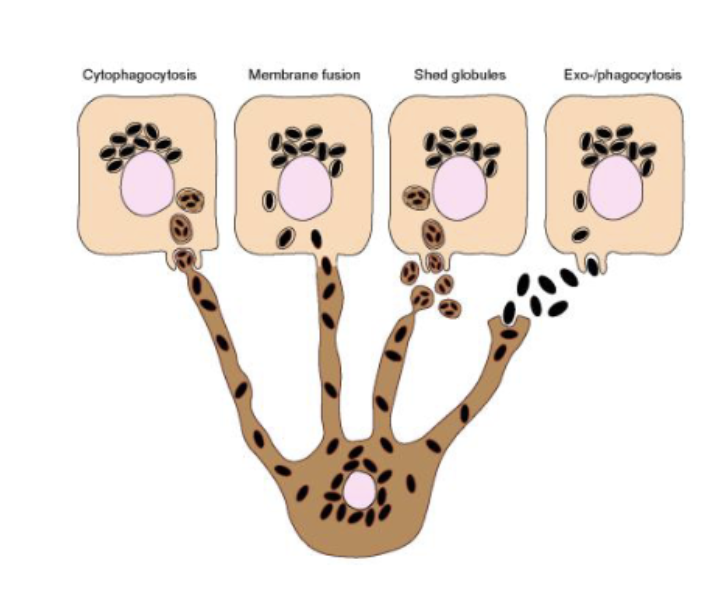
Desmosomes
strong connections that hold cells together
The Journey of Keratinocytes
Originate in basale layer, plenty of nutrients to grow
Keratinocytes connect to each other using desmosomes and produce keratin
More tightly connected with visible molecules in the cell’s cytoplasm
Cells are too far away from nutrients, no longer alive
Dead cells are filled with keratin pushed to the surface

The Dermis
The underlying connective tissue layer
Contains blood, lymph vessels, nerves, follicles and sweat glands
Composed of 2 layers of connective tissue proper, with interconnected elastin and collagenous fibers (from fibroblast)
Collagen fibers
provide structure and tensile strength
Binds water
Elastin fibers
provide elasticity to the skin
Dermis: Stretch Marks
Skin affected by rapid growth
Stretch marks result when the dermis is stretched beyond its limits of elasticity
“Marks” are due to collagen fibers breaking

Dermis: Papillary Layer
Made up of loose areolar connective tissue
Forms the papillae that project into the stratum basale
Papillary layer contains
Fibroblasts, blood vessels, nerve fibers, fine collagen fibers
Dermis: Reticular Layer
Much more substantial than papillary layer
Composed of dense irregular connective tissue
Dense weave of thick collagen fibers
Vascularized with sensory and sympathetic nerves
Hypodermis: What is Fat?
Adipose tissue (fat) is found in the hypodermis
Thickness varies greatly from one person to another
Aids in thermoregulation
Adults have white fat
Infants have brown fat
Excess calories are stored as subcutaneous fat (hypodermis) or surrounding our organs (visceral)
Hair shaft
Long shaft of keratin
Exposed at the skin’s surface
Hair root
Below surface section
Arrector pilli
Muscle that moves hair shaft
Hair bulb
Epithelial cells, similar to stratum basale
Rather than grow, keratinocytes fill themselves with keratin until they die
Hair papilla: connective tissue with blood vessels and nerves
Unique Hair Love
Hair grows from the bulb within follicle
The hair we see is just the outer layer of the cuticle
Pigments are produced in the bulb
Textures across ethnicities/regions, determined by medulla

Hair Damage
Healthy cuticle is smooth, shiny, and closed with intact scales
Damaged cuticles are rough, with raised scales, allows water to enter
Frizz is caused by swelling in the hair due to excess moisture entering hair
