Lab Exam 1 for A&P2
 Call Kai
 Learn
 Practice Test
 Spaced Repetition
 Match
 Flashcards
 Knowt Play
1/93

There's no tags or description
Looks like no tags are added yet.
94 Terms
Endocrine glands (purely endocrine function)
anterior pituitary gland, thyroid, adrenals, parathyroids
Endocrine organs (mixed function)
pancreas, hypothalamus, gonads, thymus
Anterior Pituitary Gland
“master endocrine gland” (GH,PRL,ACTH,TSH,FSH,LH)
Posterior Pituitary Gland
labor and blood pressure (oxytocin and ADH)
Thyroid
metabolism and calcium levels (T4, T3, calcitonin, PTH)
Adrenal Cortex
long-term stress (aldosterone, cortisol, androgens)
Adrenal Medulla
short-term stress (epinephrine and norepinephrine)
Pancreas
blood sugar (insulin and glucagon)
Pineal Gland
circadian cells (melatonin)
Thymus
immune (thymosin)
Ovaries and Testes
puberty and reproduction (estrogen, progesterone, testosterone)
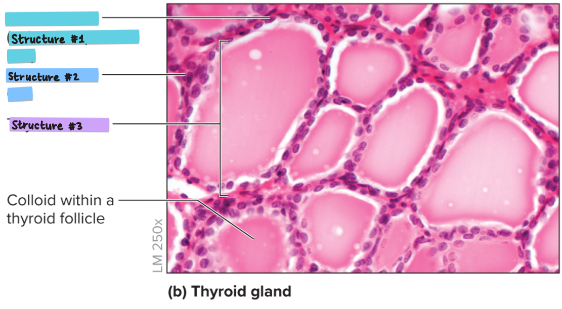
What is structure #3?
thyroid follicle
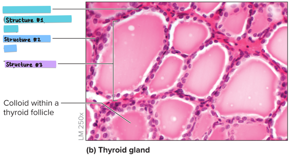
What is structure #1?
follicular cells (secretes TH like T3 and T4)
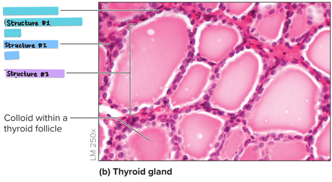
What is structure #2?
parafollicular cells (secretes calcitonin)
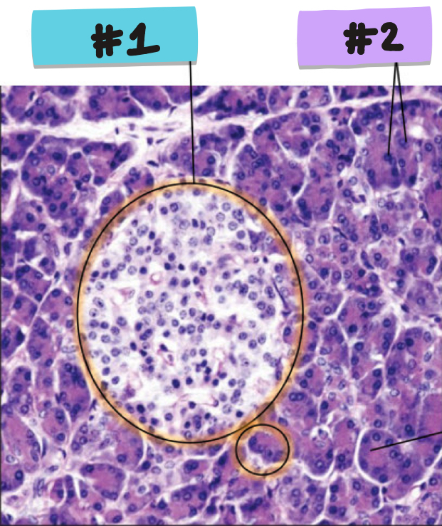
What is #2?
acinar cells
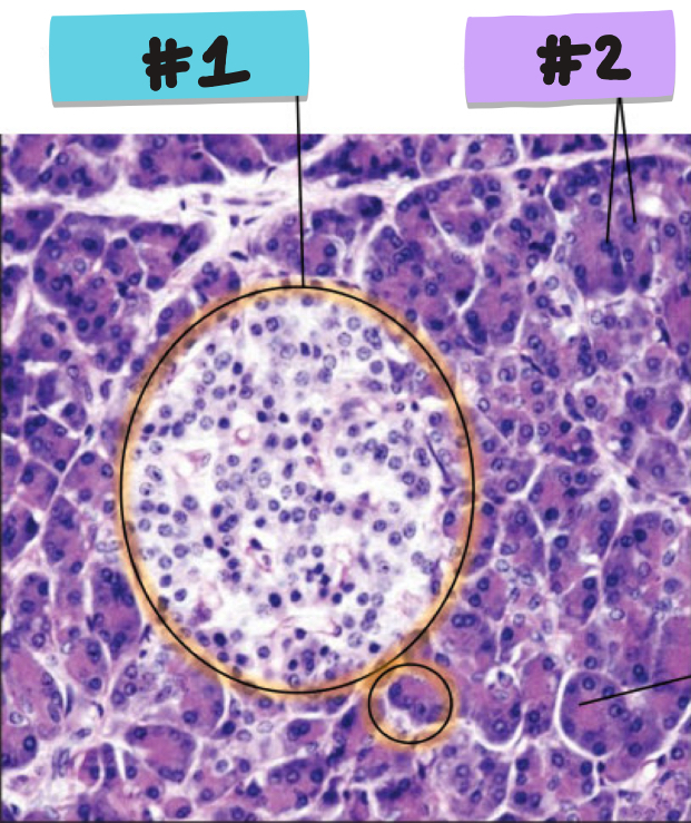
What is #1?
pancreatic islet (contains alpha and beta cells)
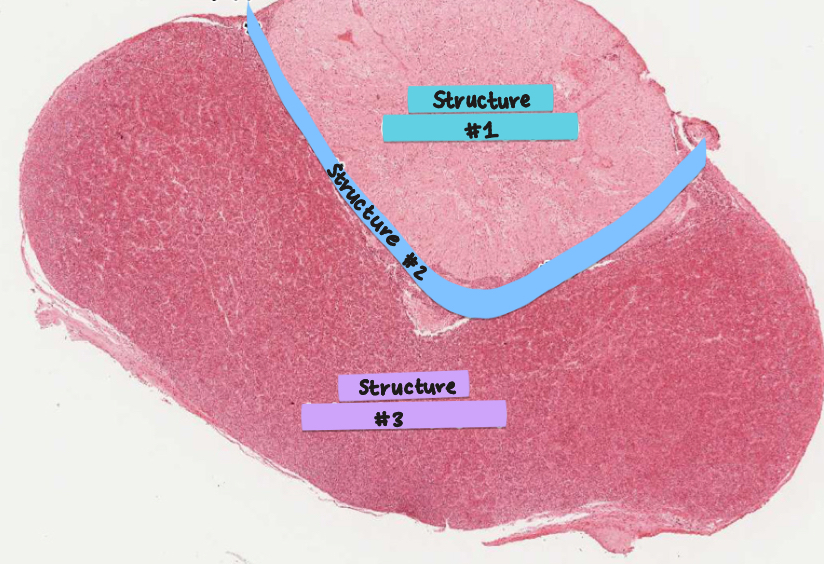
What is #1?
Pars nervosa (neurohypophysis)- nervous tissue, posterior pituitary gland, releases ADH and oxytocin from hypothalamus
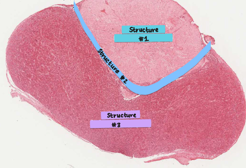
What is #2?
Pars intermedia (between 2 parts of pituitary, may not be visible) – produces melanocyte stimulating hormone (MSH) and ACTH
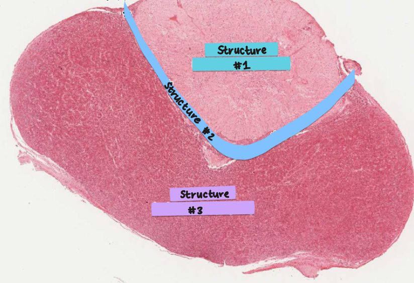
What is #3?
Pars distalis (adenohypophysis) - glandular tissue, anterior pituitary gland, produces GH, LH, FSH, Prolactin, TSH, ACTH
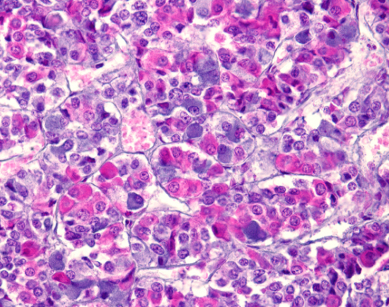
What histology is this?
anterior pituitary histology
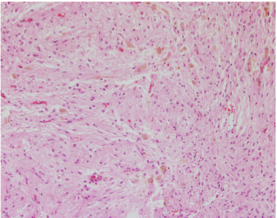
What histology is this?
posterior pituitary histology
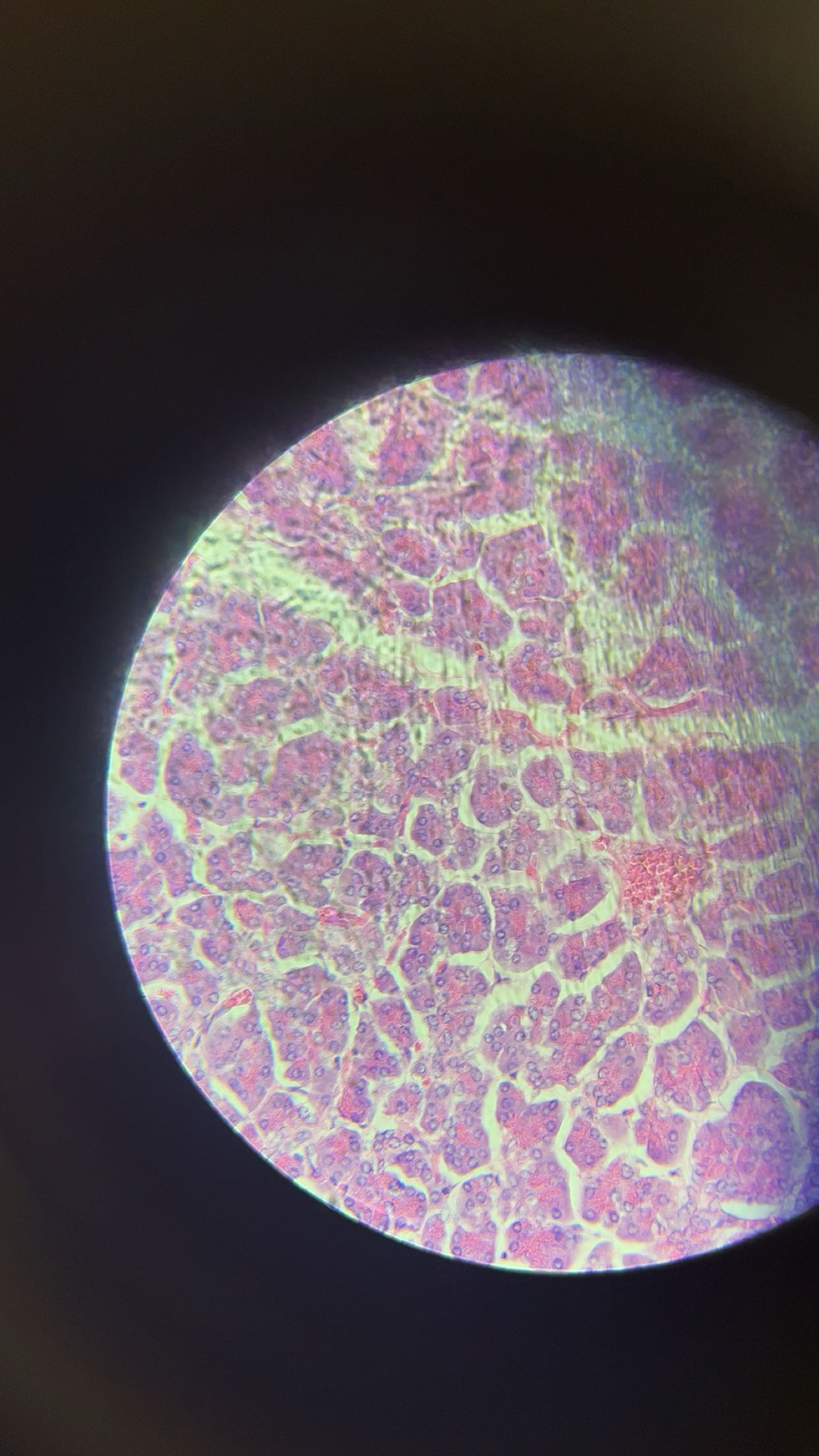
The structure at the tip of the pointer in microscope #1 produces:
insulin and glucagon
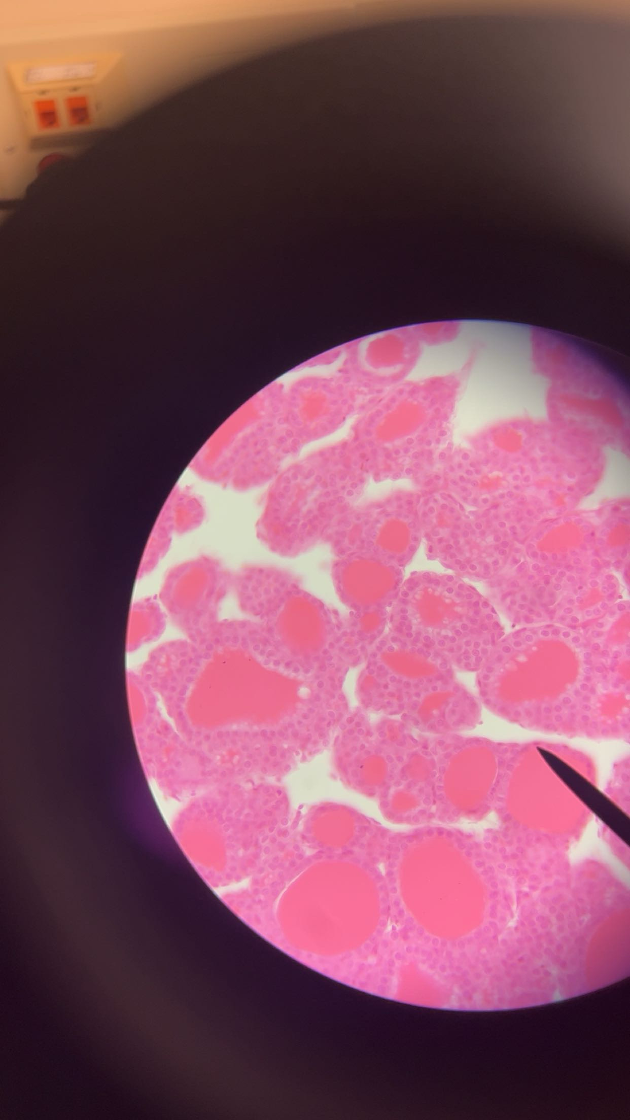
The structure at the tip of the pointer in microscope #2 produces:
thyroid hormone
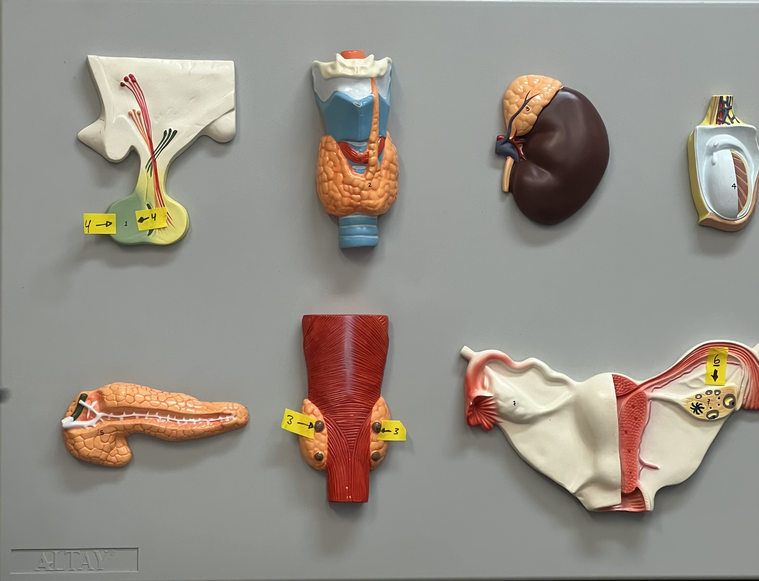
Identify the structure labelled “3”
parathyroid hormone
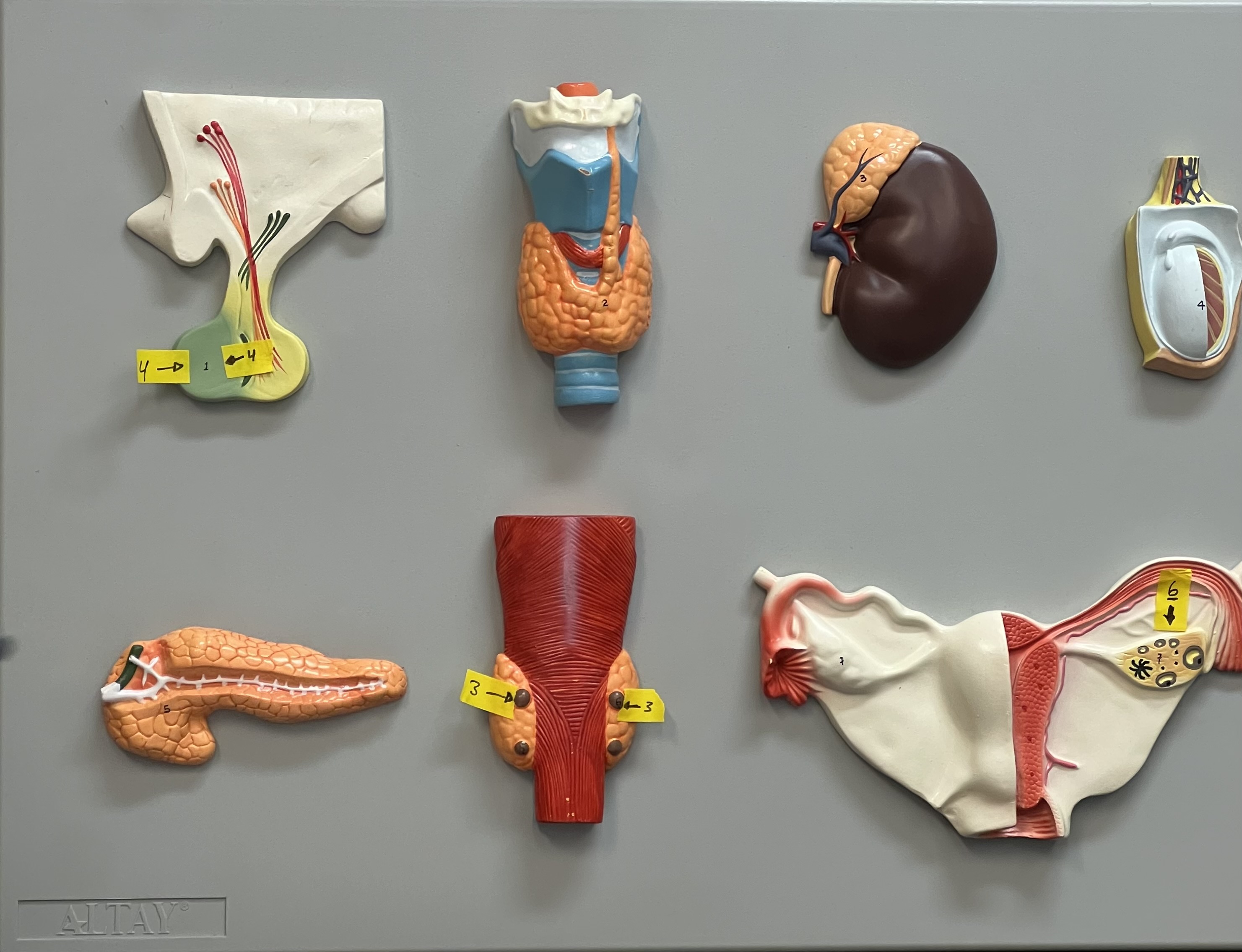
Describe the function of the structure labelled “4”
produces thyroid stimulating hormone
Which one of the following glands is purely endocrine?
thyroid gland
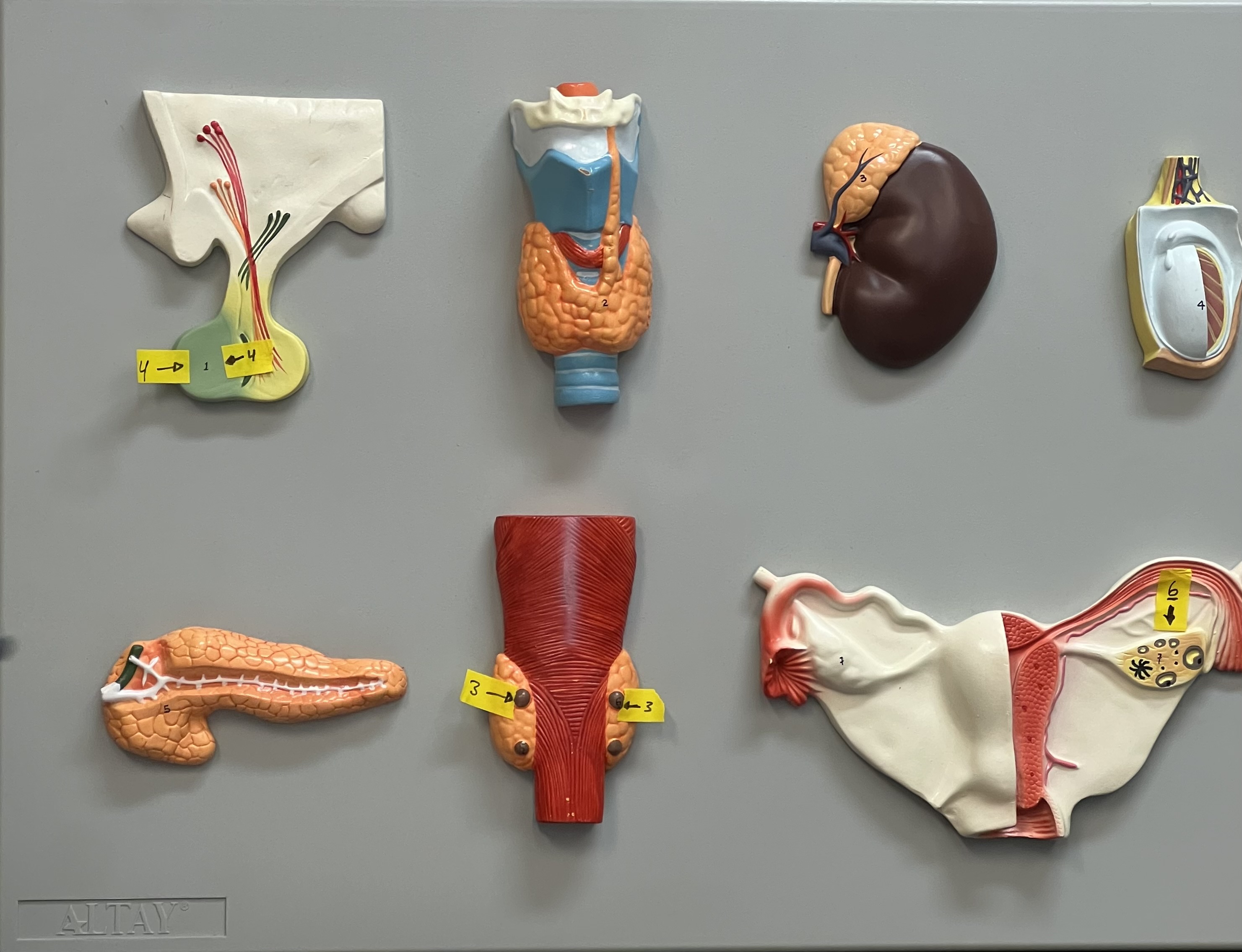
Identify the structure labelled “6”
ovary
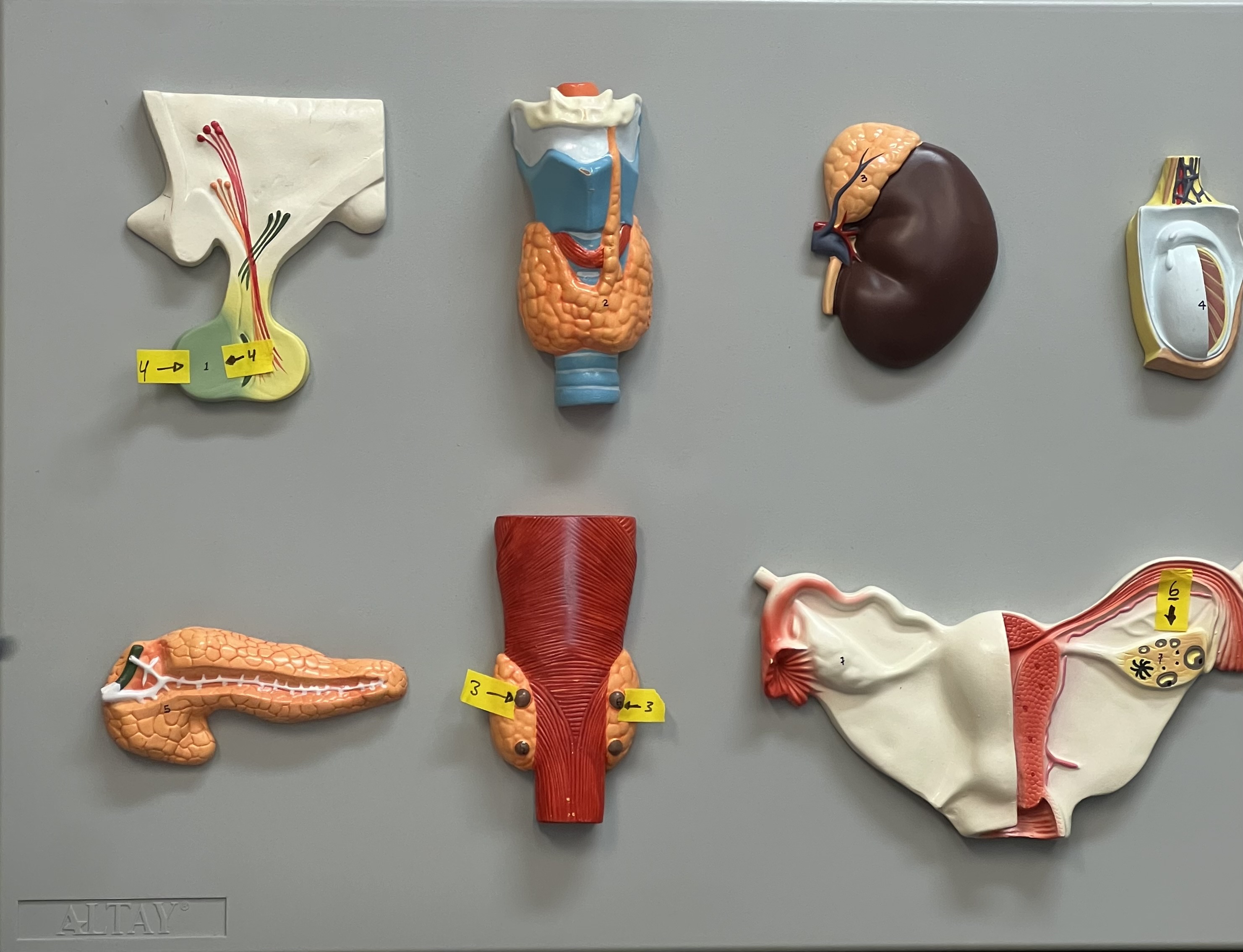
Describe the function of the structure labelled “6”
produces estrogen
Plasma
non-living liquid (55% of blood)
Formed elements
include leukocytes and erythrocytes (~45% of blood)
Plasma contains:
albumin, fibrinogen, globulins,
Albumin
major plasma protein used for osmotic balance
Fibrinogen
precursor protein for clotting
Globulins
liver function, blood clotting, and fighting infection.
Buffy coat
white blood cells and platelets, less than 1% of blood
Erythrocytes
red blood cells, approximately 44% of blood, responsible for oxygen transport
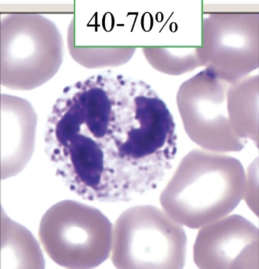
What leukocyte is this?
neutrophil: fighting bacterial infections
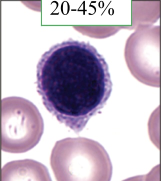
What leukocyte is this?
lymphocytes: immune response to infections
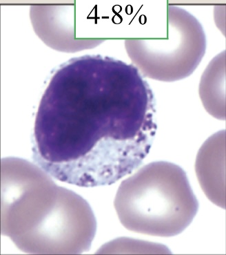
What leukocyte is this?
monocytes: large phagocytic cells
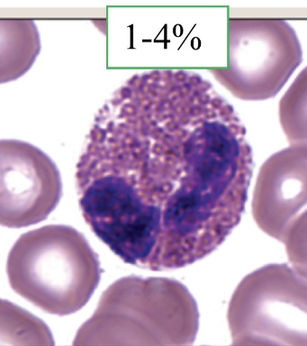
What leukocyte is this?
eosinophils: parasitic infections
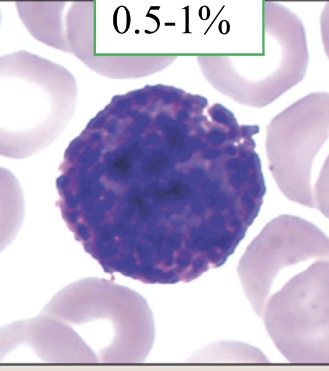
What leukocyte is this?
basophils: release histamine and heparin; involved in inflammatory response
Antigens
are protein labels that are located on the red blood cells.
Antibodies
are the “recognizer” proteins that are located in blood plasma.
If a patient with type B blood receive a transfusion of AB blood, which of the following would occur?
The patient’s anti-A antibodies (agglutinins) will agglutinate with the A antigens (antigultinogens) in the donor blood
When neither anti-A serum nor anti-B serum clot on a blood plate with donor blood, the blood type is
O
Would you expect the hematocrit level in anemic patients to be:
low
Pericardial sac
connective tissue sac external to epicardium
Epicardium
connective tissue layer that contains blood vessels, lymphatic vessels, and nerves
Myocardium
cardiac muscle, contractile tissue of the heart
Endocardium
epithelial layer that lines the chambers and valves
Tricuspid valve (Right AV)
prevents backflow of blood into right atrium
Pulmonary semilunar valve
prevents backflow of blood into right ventricle
Bicuspid valve (Left AV)
prevents backflow of blood into left atrium
Aortic semilunar valve
prevents backflow of blood into left ventricle
Coronary arteries
branch from aorta, supply oxygenated blood to heart when heart is relaxed
Cardiac veins
return deoxygenated blood to right atrium for recirculation
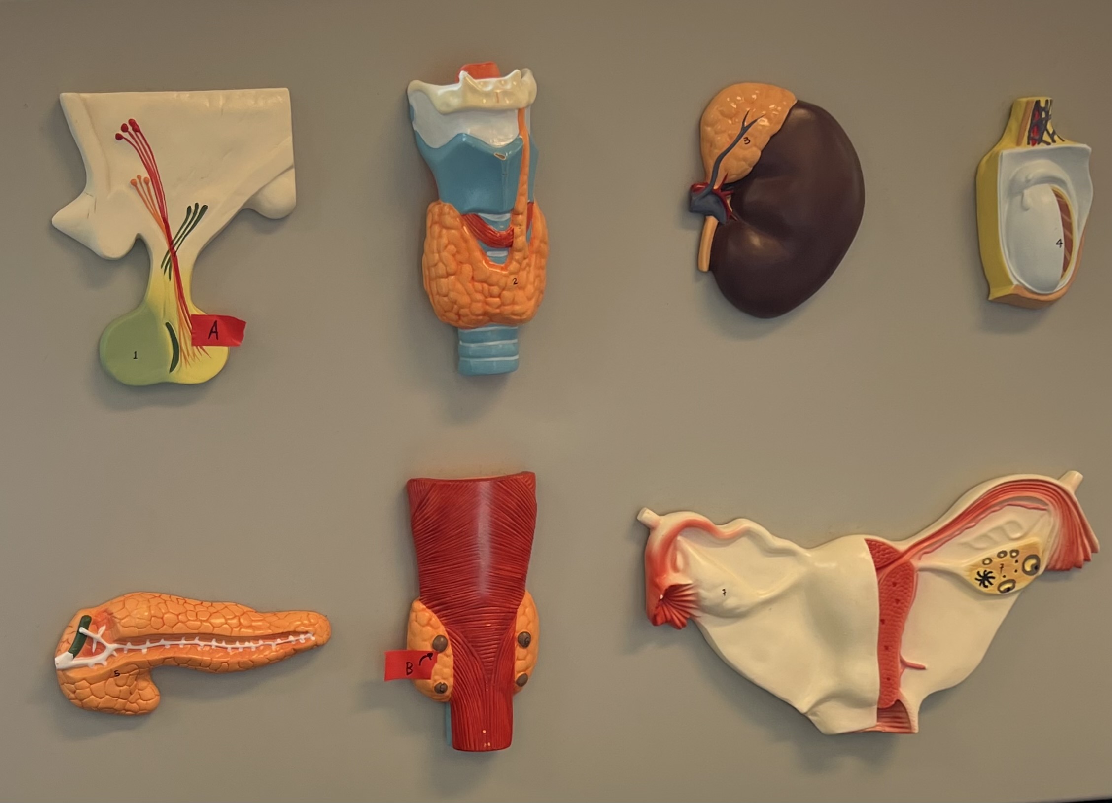
Structure “A” is best described as:
Posterior pituitary
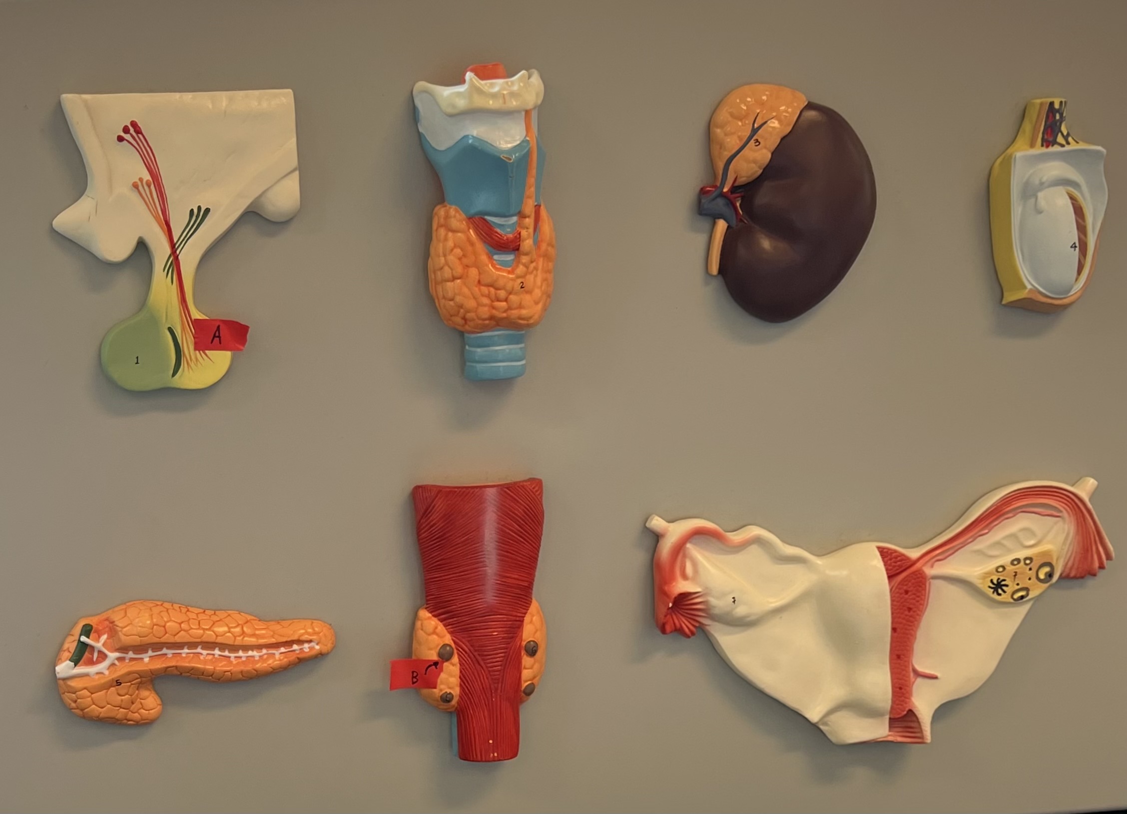
Structure “B” is best described as:
PTH
The adrenal medulla is responsible for which of the following?
short term stress (fight or flight)
A person with O+ blood would be able to accept blood from with of the following donors?
O+ and O- only
Your blood type is B-, which of the following antibodies do you have in your body plasma?
anti-A and anti-Rh antibodies
which of the following is a function of platelets?
blood coagulation
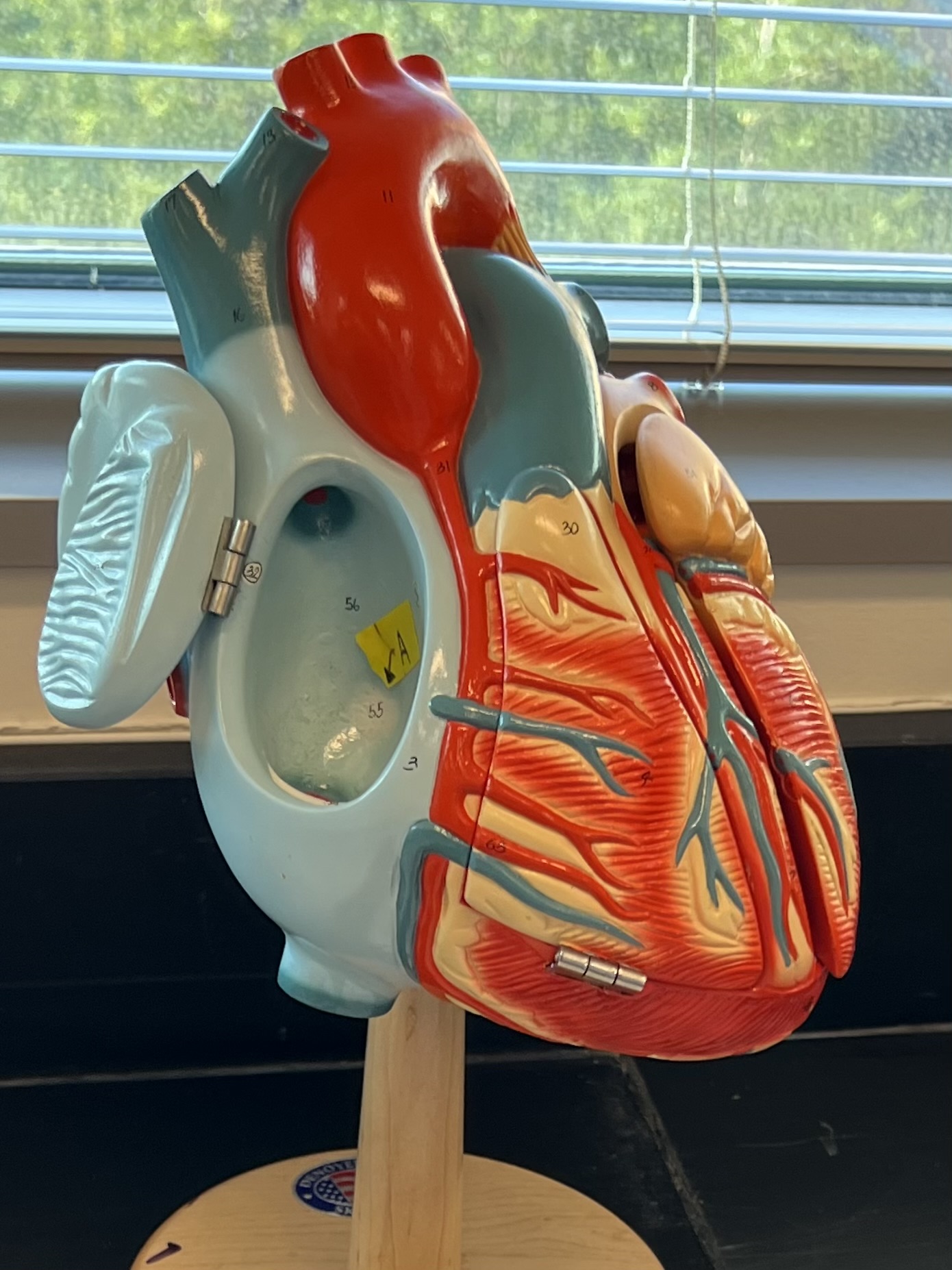
Structure “A” is best described as:
fossa ovalis
The pathway of blood from the right ventricle to the left ventricle
R ventricle → Pulmonary SL valve → pulmonary trunk → pulmonary arteries → lungs → pulmonary veins → Latrium
→ bicuspid valve → L ventricle
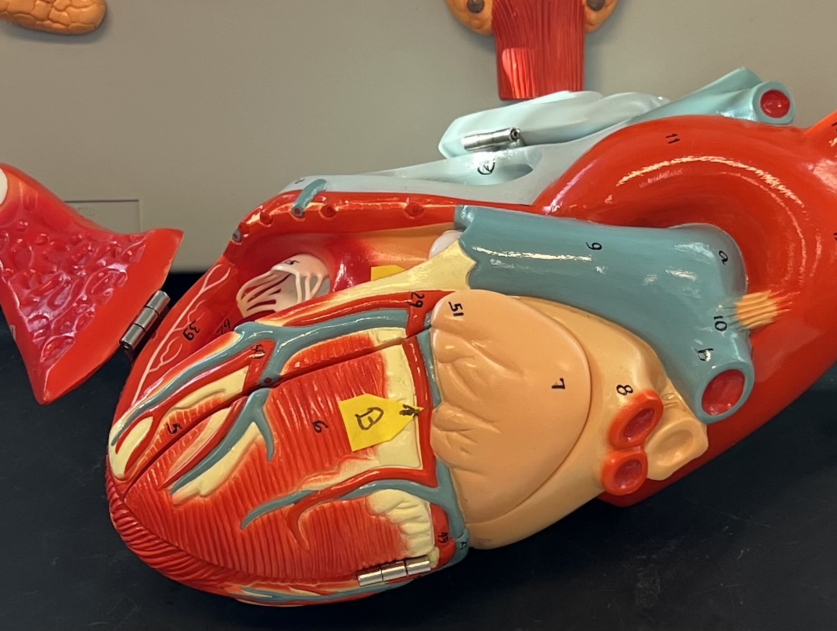
Structure “B” is best described as:
circumflex artery
the muscular layer of the heart that actually contracts is called:
myocardium
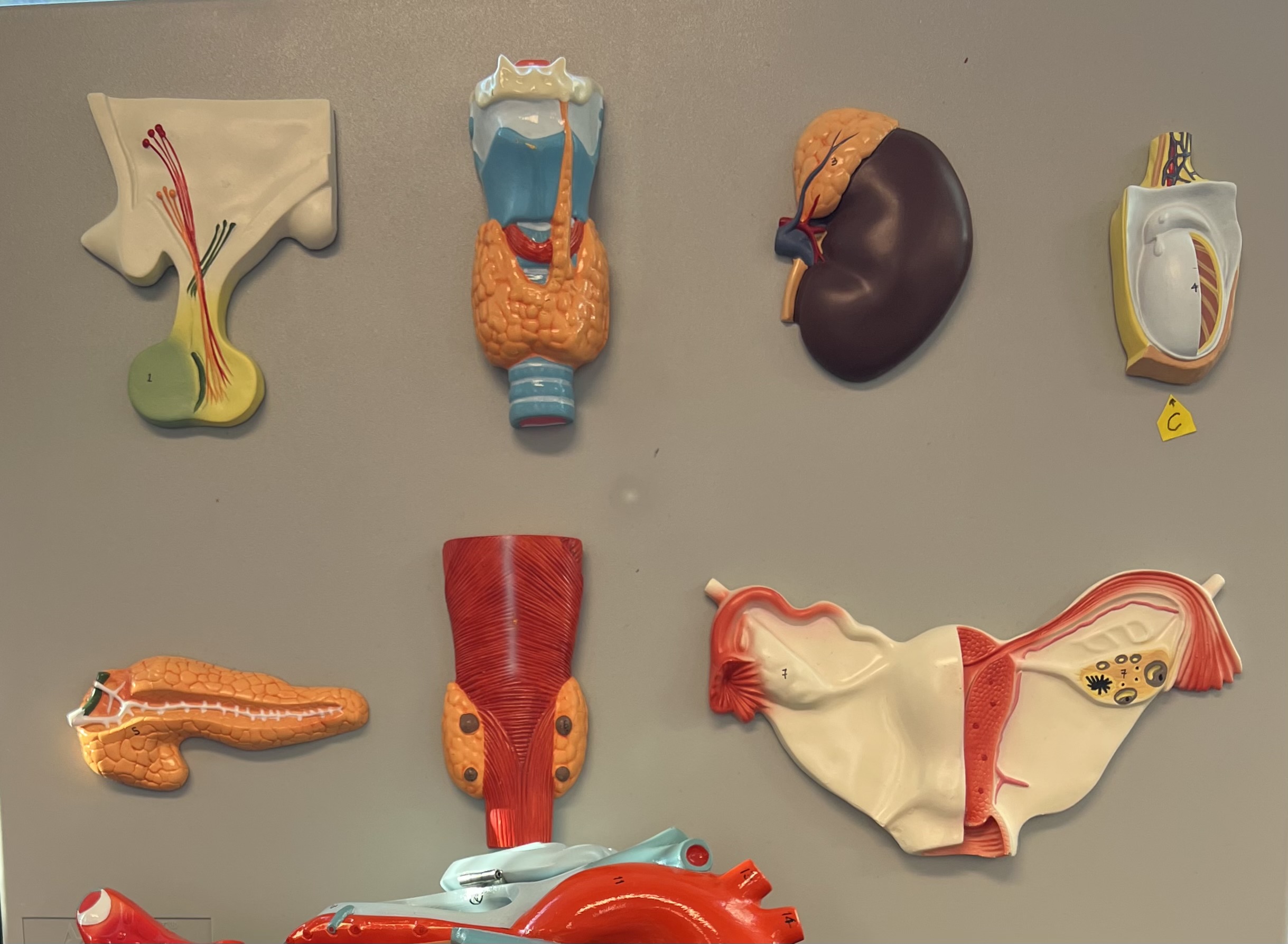
Structure “C” is best described as:
Testis
The posterior pituitary releases which of the following hormones? Select all that apply
oxytocin and ADH
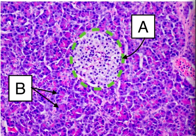
Structure B is called:
acinus
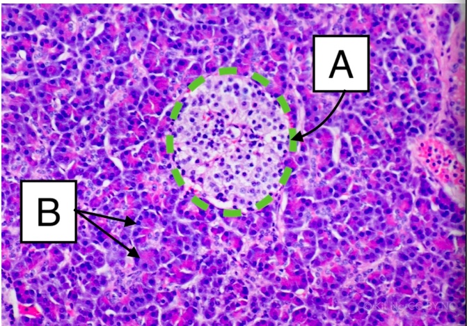
Structure A is called
pancreatic islet
Which of the following best describes the function of growth hormone?
Regulates tissue building of muscles and bones
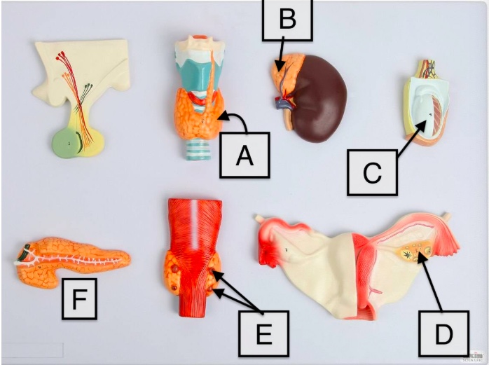
Structure "F" releases which of the following hormones?
Insulin
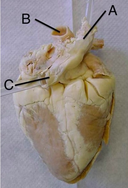
Letter C in the image below is indicating
Inferior vena cava
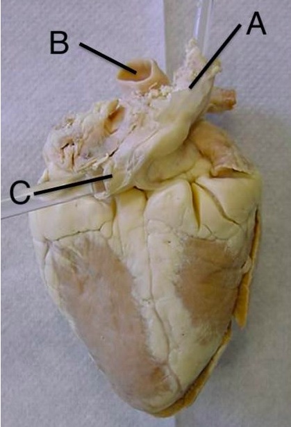
Letter B in the image below is indicating:
aorta
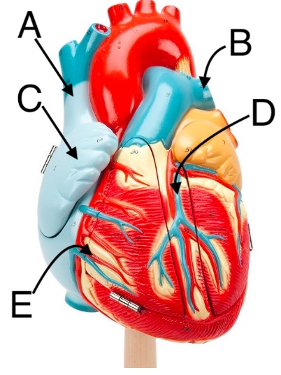
Structure "A" is:
superior vena cava
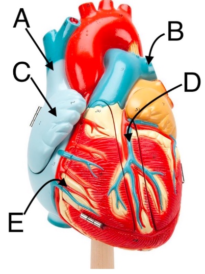
Structure "D" is:
great cardiac vein
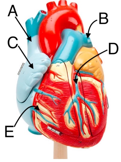
Structure "C" is:
right auricle
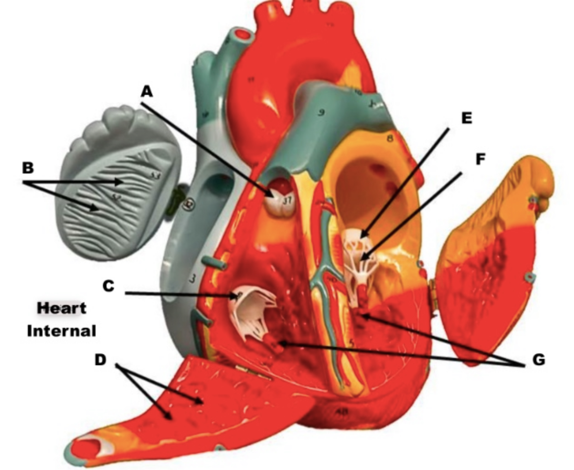
Letter "F" in the image below is indicating which of the following structures?
Chordae tendinae
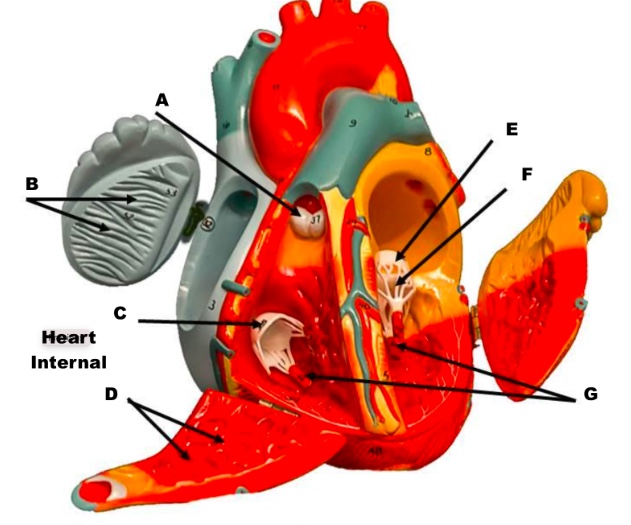
Which letter on the image below is indicating the pulmonary semilunar valve?
A
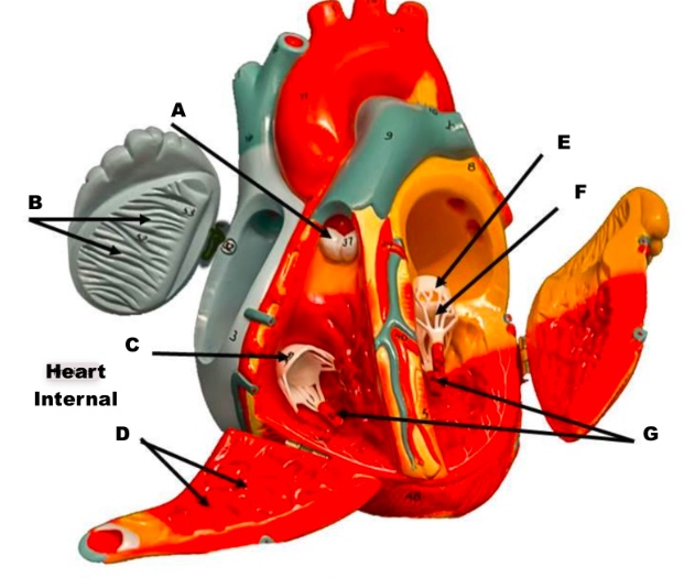
Which letter on the image below is indicating the mitral valve?
E
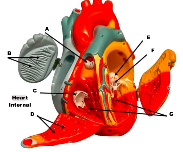
Letter "D" in the image below is indicating which of the following structures?
rabeculae carneae
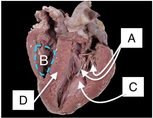
Structure "A" is:
Chordae tendinae of bicuspid valve
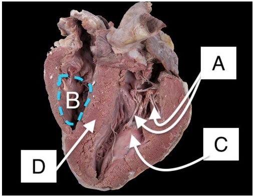
Structure "D" is:
Interventricular septum
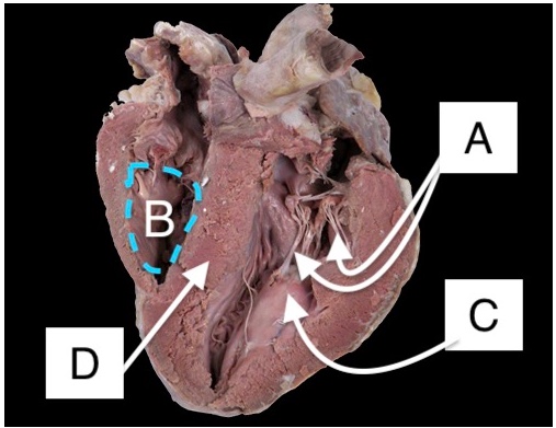
Structure "C" is:
Papillary muscle of bicuspid valve
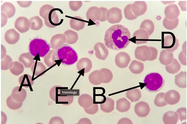
Cell "A" can do which of the following?
Produce antibodies
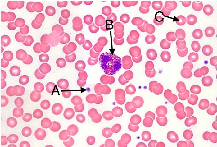
Which of the following is a function of cell "C" in the image below?
Oxygen transport to tissues
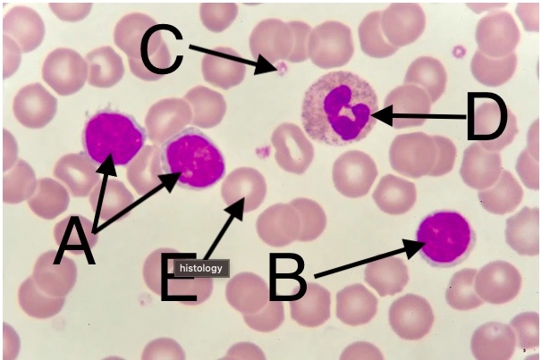
Cell "A" is best described as:
A lymphocyte
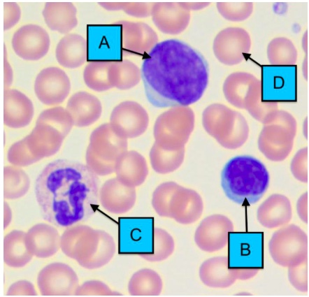
Cell "B" is a/an:
lymphocyte
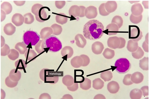
Cell "D" is best described as:
neutrophil
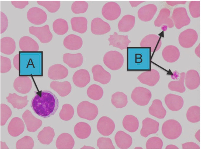
Cell "A" is a/an:
Lymphocyte
The structures marked "B" are:
platelets
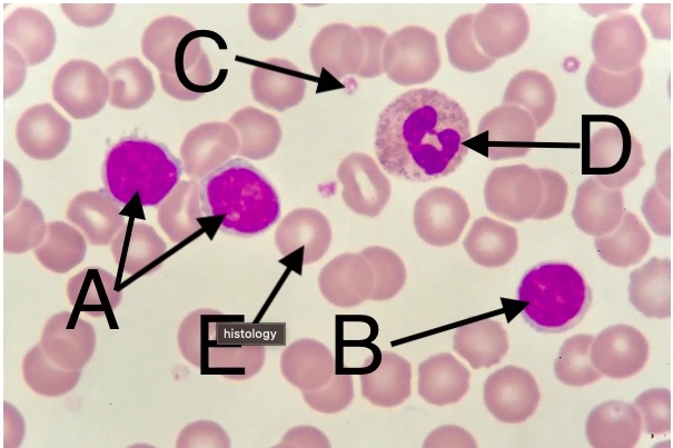
Cell "E" is best described as:
An erythrocyte
T or F: Someone with O- (O negative) blood would be able to safely donate to anyone.
True
If your blood type is AB+, which of the following is true of your blood?
Your blood plasma does not contain antibodies against A, B, or Rh.