Histology VIBS 911
 Call Kai
 Learn
Learn Practice Test
 Spaced Repetition
 Match
Match Flashcards
 Knowt Play
1/57

There's no tags or description
Looks like no tags are added yet.
Name | Mastery | Learn | Test | Matching | Spaced | Call with Kai | Chat |
|---|
No analytics yet
Send a link to your students to track their progress
58 Terms
simple squamous epithelium
one layer of flattened cells
Typical locations: organs
blood vessels, lymph vessels, body cavities and outer surface of many

Simple Cuboidal Epithelium
one layer of cuboidal cells
Typical locations: follicles of thyroid gland, kidney tubules, some sweat glands, and duct systems

Simple Columnar Epithelium
one layer of tall columnar cells Typical locations: luminal epithelium of intestines and gall bladder

Stratified Squamous Epithelium
nuclei present in all layers of cells including
the apical or free surface. Typical locations: mouth, esophagus, vagina, anal canal

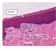
Keratinized stratified squamous
surface layers devoid of nuclei, called keratin. Amount of keratin can vary. Typical locations: integument (dry surfaces mostly), hard palate, areas of high "wear and tare".
Stratified Cuboidal Epithelium
more than 1 layer of cells; apical layer is cuboidal, not common.
Typical locations: duct systems

Pseudostratified Columnar Epithelium
Looks stratified but all cells will reach the basement membrane. Typical locations: Respiratory System, some in reproductive system

Stratified Columnar Epithelium
more than 1 layer of cells; apical layer is columnar Typical locations: duct systems

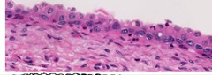
Transitional Epithelium/Urothelium
multiple layers; apical cells are rounded but can look more flattened when stretched. Apical cells are larger than basal cells.
Typical location: Urinary System
Microvilli
cytoplasmic projections from apical surface; composed of microfilaments covered by cell membranes;
Function- increase surface area to increase absorption (NOT MOTILE);
Location- intestinal tract; proximal renal tubules

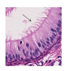
Cilia
cylindrical structures composed of microtubules; Function- motile, move in specific direction to move fluid or particles;
Location- pseudostratified columnar epithelium of respiratory system; parts of reproductive tract.

Stereocilia
Not true cilia- they are long microvilli, composed of microfilaments;
Function- increase surface area for absorption; Location- parts of male reproductive tract
Goblet Cells
unicellular glands that secrete mucus; found in respiratory tract;
small/large intestine

How many layers of cells are present in simple epithelium?
One
How is pseudostratified epithelium classified?
It appears stratified - however, all cells are touching the basement membrane and not all reach the apical (free) surface.
How is stratified epithelium classified?
2 or more cell layers
When classifying the shape of cells, the cells on the __________ (apical/basal) surface determined it's classification.
Apical
Where is simple squamous epithelium typically found?
Blood vessels, lymph vessels, body cavities and outer surface of many organs
Where is simple cuboidal epithelium typically found?
Follicles of thyroid gland, kidney tubules, some sweat glands, and duct systems
Where is simple columnar epithelium typically found?
Luminal epithelium of intestines and gall bladder
Where is non-keratinized stratified squamous epithelium typically found?
Mouth, esophagus, vagina, anal canal
Where is keratinized stratified squamous epithelium typically found?
Integumentary (dry surfaces mostly), hard palate, areas of high "wear and tare"
Where is stratified cuboidal epithelium typically found?
Duct systems
Where is pseudostratified columnar epithelium typically found?
Respiratory system, some in reproductive system
Where is transitional epithelium typically found?
Urinary system
What is the function of microvilli and where are they typically found?
To increase surface area in order to increase absorption. Located in intestinal tract and proximal renal tubules
Function and location of cilia
Motile, move in specific direction to move fluid or particles. Found on pseudostratified columnar epithelium of respiratory tract; parts of reproductive tract
Description, function and typically location of stereocilia?
They are not true cilia, they are long microvilli composted of microfilaments. They function to increase surface area for absorption. Located in parts of male reproductive tract.
Basic tissues of the body
Epithelium, Connective Tissue, Muscle, Nervous Tissue
General Features of Epithelium
Very cellular, little intercellular material, typically avascular, cells arranges in sheets/layers (membranes) or irregular masses or groups (glands)
Why is it clinically relevant to understand the general features of epithelium?
To identify the type of epithelium and knowing where its typically found, as well as determining the origin of a tumor. A tumor arising from epithelial tissue is an epithelial neoplasia
What causes the polarity of cells?
Junctional complexes (tight junctions, desmosomes, connexons)
What are clinical implications of identifying normal polarity or lack of polarity in intestinal epithelial cells on cytology samples?
If epithelial cells don't exhibit normal polarity, they wont be as clumped together or continuous between one cell to the next. This might suggest that something (i.e. virus, tumor, etc) has destroyed these intercellular connections.
What would be the consequence if the stratified squamous cell epithelium of our skin did not possess the keratinized layer?
What would be the consequences if cilia of the respiratory tract did not function properly?
Decreased ability to sweep pathogens/mucous out of respiratory tract leading to increased risk of infection
What would the consequences be if microvilli in the intestines were non functional or structurally damaged?
Decreased absorption of nutrients in the intestines leading to possible malnutrition, dehydration, and lethargy.
What would be the result of non-functioning stereocilia?
Male infertility - no bbs
Fibroblasts/cytes

What is the difference between a fibroblast and fibrocyte?
Fibroblasts are active, while fibrocytes are inactive.
Lamina propria
Collagenous connective tissue found underlying epithelium

Adipose - unicellular adipose tissue

Multiocular adipose tissue

Osteocyte

Osteoclast

Osteoblast

Osteoclast

Osteoblast

What are the main differences between epithelium and connective tissue?
connective tissue has relatively few cells, abundant intercellular material, usually well vascularized. Arrangement can be regular or irregular.
Function of connective tissue?
Support and protection
Connective tissue arises from what embryonic cells?
Mesenchymal
Components of connective tissue?
Cells, Extracellular matrix (fibers, water, ions)
Cellular component of Collagenous Connective Tissue
Fibroblasts/cytes
Adipose tissue cellular morphology
Cytoplasm and nucleus pushed to periphery, appear empty, looks like chicken wire
What is the best technique for sampling a suspected fatty mass and why?
Coring because it reduces the amount cell rupturing - adipose cells rupture easily.
Function osteocytes
Maintain the bone territorial matrix, embedded within the mineralized matrix
Function osteoblasts
Synthesize the organic portion of bone.
Function osteoclasts
Responsible for bone resorption and remodeling and result in raising blood calcium levels.
Microscopic Features of Osteoclasts
On surface of bone, derived from a fusion of macrophages and are multinucleated