Central nervous system
 Call Kai
 Learn
 Practice Test
 Spaced Repetition
 Match
 Flashcards
 Knowt Play
1/107

There's no tags or description
Looks like no tags are added yet.
108 Terms
Cranial meninges include
dura mater
arachnoid mater
pia mater
subarachnoid space
choroid plexuses
arachnoid villi
Dura mater
outermost, tough membrane
–outer periosteal layer of dura is against bone
Arachnoid mater
is “spider web” filamentous layer
pia mater
is a thin vascular layer attached to the brain surface
Meningeal layers of the superior sagital sinus
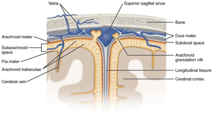
subarachnoid space
•Space between Arachnoid and Pia Mater. Contains cerebrospinal fluid.
choroid plexuses
•Capillaries where fluid flows from the blood into the ventricles of the brain (formation of cerebrospinal fluid)
Arachnoid villi
Location where cerebrospinal fluid is absorbed into the blood.
Brain diagram

rostral
toward the forehead
Caudal
toward the cord
The major parts of the brain include
cerebrum, cerebellum, diencephalon, brainstem
cerebrum is 83% of
brain volume
cerebellum contains 50% of
the neurons
the brain weighs
3 to 3.5 lbs
Longitudinal fissure separates
2 cerebral hemispheres
gyri
are the ridges of the folds
sulci
the grooves
cortex
surface layer of gray matter is called
deeper masses of gray matter are called
–nuclei (or ganglia)
bundles of myelinated axons (white matter) are called
tracts
gray matter
•unmyelinated dendrites and cell bodies (cortex and nuclei)
white mater
myelinated axons (tracts & nerves)
cerebral cortex is
a layer of gray matter with extensive folds to increase surface area – divided into lobes
frontal lobes
contains voluntary motor functions and areas for planning, mood, smell and social judgment
Parietal Lobe
contains areas for somatosensory reception, taste and integration of sensory information
Occipital lobe
•is the visual center of brain
Temporal Lobe
contains areas for hearing, smell, learning, memory, emotional behavior
Thalamus
•Receives nearly all sensory information on its way to cerebral cortex (except olfactory)
–integrate and directs incoming information to appropriate area “relay station”
Hypothalamus
•Inferior to thalamus
•Functions
–hormone secretion & pituitary
–autonomic NS control
–thermoregulation (thermostat)
–food and water intake (hunger and satiety)
–sleep and circadian rhythms
–memory
emotional behavior
The brainstem consists of
the midbrain, pons, and medulla
Midbrain consists of the
cerebral aquaduct, corpora Quadrigemina, and fiber tracts
Cerebral Aqueduct
Connects 3rd and 4th ventricles.
Corpora Quadrigemina:
–Visual and auditory reflexes.
fiber tracts
Ascending and Descending.
Pons
•Bulge in the brainstem
•Ascending sensory tracts
•Descending motor tracts
•Pathways in and out of cerebellum
•Nuclei concerned with sleep, hearing, balance, taste, eye movements, facial expression, facial sensation, respiration, swallowing, bladder control and posture
Medulla Oblongata
•Ascending and descending nerve tracts
•Cardiac center adjusts rate and force of heartbeat
•Vasomotor center adjusts blood vessel diameter
•Respiratory centers control rate and depth of breathing
•Reflex centers for coughing, sneezing, gagging, swallowing, vomiting, salivation, sweating, movements of tongue & head
Cerebellum
•Located under the cerebral hemispheres
•Functions:
–Produces smooth and coordinated movements
–Controls balance and equilibrium.
Ventricles
•Internal chambers within the CNS
–lateral ventricles
found inside cerebral hemispheres
–third ventricle
is single vertical space under corpus callosum
–cerebral aqueduct
runs through midbrain
–fourth ventricle
is small chamber between pons and cerebellum
–central canal
runs down through spinal cord
Ventricle diagams

Cerebrospinal Fluid
•Ventricles lined with ependymal cells
•Contain choroid plexuses (derived from pia) that produces CSF
•Fills ventricles and canals and bathes its external surface (in subarachnoid space)
•about 500 ml/day
–Functions
•buoyancy -- floats brain
•protection -- cushions from hitting inside of skull (water absorbs impacts well)
chemical stability -- rinses away wastes
Spinal Cord
•Information highway between brain and body
•Extends through vertebral canal from foramen magnum to L1
–Extends past L1 as medullary cone and cauda equina
Spinal Cord- Each pair of spinal nerves receives sensory information and issues
motor signals to muscles and glands
Spinal cord is a component of the
CNS while the spinal nerves are part of the PNS
Spinal Cord Functions
Conduction
Locomotion
Reflexes
conduction
bundles of fibers passing information up and down
locomotion
neurons providing control of flexors and extensors
reflexes
–involuntary, stereotyped responses to stimuli (remove hand from hot stove)
Spinal cord consists of:
•31 pairs of spinal nerves
•Medullary cone is tapered tip of spinal cord
•Cauda equinae is L2 to S5
–nerve roots resemble horse’s tail
Anatomy of the Spinal cord can be described as
•Cylinder of nerve tissue within the vertebral canal
•Central area of gray matter shaped like a butterfly and surrounded by white matter

gray matter
neuronal cell bodies
–Resembles letter H in cross section.
white matter
myelinated axons (ascending or descending tracts)
Dorsal/posterior horn:
–Contains cell bodies of interneurons.
Ventral / anterior horn
–Contain cell bodies of motor neurons.
Central canal
–Continuous with the ventricles of the brain.
Cross section of spinal cord diagram

nerve
•a bundle of nerve fibers (axons)
Spinal nerves have
•dorsal and ventral roots
–dorsal → sensory input
–ventral → motor output
•Dorsal root ganglion
–contains sensory neuron cell bodies

receptors and effectors dagram

Connective tissue wrappings include
Epineurium
Perineurium
Endoneurium
•Endoneurium:
Connective tissue sheath around each axon.
•Perineurium:
Coarser connective tissue wrapping surrounding fascicles (bundle).
Epineurium
•Tough fibrous sheath around the entire nerve.
sensory nerves
•Carry sensory fibers only
Motor nerves
•Carry motor fibers only
Mixed nerves
Contain both sensory and motor fibers
Cranial Nerves:
•12 pairs
•Some are motor, some are sensory and some are mixed
•Primarily serve the head and neck
•Are designated by name and Roman numerals.
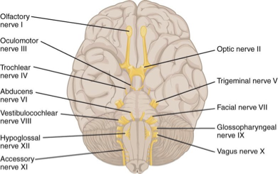
I Olfactory
Sensory | Smell |
II Optic
sensory, vision
III Oculomotor
Motor, moves eyes, pupillary constriction
IV Trochlear
Motor, Move eyes
V Trigeminal
Mixed, Sensory input from face, mastication
VI Abducens
Motor, Move eyes
VII Facial
Mixed, Facial muscle movements, taste, saliva production
VIII Vestibulocochlear
Sensory, hearing and balance
IX Glossopharyngeal
Mixed, Taste, swallowing, saliva production
X Vagus
Mixed, Taste, control organs of thorax and abdomen
XI Accessory
Motor, Control muscles of neck and shoulders
XII Hypoglossal
Motor, Tongue movements
For nerve type: Some Say Marry Money But My Brother Says Big Brains Matter Most
S = sensory
M = motor
B = mixed (both)

Spinal Nerves: 31 pairs of nerves that emerge from the spinal cord through the
•intervertebral foramina.
•All spinal nerves are mixed nerves.
•Formed by the merging of the ventral (motor) and dorsal (sensory) roots.
Reflexes
•Rapid, predictable, involuntary response to a stimulus.

Neural Pathways for Reflexes = Reflex Arc
•Sensory Receptor
•Sensory Neuron
•+/- Interneurons
•Motor Neurons
•Effector: Muscle or glands
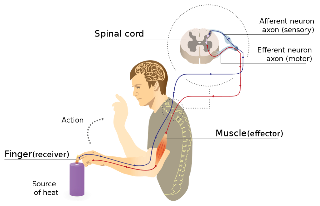
Autonomic reflexes
•Regulate smooth muscle, heart, or glands.
–Ex: Blood pressure, sweating.
Somatic Reflexes
•Reflexes that stimulate skeletal muscle.
–Ex: Knee Jerk reflex, withdrawal reflex.
What is an aneurysm
•is a weak spot in an arterial wall that swells and expands (and can rupture).
Cerebral Aneurysm Risk groups:
smoking, family history, high blood pressure, over age of 40, women slightly more than men
Cerebral Aneurysm Symptoms
•: often asymptomatic unless the aneurism gets large, leaks or puts pressure on a nerve
–Vision changes, pain near eyes, facial muscle weakness or numbness

Stroke
•When blood flow to the brain stops
Hemorrhagic stroke
blood vessel breaks and bleeds
Ischemic stroke
blood clot blocks blood from entering brain tissue
Stroke risk groups
having had prior strokes, high blood pressure, heart disease, diabetes, smoking
Stroke symtpoms
Sudden vision loss, confusion, speech problems, numbness, balance problems, severe headache.
Subdural hematoma
•Damage to veins below the Dura mater; blood collects on the surface of the brain
–Acute – result of a head injury
–Chronic – minor head injury, symptoms occur over a longer period of time (typically seen in elderly)