Zoonotic Diseases Part 2
 Call Kai
 Learn
 Practice Test
 Spaced Repetition
 Match
 Flashcards
 Knowt Play
1/12

There's no tags or description
Looks like no tags are added yet.
13 Terms
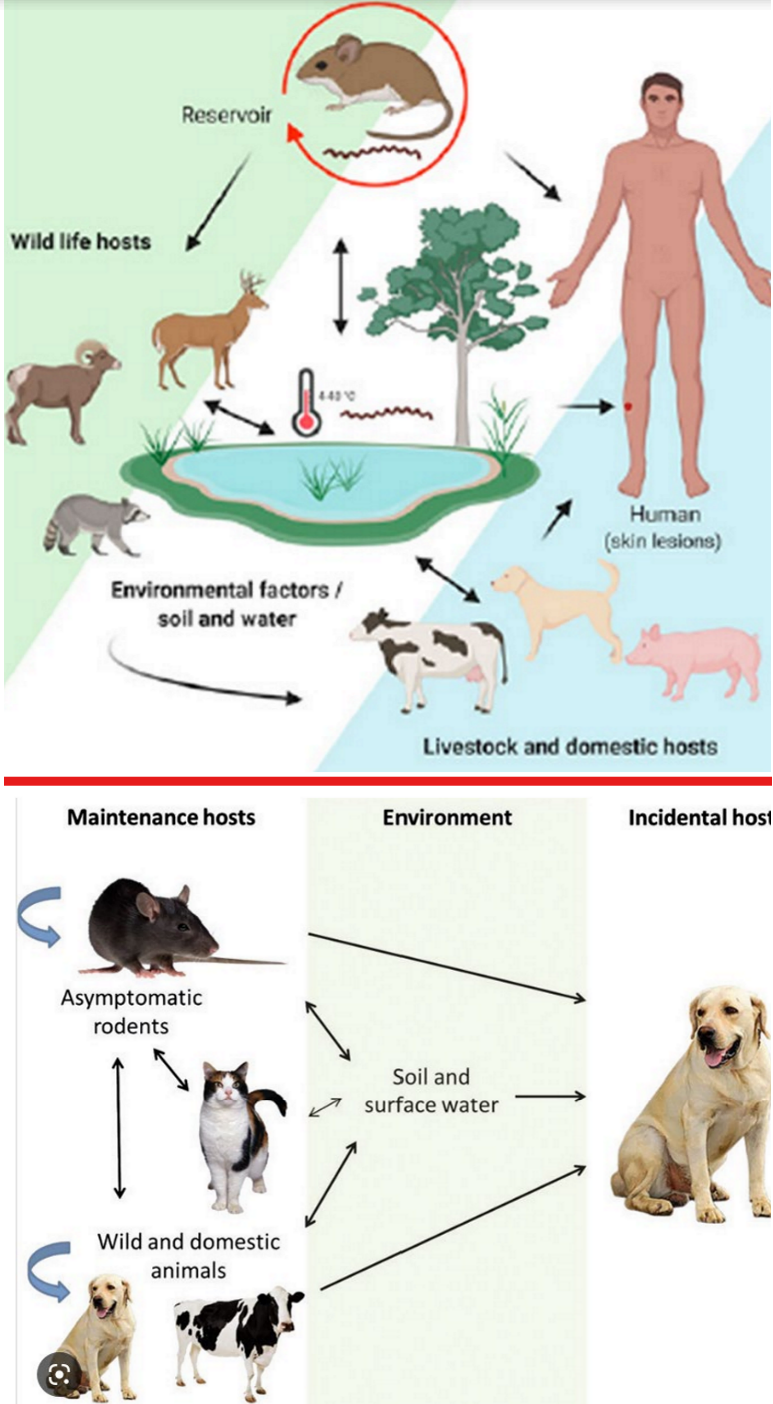
leptospirosis caused by Leptospira spp…
G- spirochete, highly motile, aerobic
.
list some reservoir hosts (natural host thats asymp & sheds it)
list some incidental hosts (pt gets super sick)
how is this bacteria shed?
route of transmission in all species? (2)
besides inside the host, where does this bacteria replicate?
mainly rodents — possibly cat, dog, & cow
dog, ppl
thru infected ani urine, blood, semen, milk, body tissues
direct contact of contaminated water, grass, etc. w/ any MM or open wound or ingesting infected animal or contaminated feed (e.g. rodent poops in it, urine splashes into cows eyes)
enviro (esp waterlogged grass) → its VERY hardy, forms biofilms, & can live in enviro for months
leptospirosis caused by Leptospira spp…
G- spirochete, highly motile, aerobic
.
1: walk thru the progression of dz (start w/ entering body), include the anatomic region it loves to hang out in
.
2: where in the USA is this endemic in cats & dogs?
.
3: this occurs worldwide in ppl.
which country has the highest rate of transmission & why?
list things that inc a persons risk of exposure (consider occupation, hobbies)
1: enters body → burrows thru tissue (bc spirochete) → replicates in circulation which triggers imm system → cytokine storm → clin signs (fever + multiorgan failure as it travels to other places in body)
LOVES lumen of PCT in kidney (also can go other places)
.
2: all over! (70% of outdoor cats are subclinical carriers!)
.
3:
sri lanka bc rice farmers stand in waterlogged areas
dairy farmer, vets, triathletes, kayaking, hiking, & unhoused ppl w/ dogs (bc dog drinks water on street, chases rats, etc. then transmits to O)
.
note: many ppl go undiagnosed if signs are mild; map darker areas = more cases

leptospirosis caused by Leptospira spp…
G- spirochete, highly motile, aerobic
.
dz in ppl ranges from mild & self limiting (esp if healthy) to life threatening (esp if immcomp).
typical clinical signs of mild dz?
typical manifestations of severe dz?
non-specific… high fever, headache, chills, muscle ache, V/D, abd pain, etc.
AKI in kidney, hepatic injury (→ jaundice), ± pulmonary hemorrhage/ meningitis/ pancreatitis (AKA it destroys your insides), death
.
note: incub period ~5-14 days; this is reportable dz in ppl!
leptospirosis caused by Leptospira spp…
G- spirochete, highly motile, aerobic
.
1: list species that can get this
.
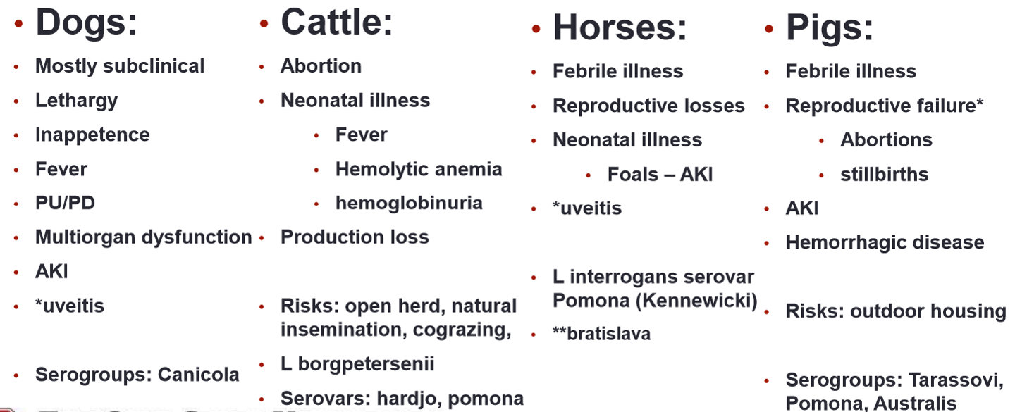
2: most dogs are subclinical but if they become clinical, it can present in one of four ways.
describe the signs of each…
peracute (lasts <24 hrs)
acute (1-2 d)
subacute (2-7 d)
chronic (>7 d)
.
3: what dz manifestation is unique to dogs & horses?
1: dog, cow, SR, horse, pig, camelid, wildlife
.
2:
sudden death
lethargy, depressed, fever, myalgia, anorexia, vomit → death
same ^ but less severe
lethargy, depression, fever or hypothermia, dehydration, PU/PD, abd pain, diarrhea
.
3: uveitis (more in horses)
leptospirosis caused by Leptospira spp. IN DOGS…
.
1: name the gold standard diagnostic test for definitive dx
whats a (+) result?
.
2: describe why each of these aren’t great diagnostic tools for lepto…
UA
urine culture
kidney or liver biopsy
.
3: how are the point of care tests useful? (idexx SNAP or zoetis WITNESS)
1: microscopic agglutination test (MAT; Ab serology test)
(+) = take two serum samples ~1-2 weeks apart & get 4-fold or greater inc in Ab titer
can make presumptive dx if initial titer is ≥1:800 & pt is unvaxed & has clin signs
.
2:
FN possible if pt isn’t currently shedding
WAY too slow (takes 3-4 weeks)
not practical if pt is super sick
.
3: vax won’t interfere w/ result & can r/o if (-) bc NPV is high
BUT cannot r/i if (+) bc PPV is low
.
review: bac is in blood for 4-6 days → form Ab’s 3-10 days after that → hangs out in kid (± liver) → pt intermittently sheds in urine
leptospirosis caused by Leptospira spp. IN HORSES…
.
name the serovar that 45-50% of lepto horses get which leads to asymp infection
name the serovar that causes dz in horses
list the 3 ways this dz can manifest
.
4: like dogs, MAT is the gold standard diagnositc (cannot differentiate vax status).
whats another good diagnostic option to dx pt’s w/ the ___ presentation?
abortion/sick neonate
equine recurrent uveitis
Bratislava serovar & asymp
Pomona type kennewicki
.
3: AKI
or abortion if infected early in gestation or sick neonate (weak, jaundice, hemolytic anemia, etc.) if late in gestation
or chronically get equine recurrent uveitis bc of autoimmune rxn (esp in appaloosa & warmbloods)
.
4:
combo of PCR (urine, ocular, fetal, blood) & fluorescent Ab test
culture serum AND aqueous fluid → (+) = 4x higher in aqusous fluid than serum
.
note: can also do UA & see RBC & WBC ± presence of spirochetes

leptospirosis caused by Leptospira spp. IN COWS…
.
1: why is the prevalence higher in dairy compared to beef?
.
2: clinical signs that are unique to cows (compard to dogs)?
.
3: describe the best way to diagnose a herd w/ lepto
.
4: why isn’t MAT gold standard?
1: condensed housing makes transmission easier
other things that inc cows risk of dz: open herd, natrual insemination, cograzing
.
2: sim to horse (except uveitis)… abortion, infertility, neonate illness (fever, hemolytic anemia, hemoglobinuria)
.
3: test minimum of 15 urine samples → assume entire herd is (+) if one is (+) bc FN’s common
.
4: cows dont form strong imm response so FN’s possible & vax interferes (= FP)
.
note: 50% of herds in SE USA have lepto & one (+) bull can trans to entire herd via infected semen!
leptospirosis caused by Leptospira spp.: prevention & control…
.
1: list species we have vaccinate
.
2: list specific ways to dec risk of dz if you’re in a high risk enviro (e.g. rice farmer)
.
3: list other general prevention & control strategies
1: dog, cow, ± horse (not regularly used)
human vax avail but too serovar specific to be helpful
.
2: wear boots & long sleeves (so water doesn’t get on ankles), cover wounds w/ bandages, shower afterwards
.
3: drink clean water, wash hands before eating, rodent control, wear PPE when handling (+) pt’s, isolate (+) pt’s, take prophylactic doxycycline
zoophilic dermatophytosis caused by Microsporum canis in SA’s…
AKA ringworm
.
transmission?
typical appearance of lesion?
other ddx to consider?
list the 4 big diagnostic tests used for ringworm & which is the gold standard for definitive dx
1: direct contact w/ infected ani (fur, lesion, etc.) or fomites (e.g. bedding, grooming tools)
spores can penetrate microtrauma in skin (e.g. scratch)
.
2: circular alopecia, scaling, crusting
.
3: flea infestation, otitis externa, ear mites, cheyletiella, feline scabies, feline demodicosis
instead of doing diagnostics for these ^, why can’t we just give revolution & see what happens?: it doesn’t cover ringworm & dont want it spreading all over a shelter for e.g.
.
4: woods lamp, trichogram, real time PCR, culture (gold standard)
how to diagnose ringworm…
.
1: woods lamp…
how do we perform this?
limitations?
.
2: trichogram…
how do we perform this?
whats a (+) result?
limitations?
1:
wave lamp over entire body (face, feet, belly, inside eats, etc.) & look for glowing/fluoresce
cannot r/o if (-) bc FN common; dead skin cells, disinfectant, tetracycline, doxycycline will glow & cause FP (these are typically on ends of hair so look at base of shaft)
note: electric powered preferred over battery bc light fades as battery dies; takes ~5 min
.
2:
pluck a few piece of glowing hairs → suspend in mineral oil → look under microscope
hair looks frayed/fuzzy on the edges (bc keratins breaking down)
can’t r/o if (-), FP possible if unexperienced

how to diagnose ringworm…
.
1: real time PCR of hair sample…
pros?
cons?
.
2: culture & look under microscope…
describe how we collect the sample for this
we have 2 options for media. whats the major diff between how we interpret them: dermatophyte test medium (DMT) vs. sabourauds media
1:
VERY sensitive (can r/o if (-)) & faster than culture
more FN’s than culture & cant use to assess if pt is responding to tx bc doesn’t tell us if fungus is alive, just tells us DNA is present
.
2:
use new toothbrush to rub over lesion → gently brush onto culture media → use sterile hemostat to remove debris/hair deeper in bristles & place on culture → after growth, look under scope for conidia & hyphae
DMT turns red when (+) (still must confirm under scope), sabouraud’s doesn’t

how do we handle ringworm in a population of shelter cats…
.
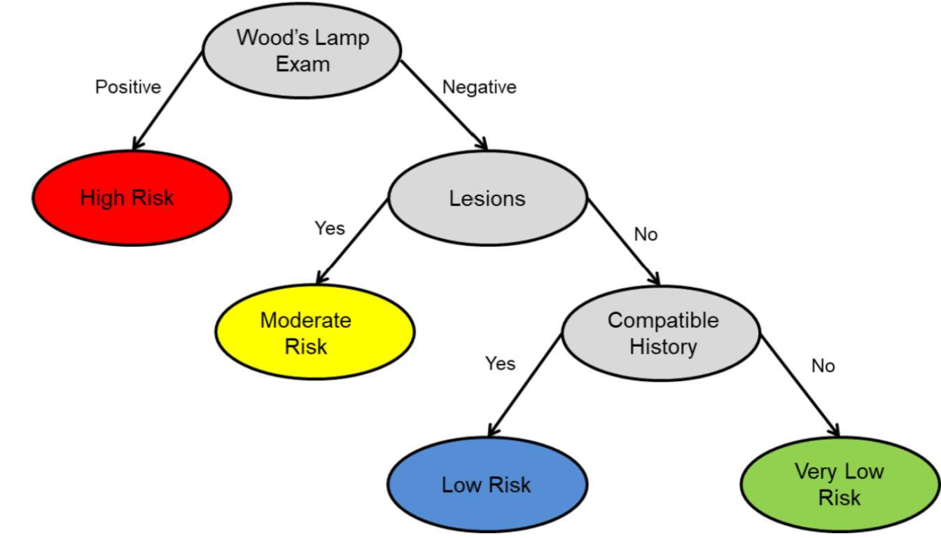
1: we do woods lamp on all cats to categorize their level of risk.
is the pt low, moderate, or high risk if…
woods lamp is (+)
wood lamp (-), pt doesn’t have lesions, but hx is compatible w/ being exposed
woods lamp (-) but pt has lesions
.
2: whats our next step for ___ risk cats…
very low/low
moderate
high
1:
high
low (even lower if hx isn’t compatible)
moderate
.
2:
send to adoption floor (± recheck in 1 week)
trichogram, ± culture or PCR (but $$)… if (+), start tx!, if (-), do risk assessment, ± more testing
START TX NOW!, ± cultute or PCR
how do we handle ringworm in a population of shelter cats…
.
1: name our three big goals
.
2: describe an appropiate isolation & PPE protocol
.
3: describe an appropiate C&D protocol for surfaces, floors, & soft items (e.g. bedding)
1: identify all cases, isolate cases to prevent further transmission to ani & ppl, tx as fast as possible
.
2: restrict access to only a few staff members, dedicate supplies to only use in isolation ward, take care of these pt’s last
wear full PPE, cover all exposed skin & hair bc cat hair goes EVERYWHERE! & carries spores w/ it
.
3: soap & water first then disinfect (e.g. bleach), use swiffer/damp mop to catch hair, (NO SWEEPING!) vacuum
soft items: wash & dry (dryer kills spores)
C&D is huge priority bc spores persist for months