Human Skeletal Anatomy Bone and Landmark Identification
 Call Kai
 Learn
 Practice Test
 Spaced Repetition
 Match
 Flashcards
 Knowt Play
1/61

Earn XP
Description and Tags
Comprehensive vocabulary flashcards covering anatomical bones, landmarks, processes, and structures listed across the human skeletal system.
62 Terms
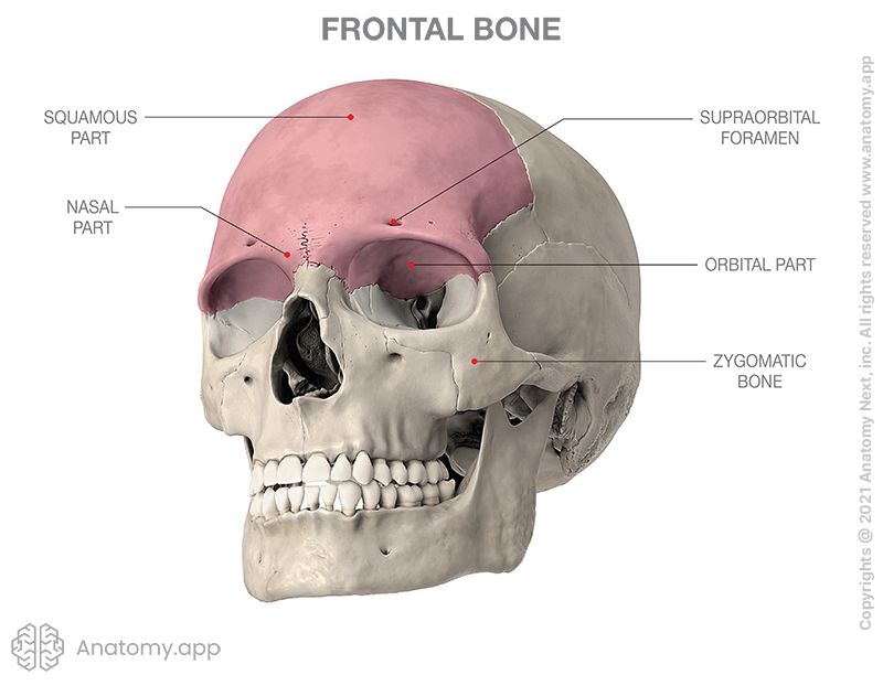
Frontal bone
The cranial bone forming the forehead, the superior margins of the orbital cavities, and the anterior cranial fossa.
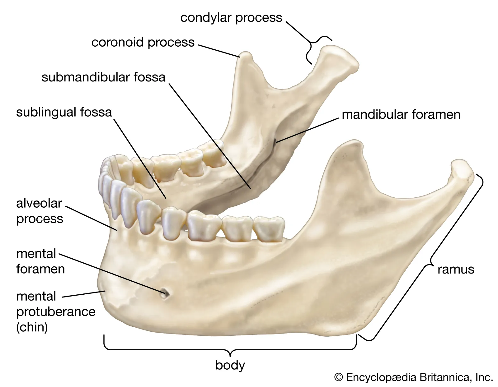
Mandible
The lower jawbone; the largest, strongest, and only movable bone of the human facial skeleton.
Mastoid process
A conical bony prominence projecting from the inferior surface of the temporal bone behind the ear, serving as a site for muscle attachment.

Maxilla
The upper jawbone formed by the fusion of two bones, supporting the upper teeth and forming parts of the orbit, nasal cavity, and hard palate.

Occipital bone
The bone forming the posterior and inferior base of the cranium, featuring the foramen magnum and occipital condyles.

Parietal bone
A paired cranial bone forming the central upper sides and roof of the skull.

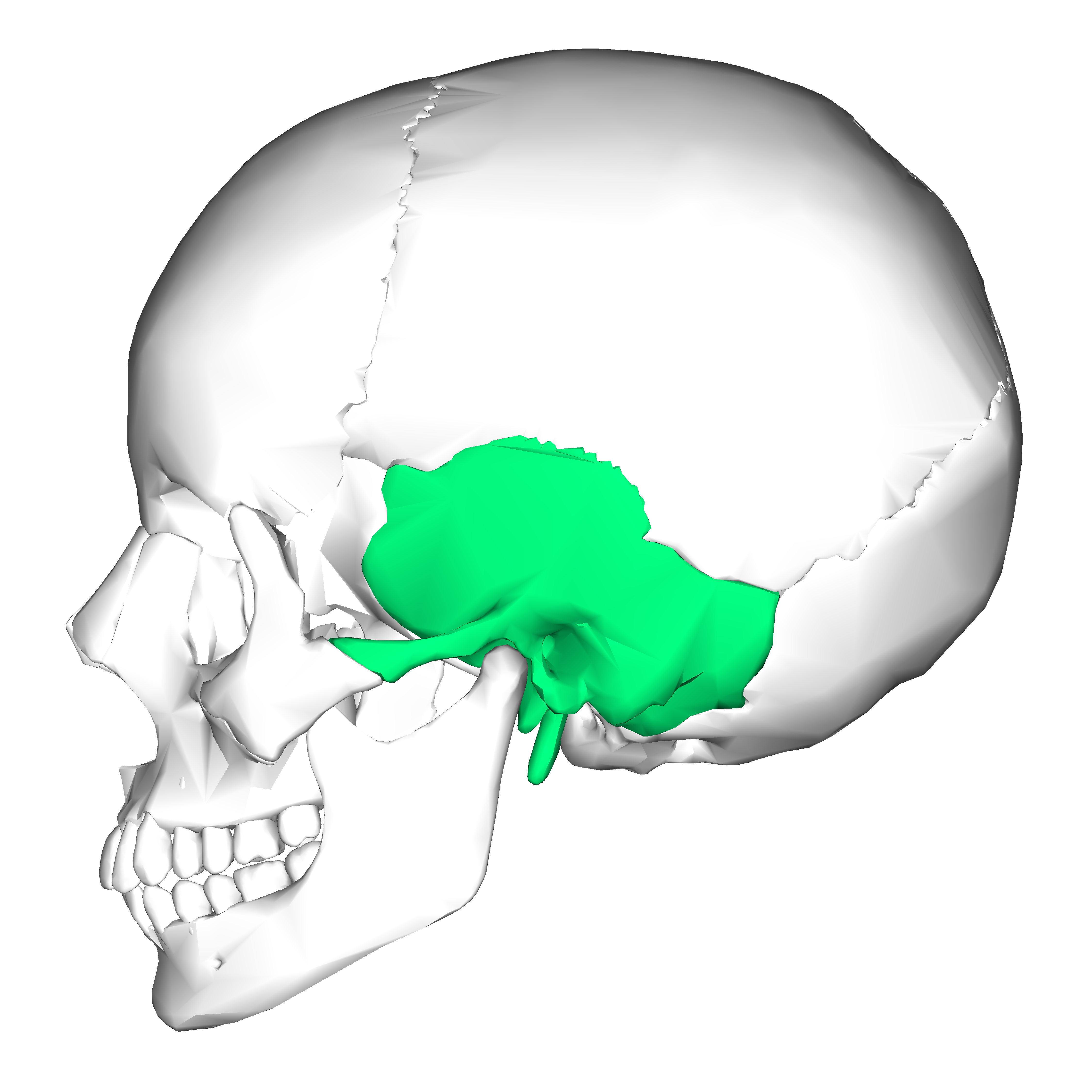
Temporal bone
A paired bone located at the sides and base of the skull, housing the structures of the inner and middle ear.
Zygomatic bone
The paired facial bone commonly known as the cheekbone, forming the prominence of the cheek and the lateral orbital wall.

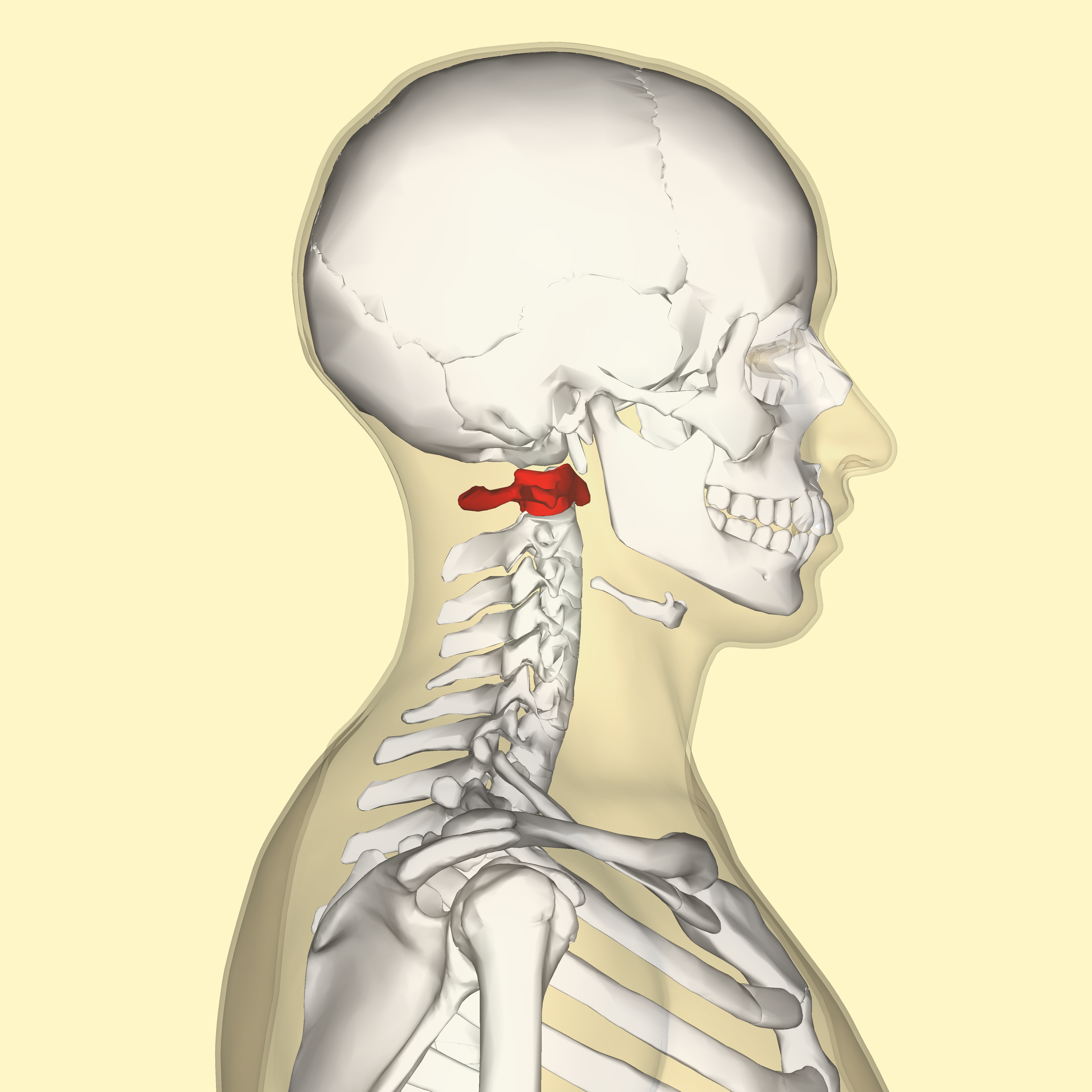
Atlas
The first cervical vertebra (C1), which articulates with the occipital condyles to support the skull and permit nodding motions.
Axis
The second cervical vertebra (C2), distinguished by the dens (odontoid process) that serves as a pivot point for skull rotation.

Cervical spine
The most superior segment of the vertebral column, consisting of seven vertebrae (C1 to C7) in the neck region.

Coccyx
The small triangular bone at the inferior terminus of the vertebral column (tailbone), formed by the fusion of several rudimentary vertebrae.

Lumbar spine
The lower back segment of the vertebral column, composed of five large, weight-bearing vertebrae (L1 to L5).

Sacrum
A large, triangular wedge-shaped bone situated at the inferior base of the vertebral column, formed by the fusion of five sacral vertebrae.

Thoracic spine
The intermediate segment of the vertebral column, comprising twelve vertebrae (T1 to T12) that articulate with the ribs.

Clavicle
The collarbone; an S-shaped slender bone that serves as a strut between the manubrium of the sternum and the acromion of the scapula.

Manubrium
The superior, broad segment of the sternum articulating with the clavicles and the first pair of ribs.

Rib cage
The bony and cartilaginous cage of the thoracic cavity enclosing and protecting the thoracic organs, formed by ribs, costal cartilages, thoracic vertebrae, and the sternum.

Sternum
The breastbone; a flat bone positioned in the central anterior region of the thorax, consisting of the manubrium, body, and xiphoid process.

Xiphoid process
The small cartilaginous and bony projection that forms the most inferior aspect of the sternum.

Acromion process
A lateral bony projection on the spine of the scapula that articulates with the clavicle to form the acromioclavicular joint.

Coracoid process
A hook-like bony process originating from the superior and lateral border of the scapula, pointing anteriorly and laterally.

Scapula
The shoulder blade; a flat triangular bone located on the posterolateral aspect of the thoracic cage.

Scapular spine
A prominent, transverse bony ridge on the posterior surface of the scapula that separates the supraspinous and infraspinous fossae.

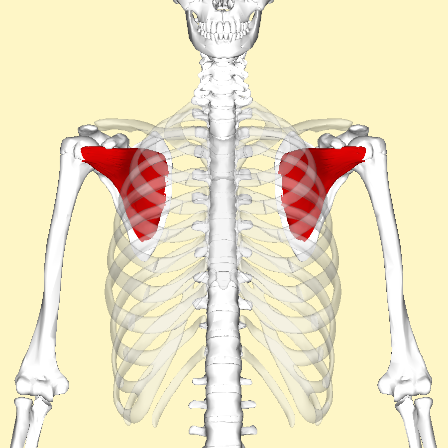
Subscapular fossa
The broad, shallow concave surface occupying the anterior (costal) aspect of the scapula.
Capitulum
A smooth, rounded articular prominence on the lateral aspect of the distal humerus that articulates with the head of the radius.

Deltoid tuberosity
A rough, triangular elevation on the lateral midshaft of the humerus that serves as the insertion site for the deltoid muscle.

Greater tubercle
A prominent bony protrusion on the lateral aspect of the proximal humerus that serves as an attachment site for rotator cuff tendons.
Humerus
The single long bone of the upper arm, extending between the shoulder and the elbow joint.
Radial tuberosity
An oval prominence on the medial aspect of the proximal radius, acting as the primary insertion point for the biceps brachii muscle.
Radius
The lateral bone of the forearm (aligned with the thumb), running parallel to the ulna.
Ulna tuberosity
A roughened prominence situated on the proximal ulna inferior to the coronoid process, providing an insertion site for the brachialis muscle.
Ulna
The medial and longer bone of the forearm (aligned with the pinky finger), articulating proximally with the humerus and radius.
Capitate
The largest carpal bone, situated centrally in the distal row of the wrist.
Carpals
The set of eight small, irregular bones that comprise the wrist.
Hamate
A wedge-shaped carpal bone on the medial side of the distal carpal row, identified by a distinct hook-like projection (unciform process).
Lunate
A crescent- or moon-shaped carpal bone situated in the center of the proximal carpal row between the scaphoid and triquetrum.
Metacarpals
The five intermediate long bones of the hand, located between the carpal bones of the wrist and the proximal phalanges.
Phalanges
The small bones that form the fingers and toes (singular: phalanx).
Pisiform
A small, pea-shaped sesamoid carpal bone found on the anterior surface of the triquetrum within the proximal row of the wrist.
Scaphoid
A boat-shaped carpal bone located on the lateral (radial) side of the proximal carpal row.
Sesamoid
A bone that develops embedded within a tendon where it passes over a joint, functioning to protect the tendon and improve mechanical leverage.
Trapezium
The most lateral carpal bone in the distal row of the wrist, articulating with the first metacarpal of the thumb.
Trapezoid
A small, wedge-shaped carpal bone located in the distal row between the trapezium and capitate bones.
Triquetrum
A pyramidal carpal bone located in the proximal row on the medial (ulnar) side of the wrist.
Illium
The uppermost and largest broad bone of the pelvic girdle, forming the superior region of the hip bone.
Ischial tuberosity
A large, swollen bony protuberance on the posteroinferior part of the ischium that supports body weight when sitting.
Pelvis
The basin-shaped skeletal ring formed by the hip bones, sacrum, and coccyx, connecting the axial spine to the lower extremities.
Femur
The thigh bone; the longest, heaviest, and strongest long bone in the human body.
Patellar groove
The smooth, anterior trochlear depression on the distal end of the femur where the patella glides during flexion and extension.
Fibula
The slender, lateral bone of the lower leg running parallel to the tibia, functioning primarily for muscle attachment.
Lateral malleolus
The bulbous expansion at the distal end of the fibula that forms the outer ankle prominence.
Medial malleolus
The rounded bony process at the distal medial end of the tibia forming the inner ankle prominence.
Tibia
The shinbone; the larger, stronger, and medial weight-bearing bone of the lower leg.
Tibial tuberosity
An oblong bony elevation situated on the proximal anterior surface of the tibia, providing the insertion point for the patellar ligament.
Calcaneus
The heel bone; the largest of the tarsal bones located at the posterior aspect of the foot.
Cuboid
A cube-shaped tarsal bone positioned on the lateral side of the foot anterior to the calcaneus.
Intermediate cuneiform
The middle, wedge-shaped tarsal bone of the tarsus, positioned between the medial and lateral cuneiforms.
Lateral cuneiform
The outer wedge-shaped tarsal bone situated between the intermediate cuneiform and the cuboid.
Medial cuneiform
The largest of the three cuneiform bones, located on the medial aspect of the foot articulating with the first metatarsal.
Navicular
A boat-shaped tarsal bone positioned on the medial side of the foot between the talus and the three cuneiform bones.
talus
The large ankle tarsal bone that articulates with the tibia and fibula superiorly to form the talocrural joint, transmitting body weight to the foot.