Lab final rad 101 positions
 Call Kai
 Learn
 Practice Test
 Spaced Repetition
 Match
 Flashcards
 Knowt Play
1/35

There's no tags or description
Looks like no tags are added yet.
36 Terms
CR angling positioning trick
every 5 degrees you angle you should move your tube down 1 inch
Example:
Plantodorsal calcaneus CR angle is 40 degrees.
SID is 40 inches.
To ensure proper SID go down 8 inches from 40 (40/5=8) before angling your tube

Hand Positioning PA
IR on table top
Patient seated hand on IR
Hand fully pronated with digits slightly separated
align long axis of hand and forearm along long axis of IR
CR to 3rd MCP joint
tilt thumb side of hand upwards about 30-40 degrees for the oblique image

Hand lateral Oblique
IR on table top
from PA hand position tilt thumb side of hand upwards about 30-40 degrees for the oblique image
CR at 3rd MCP joint

Wrist PA positioning
IR on table top
Patient seated with hand and forearm extended
Lower shoulder so that elbow and wrist are on same horizontal plane
Long axis of hand goes with long axis of IR
Hand pronated fingers flexed to place wrist bones closer to IR
CR to midcarpals

Wrist Axial scaphoid positioning
IR on table top
Axial Scaphoid:
From PA wrist position turn the hand towards the ulnar side
CR angled 10-15° proximally towards elbow centered to scaphoid

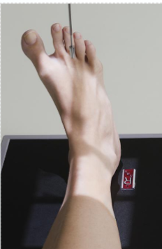
Ankle AP positioning
IR on table top
Patient supine or seated on table, leg extended. Support under knee as needed
Center and align leg and ankle joint parallel to edge of IR
To ensure true AP make sure there is no rotation of the lower leg long axis of foot is vertical parallel to CR
CR midway between malleoli
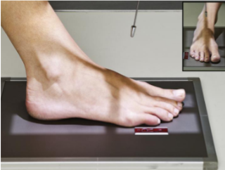
Ankle mortise positioning
IR on table top
From AP ankle position, rotate entire leg and ankle in 15-20 degrees
So that intermalleolar plane is parallel to IR
CR mid malleoli
Foot AP positioning
IR on table top
patient supine or seated with plantar surface flat on IR
Align foot long axis to long axis of IR
CR angled 10 degrees posteriorly to the base of the third metatarsal

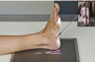
Foot medial oblique positioning
IR on table top
From Ap foot position rotate foot internally 30-40 degrees and support as needed
Higher arches = around 40 degrees
low arches = around 30 degrees
CR centered to base of the third metatarsal
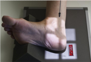
Calcaneus Plantodorsal axial positioning
IR on Table top
patient supine or seated. Dorsiflex foot 90 degrees till its near perpendicular to IR.
CR angled 40 degrees directed at the base of the 3rd metatarsal to emerge just distal and inferior to ankle joint
Calcaneus lateral positioning
IR on table top
Patient should be recumbent on affected side, knee flexed about 45 degrees with unaffected limb behind patient to prevent over rotation
place support under knee and leg as needed for true lateral
You can place a sponge under the lateral side of the foot as well to prevent over rotation
dorsiflex foot near 90 degrees to leg
CR Mid calcaneus 1in inferior to medial malleolus

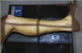
Lower leg(Tib Fib) AP positioning
IR on table top
Patient supine leg extended. Ensure no rotation of knee, lower leg, or ankle
include 1 - 2in minimum beyond knee and ankle joints
if limb is too long place the lower leg diagonally across the IR
make sure you have the lateral malleolus on the IR and a 1-2 inches of the knee joint
CR to mid Tib Fib
May increase SID to 44-48
Lower leg(Tib Fib) Lateral positioning
IR table top
patient recumbent affected side down. Place unaffected limb behind patient.
place support under foot as needed to achieve true lateral of foot ankle and knee
1-2 inches of knee and ankle joints required
If limb is too long place IR diagonally
CR mid Tib Fib
may increase SID to 44-48
Knee intercondylar fossa positioning (Camp coventry and Holmblad Choose one)
IR in table bucky for both
(If holmblad is performed standing place IR underneath knee)
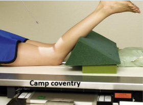
Camp coventry:
patient prone knee flexed 40-50 degrees with sponge underneath lower leg for support
Match CR angle to knee flexion 40-50 degrees caudad
Center CR at knee joint emerge at distal margin of patella
Holmblad:
Patient kneeling all fours on table or partially standing straddling table or chair
lean patient forward 20-30 degrees results in 60-70 degree knee flexion
CR perpendicular to IR center to popliteal crease


Knee AP positioning
IR in table bucky
Patient supine or seated on table with leg extended.
Rotate leg internally 3-5 degrees for true AP
angle 5° as needed(5°cephalic for big booty(>24cm), 5° caudad for small booty(<19cm) )
Center IR to CR
CR centered ½ in distal to apex of patella

Knee lateral positioning
IR in table bucky
May be taken crossfire or lateral recumbent position affected side down
Flex knee 20-30 degrees if patient is able.
unaffected leg placed behind patient
place support under foot and or ankle as needed for a true lateral
CR angled 5-7° cephalad centered 1in distal to medial epicondyle

Patella PA positioning
IR in table bucky
Patient prone knee centered to CR and midline of table or IR
rotate anterior knee 5° internally. Make sure epicondyles are parallel to the plane of the IR for true PA
CR centered to midpatella area
Patella Tangential positioning (settegast prone)
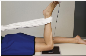
IR on table top
patient prone knee flexed 90°
plantar flex patients foot(pull heel back to point toes out)
Angle CR 15-20° centered to mid patellofemoral joint
SID 40-48

Forearm AP positioning
IR table top
Patient seated with hand and arm extended hand supinated
Drop shoulder to place upper limb on the same horizontal plane
have patient lean laterally as needed to get true AP
CR to midpoint of IR
SID 40
Forearm lateral positioning
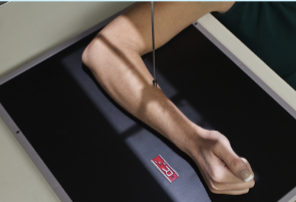
IR table top
Patient seated with elbow flexed 90°
drop shoulder to place entire upper limb on same horizontal plane
rotate hand and wrist into true lateral position(thumb up)
ensure that both wrist and elbow joints are included
CR to midpoint of forearm
SID 40

Elbow AP positioning
IR table top
Patient seated elbow extended hand supinated
Lean laterally as needed to get true AP
CR centered to midelbow joint
SID 40
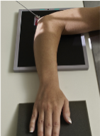
Elbow Coyle (radial head) positioning
IR table top
Patient supine or seated
elbow flexed 90° hand pronated if possible
CR angle 45° toward shoulder centered to radial head (about 1.5 inches from olecranon)

Shoulder internal rotation positioning
IR in wall bucky
Patient erect or supine
rotate body toward affected side as needed to ensure contact with IR
center of IR to scapulohumeral joint and CR
abduct arm slightly; internally rotate arm and hand until epicondyles of distal humerus are perpendicular to IR
CR directed 1in inferior to coracoid process
SID 40
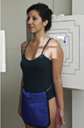
Shoulder External positioning
IR in wall bucky
Patient erect or supine
rotate body toward affected side as needed to ensure contact with IR
center of IR to scapulohumeral joint and CR
abduct arm slightly; Externally rotate arm until epicondyles are parallel to IR(supinate hand)
CR directed 1in inferior to coracoid process
SID 40

Shoulder Grashey positioning
IR in wall bucky
Patient erect or supine
rotate body 35-45° toward affected side into a LPO or RPO
Hand and arm in neutral position
CR to scapulohumeral joint approximately 2in inferior and 2in medial from the supralateral border of shoulder
SID 40

Shoulder scap Y view positioning
IR in wall bucky
Patient erect
Patient starts from PA and rotates affected shoulder 45-60° into a LAO or RAO
Unaffected arm should be up in front of the patient(you can rest it on the top of the bucky or their head)
CR to scapulohumeral joint
SID 40

Scapula AP positioning
IR in wall bucky
Patient should be erect
Top of IR should be approximately 2 inches above the shoulder
Gently abudct patients arm 90° and supinate hand and place back of the hand against patients forehead
CR to midscapula 2in inferior to coracoid process and 2in medial from lateral border of patient

Scapula Lateral positioning
IR in wall bucky
Patient erect
Patient is facing wall bucky placed in an RAO or LAO 45-60° until body of scapula is perp to IR
have patient pull the affected side arm across their body and place it on the opposite shoulder
(if the area of interest is the acromion and coracoid process let the arm hang down)
CR to midvertebral border of scapula
SID 40

Humerus AP positioning
IR in wall or table bucky
Patient erect or supine
arm slightly abducted supinate hand for true AP(Epicondyles parallel to IR)
included both elbow and shoulder joint
CR to midpoint of the humerus
SID 40 inches

Humerus Lateral(lateromedial) positioning
IR in wall or table bucky
patient erect or supine
include both elbow and shoulder joints
Elbow slightly flexed arm and wrist rotated internally until epicondyles are perpendicular to IR
CR to midpoint of Humerus
SID 40

Hip - Pelvis AP positioning
IR in wall or table bucky
patient erect or supine pelvis centered to IR
Both feet should be internally rotated 15-20°
CR mid way between ASIS and symphysis pubis, approximately 2in distal to level of ASIS
SID 40

Hip - Frog lateral unilateral positioning
IR in table or wall bucky
Patient erect or Supine
flex affected knee and hip and abduct femur 45° from vertical
Center IR to CR
CR perpendicular to midfemoral neck
(1-2in medial from ASIS, 3-4in distal from there)
SID 40

Pelvis inlet positioning
IR in table bucky
Patient should be supine, align midsagittal plane to CR and to midline of table
Center IR to CR
CR angled 40° to level of ASIS
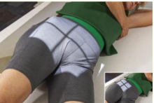
Pelvis LPO judet method (Upside) positioning
IR in table Bucky
Patient semisupine in a 45° left posterior oblique
Place 45° sponge under the right elevated side, keep arms out of the way of the light field
CR 2in distal to upside ASIS
SID 40in

Femur Ap (2 views - upper and lower) positioning
IR in table bucky
Proximal view:
patient supine, rotate affected leg internally 15-20°
CR directed at femoral neck
^(1-2in medial from ASIS, 3-4in distal from there)
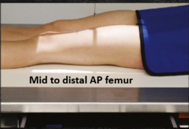
Mid to distal view:
patient supine, rotate entire limb 5° internally
lower border of IR about 2 inches below the knee to include knee joint
CR to mid femur

Femur Lateral (2 views - upper and lower) positioning
IR in table bucky CR centered perpendicular and locked into place.(MOVE TABLE NOT THE CR)
mid to Proximal view:
Lateral recumbent on affected side, flex knee 45°
unaffected leg placed behind patient
include hip joint in this view
CR perpendicular to femur, to mid IR
Mid to distal view:
move table and patient so that you can include knee in this view
CR perpendicular to femur, to mid IR
