RENAL EMERGENCIES
 Call Kai
 Learn
 Practice Test
 Spaced Repetition
 Match
 Flashcards
 Knowt Play
1/88

There's no tags or description
Looks like no tags are added yet.
89 Terms
Acute Kidney Injury (AKI)
is a rapid loss of kidney function caused by kidney damage. It can lead to life-threatening complications, including:
● Metabolic acidosis
● Fluid imbalance
● Electrolyte imbalance
50%
< 1 mg/dL
A common criterion for AKI is a (1)___ or greater increase in serum creatinine from baseline. Normal creatinine is generally (2) __ mg/dL.
>800 mL/day
<400 mL/day or <0.5 mL/kg/hour for 6 hours
<50 mL/day
AKI CHANGES IN URINE OUTPUT:
Nonoliguria: __ mL/day
Oliguria: __ mL/day or __ mL/kg/hour for __ hours
Anuria: __ mL/day
RIFLE Classification
Risk - ↑ 1.5×; ↓ ≥25%
Injury - ↑ 2×; ↓ ≥50%
Failure - ↑ 3×; ↓ ≥75%
Loss - >4
End-Stage Kidney Disease (ESKD) - >3
CLASSIFICATIONS OF ACUTE KIDNEY INJURY (AKI)
__- classifies AKI based on severity and outcomes:
R__ - Creatinine __ baseline OR GFR __
I__ - Creatinine __ baseline OR GFR __
F__ - Creatinine __ baseline OR GFR __; severe decrease in urine output
L__ - Complete loss of kidney function for __ weeks
E__ - for __ months
R, I, F
L, E
RIFLE Classification
__ = severity of AKI
__ = outcomes of kidney function loss
PRERENAL
60–70%
↓ GFR
(categories of AKI)
↓ blood flow to the kidneys
Most common (__%)
Causes: volume loss, hemorrhage, burns, GI losses, hypotension, sepsis, and shock
Results in ↓ __
INTRARENAL (Intrinsic)
glomeruli, tubules
Acute Tubular Necrosis (ATN)
↓ GFR, azotemia, and fluid/electrolyte imbalances
(categories of AKI)
damage to kidney tissue
Damage to the (1) __ or __
Most common type: (2)__
Can cause (3)__, __, & __
Associated with CKD, diabetes, heart failure, hypertension, and cirrhosis
POSTRENAL
kidney
GFR
(categories of AKI)
obstruction of urine flow
Caused by kidney stones, strictures, blood clots, BPH, malignancy, or pregnancy
Obstruction ↑ pressure in the (1)__ and (2) ↓ __
INITIATION
OLIGURIA
DIURESIS
RECOVERY; 3–12 months.
PHASES OF AKI:
__ – Begins with kidney injury and ends when oliguria develops.
__ – Urine output ↓; urea and creatinine accumulate, with risk of hyperkalemia.
__ – Urine output ↑ as kidney function begins to recover; dehydration is a risk.
__ – Renal function and laboratory values gradually return toward normal; recovery may take __ months.
↑ BUN and creatinine
↑ phosphorus
↓ calcium
Anemia
CLINICAL MANIFESTATIONS of AKI:
↑ __
↑ __
↓ __
A__
low or normal
hematuria, specific gravity
<20 mEq/L
>40 mEq/L
BUN, creatinine, electrolytes, and ECG
Ultrasound/CT
ASSESSMENT & DIAGNOSIS of AKI
Urine output may be __ or __
Possible __ and low __
Prerenal: urine Na⁺ __ mEq/L
Intrarenal: urine Na⁺ __ mEq/L, casts/debris
Monitor B__, c__, e__, and E__
__ may detect abnormalities
Hyperkalemia → cardiac arrhythmias → cardiac arrest
MAJOR COMPLICATION of AKI
H__ → C__ → C__
nephrotoxic medications; toxins
kidney function
drug levels
PREVENTION of AKI:
Identify and avoid __ and __
Monitor __ before and during treatment
Monitor __ of nephrotoxic medications when indicated
Hemodialysis (HD)
Peritoneal dialysis (PD)
Continuous renal replacement therapy (CRRT)
RRT/Dialysis may be needed for severe complications:
(HPC)
Monitor Fluid & Electrolytes
Reduce Metabolic Rate
Promote Pulmonary Function
Prevent Infection
Skin Care
Psychosocial Support
NURSING MANAGEMENT OF AKI:
__ – Monitor I&O, daily weight, electrolytes, and ECG; watch for hyperkalemia.
__ – Prevent and treat fever and infection.
__ – Encourage turning, coughing, and deep breathing.
__ – Use aseptic technique and avoid unnecessary catheters.
__ – Keep skin clean and moisturized; prevent breakdown.
__ – Explain treatments and provide emotional support to the patient and family
Chronic Kidney Disease (CKD)
glomerular filtration rate (GFR)
3
end-stage kidney disease (ESKD)
is kidney damage or a reduced (1)__ that lasts for (2)__ months or more. It can lower quality of life, increase healthcare costs, and lead to premature death. If untreated, CKD may progress to (3)__, where waste products build up in the body and renal replacement therapy (RRT), such as dialysis or kidney transplantation, is needed.
Major risk factors: cardiovascular disease, diabetes, hypertension, and obesity.

125 mL/min/1.73 m²
STAGES OF CKD CKD is classified into 5 stages based on GFR. Normal GFR is about __.

Anemia
Metabolic acidosis
calcium, phosphorus
CLINICAL MANIFESTATIONS of CKD
As kidney function declines and creatinine levels increase, symptoms may develop, including:
__due to decreased erythropoietin
M__
Abnormal __ and __ levels
Fluid retention and edema
Congestive heart failure
Electrolyte abnormalities
Worsening hypertension
GFR
Creatinine clearance; 24 hours.
GFR
ASSESSMENT AND DIAGNOSTIC FINDINGS of CKD
__ measures how much plasma is filtered by the kidneys.
__ measures how much creatinine the kidneys remove in __ hours.
__ is an important measure for assessing CKD and its progression.
Dialysis
hemodialysis (HD)
continuous renal replacement therapy (CRRT)
peritoneal dialysis (PD)
is a treatment used when the kidneys cannot adequately remove wastes, excess fluid, and electrolytes from the blood. The main types are:
HD
CRRT
PD
Acute or urgent dialysis
may be needed for:
Increasing serum potassium (hyperkalemia)
Fluid overload or impending pulmonary edema
↑ acidosis
Pericarditis, especially a pericardial friction rub indicating uremic pericarditis
Advanced uremia
Severe edema or hypertension not responding to treatment
Removal of certain medications or toxins from the blood
CHRONIC OR MAINTENANCE DIALYSIS
Used in advanced CKD and ESKD when there are:
Uremic S/Sx: such as nausea, vomiting, severe anorexia, lethargy, and mental confusion
Hyperkalemia
Fluid overload not controlled by diuretics and fluid restriction
General lack of well-being
Hemodialysis (HD)
3 times a week for 3–4 hours.
may be used: Short-term in AKI until kidney function returns, Long-term or permanently in advanced CKD and ESKD
prevents death but does not cure kidney disease or completely replace the endocrine and metabolic functions of the kidneys.
Most patients receiving intermittent HD are treated __ times a week for __ hours.
Remove excess fluid
Balance electrolytes
Help manage acidosis
blood
dialyzer
OBJECTIVES OF HD:
(RBH)
(1)__ is pumped from the patient to a (2)__ (artificial kidney), where wastes and excess fluid are removed, then the cleansed blood is returned to the patient.
1. Diffusion
2. Osmosis
3. Ultrafiltration
PRINCIPLES OF HD:
1. __ – wastes move from an area of ↑ concentration in the blood to a ↓ concentration in the dialysate.
2. __ – water moves across the semipermeable membrane.
3. __ – pressure is used to remove excess fluid more efficiently.
dialysate
The __ contains electrolytes in appropriate concentrations and helps control the patient's electrolyte levels.
Dialyzer
High-flux dialysis
contains thousands of tiny hollowfiber tubes that act as a semipermeable membrane. It allows toxins, fluid, and electrolytes to pass across the membrane while retaining larger molecules such as RBCs and proteins.
uses highly permeable membranes and higher blood-flow rates to improve removal of low- and medium-molecular-weight substances and shorten treatment time.
Vascular Access Devices
A double-lumen catheter may be inserted into the:
Internal jugular vein
Femoral vein
It provides immediate access for acute HD but may cause bleeding, hematoma, infection, thrombosis, pneumothorax, or inadequate flow.
Arteriovenous Fistula (AVF)
3
The preferred permanent vascular access.
An artery is surgically connected to a vein, usually in the forearm.
It needs about __ months to mature before use.
Patients may perform hand exercises, such as squeezing a rubber ball, to help enlarge the vessels.
Arteriovenous Graft (AVG)
A graft connects an artery and vein using biologic or synthetic material. It is used when the patient's vessels are not suitable for an AV fistula
Stenosis, Infection, Thrombosis
Arrhythmias, Air embolism, Dialysis disequilibrium syndrome
bruit and thrill
Common Complications of Dialysis (SIT)
Complications of Hemodialysis (AAD)
The access should also be checked for a __ and __.
DIALYSIS DISEQUILIBRIUM Syndrome
>150
Caused by cerebral fluid shifts. Signs include:
Headache
Nausea and vomiting
Restlessness
Decreased level of consciousness
Seizures
It is more likely in AKI or very high BUN levels (__ mg/dL).
Sodium, Potassium, Phosphorus, Fluid
1.2; 2
<4
Patients on HD commonly need restrictions on: S__, P__, P__, F__
Protein intake is about __ g/kg ideal body weight/day, with high-biologic-quality protein. Sodium is usually restricted to __ g/day.
The goal is to keep interdialytic weight gain __% of estimated dry weight.
Continuous Renal Replacement Therapy (CRRT)
used to remove excess fluid, electrolytes, and metabolic wastes from the blood.
Requires access to the patient's circulation
Blood continuously passes through the filter
Treatment can be performed in the critical care unit (CCU)
is commonly indicated for patients who:
Have acute or chronic kidney disease
Are too clinically unstable for traditional hemodialysis
Have fluid overload due to oliguria
Have kidneys that cannot handle their high metabolic or nutritional needs
Process: Blood access → Blood passes through an artificial filter → Waste and excess fluid are removed → Blood returns to the patient
Hemofilter
Extremely porous blood filter
Contains a semipermeable membrane
Allows removal of excess fluid and waste products
key component of CRRT
Continuous Renal Replacement Therapy (CRRT)
provide:
Integrated fluid warmer - warms infusion and dialysate fluids
Fluid measurement system - Helps accurately monitor fluid balance
Battery backup - allows treatment to continue when the patient is transported
Can be initiated quickly in critical-care settings
Provides continuous and gentle fluid removal
Useful for hemodynamically unstable patients.
HYPOVOLEMIA
or Fluid Volume Deficit (FVD), occurs when the body loses more fluid than it takes in.
Causes: Vomiting, diarrhea, blood loss, burns, fever, excessive sweating, GI suction, 3rd-space fluid shifts, and inadequate fluid intake.
S/Sx: Weight loss, ↓ blood pressure, ↑ heart rate, thirst, dizziness, weakness, dry skin, ↓ urine output, and confusion.
Laboratory Findings: ↑ Hgb/Hct, ↑ BUN/creatinine, ↑ urine specific gravity, ↑ osmolality, and ↓ urine sodium.
oral fluids
IV isotonic fluids: 0.9% NaCl or Lactated Ringer's
fluid challenge
1 kg weight loss ≈ 1 L fluid loss
vomiting or diarrhea.
Hypovolemia Mgmt:
For mild fluid loss, __ are preferred when the patient can drink safely.
For severe or acute fluid loss, __ such as __ or __ may be given.
__ may be used to determine whether ↓ kidney function is related to reduced blood flow
__ weight loss ≈ __ fluid loss
The nurse should also identify and manage ongoing fluid losses such as __ or __.
HYPERVOLEMIA
or Fluid Volume Excess (FVE), occurs when the body retains excessive sodium and water, ↑ extracellular fluid volume.
Causes: Kidney disease, heart failure, cirrhosis, nephrotic syndrome, excessive IV fluids, and hyperaldosteronism.
S/Sx: Weight gain, edema, ascites, distended neck veins, crackles, shortness of breath, ↑ blood pressure, and bounding pulse
Lab/Dx Findings: BUN and hematocrit may ↓ because of dilution. Chest X-ray may show pulmonary congestion.
Fluid restriction + sodium restriction + prescribed diuretics
hemodialysis, peritoneal dialysis, or CRRT
Thiazide Diuretics (Hydrochlorothiazide)
Loop Diuretics (Furosemide)
fluid retention
Hypervolemia Mgmt:
F__ + S__ + P__ are commonly used to reduce excess fluid.
If kidney function is severely impaired, H__, P__, or C__ may be needed.
Used mainly for mild to moderate fluid excess.
Used for more severe fluid overload because they cause greater sodium and water loss.
Rapid weight gain may indicate __
sodium
high-sodium
Lemon, garlic, and onion
Potassium-containing salt substitutes; spironolactone.
Hypervolemia Mgmt:
Reducing __ helps ↓ water retention and edema.
Patients should limit __ foods and check food labels.
L__, G__, and O__ may be used for flavor.
__ should be used cautiously in kidney disease or when taking potassium-sparing diuretics such as __.
fluid and electrolyte
Skin turgor
OLDER ADULT CONSIDERATIONS for Hypervolemia
Older adults are more sensitive to __ and __ changes.
__ is less reliable in older adults because skin elasticity decreases with age.
Electrolyte imbalance
occurs when the level of an electrolyte in the body becomes too low or too high. Assessment is based on the patient’s history, physical findings, and laboratory results.
Sodium (Na⁺)
135–145 mEq/L
is the major electrolyte in the extracellular fluid (ECF). It helps control fluid balance, blood volume, and osmolality and is important for nerve impulses and muscle contraction.
is mainly regulated by ADH, thirst, and the renin-angiotensin-aldosterone system (RAAS).
NV:__
HYPONATREMIA
occurs when the serum sodium level is ↓ 135 mEq/L.
Usually, it happens because of too much water compared with sodium or excessive sodium loss.
Causes: Fluid overload, excessive water intake, sodium loss through sweating/GI losses, and SIADH.
can cause neurologic problems

S/Sx of Hyponatremia
(SALT LOSS)
Serum Na⁺ <135 mEq/L; Serum osmolality ↓
SIADH
Hyponatremia Lab Findings (SS)
occurs when there is too much ADH, causing the kidneys to retain excessive water; ↑ ADH → ↑ water reabsorption → water retention → dilution of Na⁺ → hyponatremia; The problem is mainly too much water, not simply a lack of sodium.
0.9% NaCl or Lactated Ringer's
fluid restriction
hypertonic saline (3% NaCl).
slowly
confusion, lethargy, muscle twitching, & seizures.
fluid overload
Hyponatremia Mgmt:
Sodium may be given orally or through IV fluids such as __ or __ when sodium loss is the cause.
When excess water is the cause, such as in SIADH, __ is commonly used.
Seizures, severe confusion, or coma may require carefully controlled __
Sodium must be corrected __ to prevent serious neurologic injury.
Watch closely for c__, l__, m__, and s__
When giving IV fluids, monitor for __, especially in patients with heart or kidney problems.
Hypernatremia
>145 mEq/L
>300 mOsm/kg
occurs when the serum sodium level is above 145 mEq/L.
It usually occurs because of water loss greater than sodium loss or excessive sodium gain
The brain is particularly sensitive to this cellular dehydration.
The urine is usually concentrated as the kidneys try to conserve water.
Assessment:
Serum Na⁺ __ mEq/L
↑ Serum osmolality __ mOsm/kg

Intense thirst
HYPERNATREMIA SIGNS & SYMPTOMS
(FRIED SALT)
The most characteristic symptom is: __
water; sodium.
0.45% NaCl or D5W
cerebral edema
Desmopressin (DDAVP)
thirst, restlessness, confusion, disorientation, and lethargy.
HYPERNATREMIA Mgmt:
The main goal is to replace __ and gradually lower __.
Depending on the patient's condition, treatment may include: __ or __
The sodium level must be lowered gradually because correcting it too quickly can cause __
If Diabetes Insipidus is the cause, __ may be given because it acts like ADH.
Watch for t__, r__, c__, d__, and l__.
Dysnatremia
<135 mEq/L
>145 mEq/L
swelll
shrink
means an abnormal sodium level in the blood. It includes:
Hyponatremia → Na⁺ __ mEq/L
Hypernatremia → Na⁺ __ mEq/L
HYPO → H₂O goes INTO cells → cells __
HYPER → H₂O goes OUT of cells → cells __
Potassium
98%
3.5–5.0 mEq/L
is the major intracellular electrolyte.
About (1)__% of potassium is inside the cells.
It is very important for muscle contraction, nerve impulses, and especially cardiac function.
Normal Level (2)__ mEq/L
Even small changes can cause dangerous cardiac and neuromuscular problems.
Prominent U wave
digoxin toxicity.
__ = classic ECG finding of hypokalemia.
Hypokalemia can also increase the risk of __
HYPOKALEMIA
occurs when the serum potassium level is below 3.5 mEq/L.
Causes: vomiting, diarrhea, gastric suction, diuretics, excessive urination, corticosteroids, and hyperaldosteronism. It can also occur when potassium shifts into the cells, such as during alkalosis.
Pathophysiology: ↓ K⁺ in blood → ↓ muscle and nerve activity → muscle weakness + cardiac abnormalities

Hypokalemia S/Sx
(A SIC WALT ↓)
Potassium
oral potassium
ECG
muscle strength and bowel sounds
intake and output and renal function.
IV push; infusion pump
Hypokalemia Mgmt:
__-rich foods
__ supplements
Monitoring of serum potassium and __
Assess m__ and b__
Monitor i__ and r__
Never administer potassium by __. use an __ for IV replacement.
HYPERKALEMIA
Causes: Kidney disease, Potassium-sparing diuretics, Excessive potassium supplements, Metabolic acidosis Addison disease, Severe burns and crush injuries, ACE inhibitors and NSAIDs
Patho: ↓ potassium excretion or cellular damage → High serum potassium → Abnormal cardiac electrical activity → Dangerous arrhythmias and possible cardiac arrest.

Hyperkalemia S/Sx
(MURDER)
Calcium
8.8–10.4 mg/dL
↓ 8.8 mg/dL.
↑ 10.4 mg/dL.
is an essential mineral for strong bones, nerve transmission, muscle contraction, and blood clotting.
Normal total serum calcium: __ mg/dL
Hypocalcemia: ↓ __ mg/dL.
Hypercalcemia: ↑ __ mg/dL.
Hypocalcemia
Cause: Hypoparathyroidism, Vitamin D deficiency. Chronic kidney disease, Pancreatitis, Malabsorption, Magnesium deficiency, Thyroid or neck surgery, Chronic diarrhea
Patho: ↓ calcium level → ↑ nerve and muscle excitability → Muscle spasms, tingling, and tetany → Possible seizures and cardiac abnormalities.

Hypocalcemia S/Sx
(CRAMPS)
Oral calcium
IV calcium gluconate
Vitamin D
Magnesium
calcium, magnesium, & phosphate
muscle spasms, tingling, & reflexes.
laryngospasm.
milk, cheese, & leafy vegetables.
Hypocalcemia Mgmt:
__ supplements for mild cases
__ for severe or symptomatic cases
__ supplementation
__ replacement when deficient
Monitoring of c__, m__, and p__ levels
Assess m__, t__, and r__
Monitor airway and breathing for __.
Encourage calcium-rich foods such as m__, c__, and l__

HYPERCALCEMIA
↑ calcium level → ↓ nerve and muscle excitability → Muscle weakness, constipation, and lethargy → Kidney stones, cardiac abnormalities, and possible cardiac arrest.
Causes: (CHIMP)

Hypercalcemia S/Sx
(BACK ME)
IV normal saline
Calcitonin
Bisphosphonates
Loop diuretics
ambulation & mobility.
fiber; constipation.
mental status & cardiac rhythm.
digoxin toxicity
Hypercalcemia Mgmt:
__ to promote calcium excretion when appropriate
__ for rapid, temporary calcium reduction
__for selected cases
__ only when indicated, for fluid overload after adequate hydration
Promote a__ and m__.
Encourage __ intake to prevent __.
Assess M__ and C__.
Monitor for D__ when applicable
Phosphorus
2.7–4.5 mg/dL
↓ 2.7 mg/dL
↑ 4.5 mg/dL
is an electrolyte needed for strong bones and teeth, muscle function, energy production (ATP), and oxygen delivery.
Normal Value: __ mg/dL
Hypophosphatemia: __ mg/dL
Hyperphosphatemia: __ mg/dL
HYPOPHOSPHATEMIA
Causes: Malnutrition or starvation, Alcohol withdrawal, Vomiting and diarrhea, Vitamin D deficiency, DKA treatment, Low magnesium or potassium, Excessive use of antacids or diuretics
Patho: ↓ phosphorus → ↓ ATP → muscle weakness and impaired oxygen delivery → confusion, respiratory failure, and possible seizures.

Hypophosphatemia S/Sx
(RNCRB)
Oral phosphate
IV phosphate
phosphorus-rich
Vitamin D
phosphorus & calcium
muscle strength & mental status.
milk, fish, meat, beans, & nuts.
fall & seizure
Hypophosphatemia Mgmt:
__ supplements for mild cases
__ for severe cases
Encourage __ foods
__ supplementation if needed
Monitor p__ and c__ levels
Assess m__ and m__
Encourage m__, f__, m__, b__, and n__
Use f__ and s__ precautions when indicated.
HYPERPHOSPHATEMIA
Cause: Acute or chronic kidney disease, Excessive phosphorus intake, Excess vitamin D, Hypoparathyroidism, Severe muscle or tissue breakdown, Excessive use of phosphate-containing laxatives or enemas
Patho: ↑ phosphorus → ↓ calcium levels → muscle cramps and tetany.
Excess phosphorus may also combine with calcium and deposit in soft tissues, blood vessels, and organs.
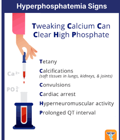
Hyperphosphatemia — Signs and Symptoms
Tweaking
Calcium
Can
Clear
High
Phosphate
phosphorus.
phosphate binders
diuretics
Hemodialysis
phosphorus, calcium, & kidney function.
muscle cramps & tingling.
phosphate-containing laxatives
Hyperphosphatemia Management:
Restrict dietary __.
Give prescribed __ with meals.
Use __ only when appropriate.
__ for severe cases, especially with kidney failure
Monitor p__, c__, and k__
Assess for m__ and t__.
Avoid __ unless prescribed.
Chloride
97–107 mEq/L
↓ 97 mEq/L.
↑107–108 mEq/L.
is an electrolyte that maintains fluid balance and acid– base balance.
Normal Value: __ mEq/L
Hypochloremia: __ mEq/L.
Hyperchloremia: __ mEq/L.
HYPOCHLOREMIA
Tremors
Cardiac arrhythmias
seizures or coma (Severe cases)
Causes: Excessive vomiting or diarrhea, Gastric suctioning, Excessive sweating, Diuretic use, Dehydration and electrolyte loss, Excessive bicarbonate intake, Chloride-deficient IV fluids
Patho: Chloride loss → bicarbonate ↑ → blood becomes too alkaline (metabolic alkalosis) → muscle cramps, weakness, and possible cardiac arrhythmias.
Signs and Symptoms: (TCS)
sodium & potassium
diuretics
chloride levels & blood pH
chloride, sodium, potassium, & ABG results.
such as milk, cheese, eggs, & broth.
Hypochloremia Management
Correct s__ and p__ imbalances.
Adjust d__ if prescribed.
Monitor c__ and b__
Check c__, s__, p__, and A__
Encourage chloride-containing foods when appropriate, such as m__, c__, e__, and b__
HYPERCHLOREMIA
Weakness
lethargy
fluid retention or swelling
Causes: Dehydration, Excessive normal saline, IV infusion, Kidney dysfunction, Severe diarrhea causing bicarbonate loss, Excessive sodium or chloride intake, Metabolic acidosis
Patho: Excess chloride or bicarbonate loss → chloride level ↑ → blood becomes too acidic (metabolic acidosis) → rapid breathing, weakness, and possible cardiac complications
S/Sx: (WLF)
fluid & acid–base
chloride-containing IV fluids
fluids, bicarbonate, or diuretics
chloride, sodium, & ABG
respiratory, neurological, & cardiac
edema; consciousness.
Hyperchloremia Management
Correct __ and __ imbalances
Adjust c__ as prescribed.
Give prescribed f__, b__, or d__ when indicated.
Monitor c__, s__, and A__
Assess r__, n__, and c__ status
Observe for __ and changes in __.
Acid–base disturbances
lungs
kidneys
occur when the blood becomes too acidic or too alkaline.
The __ regulate CO₂, while
the __ regulate bicarbonate (HCO₃⁻).
7.35–7.45
35–45 mmHg
22–26 mEq/L
80–100 mmHg
Metabolic acidosis
Metabolic alkalosis
Respiratory acidosis
Respiratory alkalosis
Normal ABG Values:
pH: __
PaCO₂: __ mmHg
HCO₃⁻: __ mEq/L
PaO₂: __ mmHg
(typical adult range at sea level)
Four Types of Acid–Base Imbalance
__
__
__
__
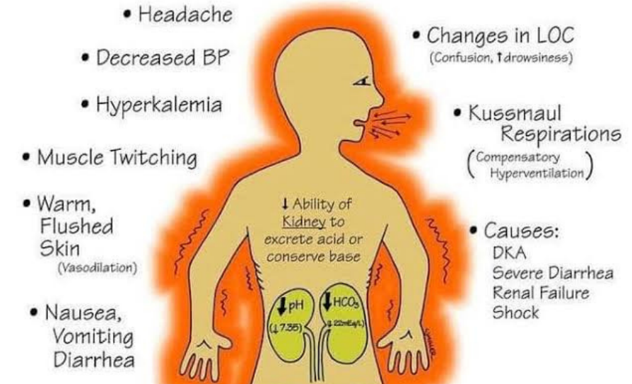
Metabolic Acidosis
Causes: DKA, Kidney failure, Severe diarrhea, Lactic acid buildup, Poisoning or toxic substances
Patho: Excess acid or bicarbonate loss → HCO₃⁻ ↓ → blood pH ↓.
IV fluids and insulin
Bicarbonate or dialysis
ABG, potassium.
breathing and cardiac rhythm.
Metabolic Acidosis Mgmt:
Give __ and __ for DKA as prescribed
B__ or D__ for selected severe cases
Monitor A__, p__, vital signs, and consciousness.
Assess b__ and c__
Respiratory Acidosis
Cause: COPD, Severe pneumonia, Airway obstruction, Respiratory muscle weakness, Sedative or opioid overdose
Patho: Hypoventilation → CO₂ retention → carbonic acid ↑ → blood pH ↓.

ventilation
bronchodilators or antibiotics
oxygen
Noninvasive or mechanical
Respiratory Acidosis Mgmt:
Improve __ and treat the cause.
Give prescribed __ or __ when indicated.
Provide __ as needed.
__ ventilation if necessary.
Respiratory Alkalosis
Causes: Anxiety or panic attacks, Pain, Fever or sepsis, Hypoxemia, Excessive mechanical ventilation
Patho: Hyperventilation → excessive CO₂ loss → carbonic acid ↓ → blood pH ↑.

hypoxemia.
breathing pattern, mental status.
reassurance, calm coaching
anxiety
Respiratory Alkalosis Mgmt:
Correct __.
Assess b__ and m__
Provide __ and __ when anxiety is the cause.
Never assume __ is the cause without assessment.
Metabolic Alkalosis
Cause: Severe vomiting, Prolonged gastric suction, Diuretic use, ↓ potassium, Excessive bicarbonate intake
Patho: Loss of stomach acid or excess bicarbonate → HCO₃⁻ ↑ → blood pH ↑.

normal saline
potassium, chloride
muscle function, respiratory status.
intake/output, cardiac rhythm.
Metabolic Alkalosis Mgmt:
Replace fluids with prescribed __ when appropriate.
Replace p__ and c__ as needed.
Assess m__ and r__
Monitor i__ and c__