Anatomical structures

0.0(0)
Studied by 0 people
call kaiCall Kai
learnLearn
examPractice Test
spaced repetitionSpaced Repetition
heart puzzleMatch
flashcardsFlashcards
GameKnowt Play
Card Sorting

1/72

encourage image

There's no tags or description

Looks like no tags are added yet.

73 Terms

1
New cards

Nasal septum

Divides the nasal cavity into left and right sides.

<p>Divides the nasal cavity into left and right sides.</p>
2
New cards

Nasal conchae (superior) (meatus nasi superior)

bony shelf

<p>bony shelf</p>
3
New cards

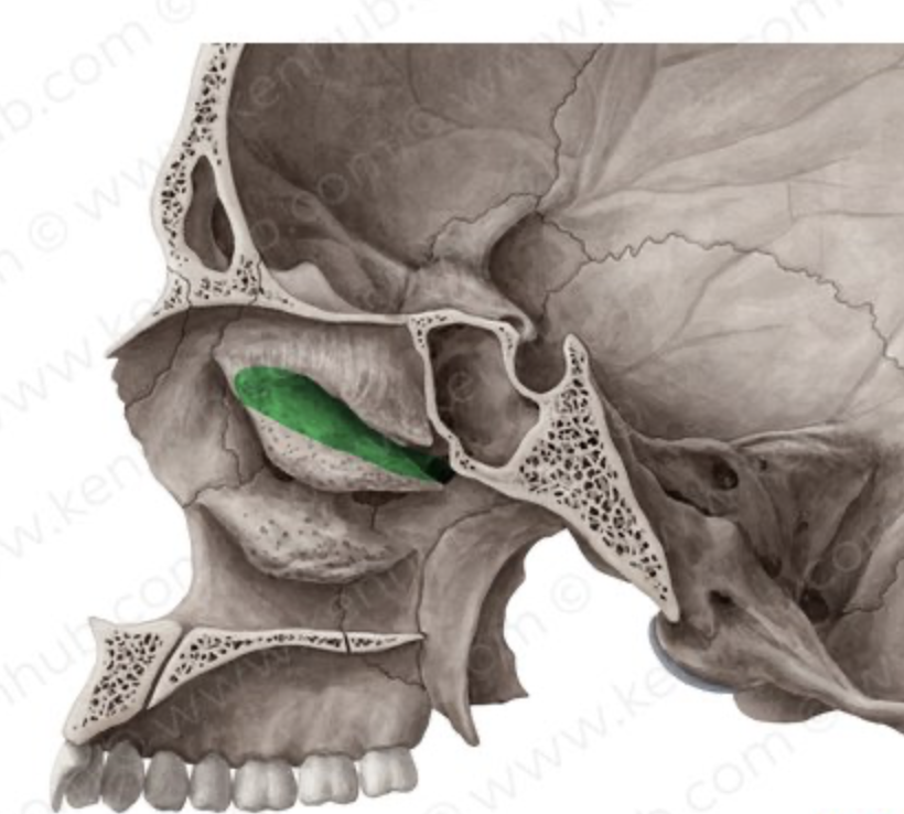
Nasal meatuses (superior)

air passage
<p><span>air passage</span></p>
4
New cards

Paranasal sinuses

Air-filled cavities connected to the nasal cavity; increase mucosal surface area.

<p>Air-filled cavities connected to the nasal cavity; increase mucosal surface area.</p>
5
New cards

Olfactory region

Contains receptors responsible for smell.

<p>Contains receptors responsible for smell.</p>
6
New cards

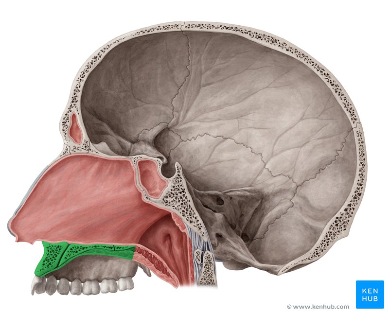
Hard palate (palatum durum)

Forms the floor of the nasal cavity (podniebienie)

<p>Forms the floor of the nasal cavity (podniebienie)</p>
7
New cards

Soft palate (palatum molle)

Elevates during swallowing to close off the nasal cavity from the pharynx.

<p>Elevates during swallowing to close off the nasal cavity from the pharynx.</p>
8
New cards

Nasopharynx

Contains the pharyngeal tonsil and openings of the auditory tubes.

<p>Contains the pharyngeal tonsil and openings of the auditory tubes.</p>
9
New cards

Oropharynx

Communicates with the oral cavity; passage for air and food.

<p>Communicates with the oral cavity; passage for air and food.</p>
10
New cards

Laryngopharynx

Shared passage leading toward the larynx and esophagus.

<p>Shared passage leading toward the larynx and esophagus.</p>
11
New cards

Pharyngeal tonsil (adenoid)

high up in the throat, behind the nasal cavity on the roof and posterior wall of the nasopharynx

<p>high up in the throat, <strong>behind the nasal cavity</strong> on the roof and posterior wall of the nasopharynx</p>
12
New cards

Tubal tonsils

in the nasopharynx

<p>in the <strong>nasopharynx</strong></p>
13
New cards

Palatine tonsils

at either side of the lateral wall of the oropharynx

<p>at either side of the lateral wall of the oropharynx</p>
14
New cards

Lingual tonsil

at the root of the tongue

<p>at the root of the tongue</p>
15
New cards

Waldeyer’s ring

Ring of lymphoid tissue providing early defense against pathogens.

<p>Ring of lymphoid tissue providing early defense against pathogens.</p>
16
New cards

Hyoid bone

Associated with the larynx and supports structures involved in swallowing.

<p>Associated with the larynx and supports structures involved in swallowing.</p>
17
New cards

Thyroid cartilage

Supports and protects the glottis and entrance to the trachea; Adam’s apple

<p>Supports and protects the glottis and entrance to the trachea; Adam’s apple</p>
18
New cards

Cricoid cartilage

Supports and protects the glottis and entrance to the trachea.

<p>Supports and protects the glottis and entrance to the trachea.</p>
19
New cards

Epiglottis

Folds backward during swallowing to help prevent food and liquid entering the respiratory tract.

<p>Folds backward during swallowing to help prevent food and liquid entering the respiratory tract.</p>
20
New cards

Arytenoid cartilages

Move and adjust vocal-fold position and tension.

<p>Move and adjust vocal-fold position and tension.</p>
21
New cards

Glottis

Opening between the vocal folds.

<p>Opening between the vocal folds.</p>
22
New cards

Vocal folds

Vibrate to produce sound; adduction closes the glottis, consists of:

  1. Mucous membrane

  2. Vocal ligament

  3. Vocalis muscle


<p>Vibrate to produce sound; adduction closes the glottis, consists of:</p><ol><li><p><span><strong>Mucous membrane</strong></span></p></li><li><p><span><strong>Vocal ligament</strong></span></p></li><li><p><span><strong>Vocalis muscle</strong></span></p></li></ol><p></p>
23
New cards

Vestibular folds

False vocal folds; lie above the vocal folds and are not responsible for normal sound production.

<p>False vocal folds; lie above the vocal folds and are not responsible for normal sound production.</p>
24
New cards

Intrinsic laryngeal muscles

Move laryngeal cartilages relative to each other

<p>Move laryngeal cartilages relative to each other</p>
25
New cards

Extrinsic laryngeal muscles

Move and stabilize the larynx as a whole.

<p>Move and stabilize the larynx as a whole.</p>
26
New cards

Superior laryngeal nerve

Branch of the vagus nerve; supplies the upper larynx.

<p>Branch of the vagus nerve; supplies the upper larynx.</p>
27
New cards

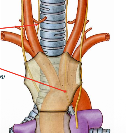
Recurrent laryngeal nerve

Branch of the vagus nerve; supplies the lower larynx and intrinsic laryngeal muscles.
<p>Branch of the vagus nerve; supplies the lower larynx and intrinsic laryngeal muscles.</p>
28
New cards

Trachea

Conducts air; supported by C-shaped cartilage rings.

<p>Conducts air; supported by C-shaped cartilage rings.</p>
29
New cards

Tracheal cartilages

Keep the trachea open; C-shaped rings leave a posterior opening for the esophagus.

<p>Keep the trachea open; C-shaped rings leave a posterior opening for the esophagus.</p>
30
New cards

Trachealis muscle

Smooth muscle spanning the posterior tracheal wall; contraction narrows the trachea.

<p>Smooth muscle spanning the posterior tracheal wall; contraction narrows the trachea.</p>
31
New cards

Carina

Internal ridge at the tracheal bifurcation separating the openings of the primary bronchi.

<p>Internal ridge at the tracheal bifurcation separating the openings of the primary bronchi.</p>
32
New cards

Right primary bronchus

Wider and descends at a steeper angle than the left; aspirated foreign bodies are more likely to enter it.

<p>Wider and descends at a steeper angle than the left; aspirated foreign bodies are more likely to enter it.</p>
33
New cards

Left primary bronchus

Main airway carrying air into the left lung.

<p>Main airway carrying air into the left lung.</p>
34
New cards

Secondary (lobar) bronchi

Supply the lung lobes; three on the right and two on the left.

<p>Supply the lung lobes; three on the right and two on the left.</p>
35
New cards

Tertiary (segmental) bronchi

Supply bronchopulmonary segments.
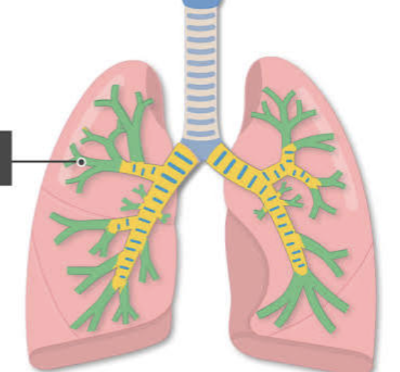
<p>Supply bronchopulmonary segments.</p>
36
New cards

Bronchioles

Smaller conducting airways; cartilage progressively decreases and is absent in bronchioles.

<p>Smaller conducting airways; cartilage progressively decreases and is absent in bronchioles.</p>
37
New cards
Terminal bronchioles
Last part of the conducting portion before the respiratory portion; contain smooth muscle but no cartilage.
38
New cards

Alveolar ducts

Conduct air toward the alveoli; part of the respiratory portion.

<p>Conduct air toward the alveoli; part of the respiratory portion.</p>
39
New cards

Alveoli

Main sites of gas exchange between air and pulmonary capillary blood.

<p>Main sites of gas exchange between air and pulmonary capillary blood.</p>
40
New cards

Respiratory mucosa

a specialized mucous membrane lining the upper and lower respiratory tracts, warms the air coming in

41
New cards

Cilia

Move mucus and trapped particles toward the pharynx.

<p>Move mucus and trapped particles toward the pharynx.</p>
42
New cards

Lamina propria

a thin layer of loose (areolar) connective tissue that lies directly beneath the surface epithelium in mucous membranes (mucosa) across the body

<p>a thin layer of loose (areolar) connective tissue that lies directly beneath the surface epithelium in mucous membranes (mucosa) across the body</p>
43
New cards

Right lung

divided into three lobes: the superior, middle, and inferior lobes, contains two fissures: an oblique fissure and a horizontal fissure

<p><span>divided into <strong>three lobes</strong>: the superior, middle, and inferior lobes, contains <strong>two fissures</strong>: an <strong>oblique fissure</strong> and a <strong>horizontal fissure</strong></span></p>
44
New cards

Left lung

divided into two lobes: the superior and inferior lobes, contains only one fissure: the oblique fissure

<p><span>divided into <strong>two lobes</strong>: the superior and inferior lobes, contains only <strong>one fissure</strong>: the <strong>oblique fissure</strong></span></p>
45
New cards

Horizontal fissure

Separates the superior and middle lobes of the right lung.

<p>Separates the superior and middle lobes of the right lung.</p>
46
New cards

Oblique fissure

Separates lung lobes; present in both lungs.

<p>Separates lung lobes; present in both lungs.</p>
47
New cards

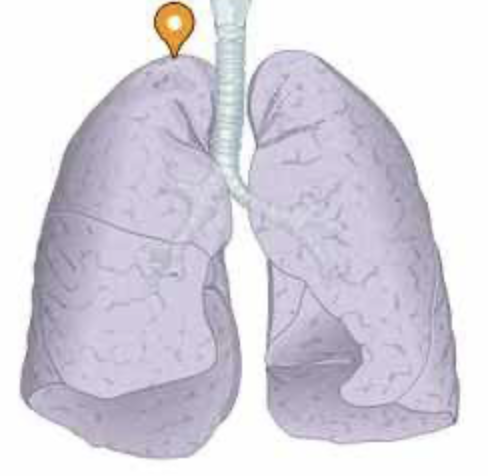
Apex of lung

knowt flashcard image
48
New cards

Hilum of lung

Passage for the primary bronchus

<p>Passage for the primary bronchus</p>
49
New cards

Visceral pleura

Serous membrane directly covering the lung. (purple on the picture)
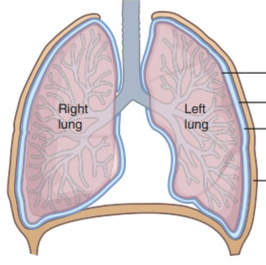
<p>Serous membrane directly covering the lung. (purple on the picture)</p>
50
New cards

Parietal pleura

Serous membrane lining the thoracic wall (navy on the picture)

<p>Serous membrane lining the thoracic wall (navy on the picture)</p>
51
New cards

Pleural cavity

Space between the visceral and parietal pleura; contains lubricating pleural fluid. (blue on the picture)

<p>Space between the visceral and parietal pleura; contains lubricating pleural fluid. (blue on the picture)</p>
52
New cards

Pleural fluid

In the pleural cavity. Reduces friction between pleural surfaces during breathing.

53
New cards
Mediastinum
Central thoracic compartment separating the lungs; contains the heart.
54
New cards

Diaphragm

Primary muscle of inhalation; contraction moves it downward and increases thoracic volume.

<p>Primary muscle of inhalation; contraction moves it downward and increases thoracic volume.</p>
55
New cards

External intercostal muscles

Elevate the ribs during inhalation.

<p>Elevate the ribs during inhalation.</p>
56
New cards

Internal intercostal muscles

Assist forceful exhalation by depressing the ribs.

<p>Assist forceful exhalation by depressing the ribs.</p>
57
New cards

Sternocleidomastoid muscle

Accessory muscle of forceful inhalation; helps elevate the sternum.

<p>Accessory muscle of forceful inhalation; helps elevate the sternum.</p>
58
New cards

Scalene muscles

Accessory muscles of inhalation; elevate the first and second ribs.

<p>Accessory muscles of inhalation; elevate the first and second ribs.</p>
59
New cards

Pectoralis minor muscle

Accessory muscle that can assist inhalation by elevating the ribs when the shoulder girdle is fixed.

<p>Accessory muscle that can assist inhalation by elevating the ribs when the shoulder girdle is fixed.</p>
60
New cards

Serratus anterior muscle

Accessory muscle that can assist inhalation when the shoulder girdle is stabilized.

<p>Accessory muscle that can assist inhalation when the shoulder girdle is stabilized.</p>
61
New cards

Transversus thoracis muscle

Assists forceful exhalation.

<p>Assists forceful exhalation.</p>
62
New cards

External oblique muscle

Compresses the abdomen during forceful exhalation.

<p>Compresses the abdomen during forceful exhalation.</p>
63
New cards

Internal oblique muscle

Compresses the abdomen during forceful exhalation.

<p>Compresses the abdomen during forceful exhalation.</p>
64
New cards

Rectus abdominis muscle

Compresses the abdomen during forceful exhalation

<p>Compresses the abdomen during forceful exhalation</p>
65
New cards
Pulmonary artery
Carries deoxygenated blood from the heart to the lungs for gas exchange.
66
New cards
Pulmonary veins
Carry oxygenated blood from the lungs to the heart.
67
New cards

Bronchial arteries

Supply the lung tissue itself; distinguish them from pulmonary vessels involved in gas exchange.

<p>Supply the lung tissue itself; distinguish them from pulmonary vessels involved in gas exchange.</p>
68
New cards

Sphenoidal sinus

knowt flashcard image
69
New cards

frontal sinus

knowt flashcard image
70
New cards

nares (nostril)

knowt flashcard image
71
New cards

nasal conchae (middle) meatus nasi medius

knowt flashcard image
72
New cards

nasal conchae (inferior) meatus nasi inferior

knowt flashcard image
73
New cards

Palatum durum

knowt flashcard image