Lesson 3.2: Dentin
 Call Kai
 Learn
 Practice Test
 Spaced Repetition
 Match
 Flashcards
 Knowt Play
1/102

There's no tags or description
Looks like no tags are added yet.
103 Terms
Where are odontoblasts located?
The bodies are located at the interface between the pulp and dentin.
The processes are in the dentin.
How thick is the dentin?
1-1.5 mm in the incisors and 3 mm in the molars
Thicker in cusps than in root.
Thicker in mature teeth than in young teeth
What are the limits of the dentin?
Crown outer surface: Enamel
Outer root surface: Cementum
Inner surface: pulp chamber and root canals
What is the function of the dentin?
Protecting the pulp
What are the components of dentin?
Mineralized matrix
Dental ducts or tubules, where the odontoblastic processes are housed
What is the embryological origin of the dentin and pulp?
Ectomesenchyme
They are embryologically and functionally related


How is the matrix of the dentin?
It is a bone-like matrix with multiple closely packed dentinal tubules running through its entire thickness
What do the dentinal tubules contain?
The cytoplasmic extensions of the odontoblasts that once formed dentin
Where are the odontoblast cell bodies attached to?
To a layer of Predentin forming the peripheral boundary of the dental pulp
How is the colour of dentin?
Yellowish-white
What determines the colour of dentin?
Degree of mineralization (white-blue, primary teeth)
Pulp vitality (gray, low vitality, pulp removal)
Age makes them yellower (physiological sclerosis of the tubules or reactive due to the deposition of intratubular calcosperides)
Pigments of endogenous origin (Hb degradation and/or fractures) or
exogenous origin (metallic fillings).
How translucent is dentin?
Dentin is less translucent than enamel and in the apical areas there is less thickness and the root canal can be seen through transparency. It decreases with age
How hard is dentin?
It depends on the degree of mineralization.
It is lower than that of enamel and higher than that of bone and cementum.
The hardness is similar to that of Ag amalgam in young people.
How elastic is dentin?
More elastic than enamel but not elastic, compensates the rigidity of enamel by cushioning impacts
Depends on organic matrix and water
How radiopaque is dentin?
Depends on mineral content
It is lower than that of enamel and higher than that of bone
How is the permeablity of dentin?
Greater than in the enamel due to the dentinal molecules, that allow the passage of molecules, solutes and microorganisms
Fluids diffuse by pressure from interstitial fluids of the pulp (depending on diameter and length of tubule) and by diffusion
Important in the clinic for the adhesion of materials

How is the composition of dentin?


How is the composition of dentin?

What is predentin and when is it created?
First deposit of the odontoblast which is a non mineralized matrix mainly consisting of collagen fibers.
It is released in the early bell stage and then mineralized to dentin in late bell stage
Does predentin disappear with time?
No it remains constant and there is always a layer of predentin.
it is thicker in the active process of dentinogenesis and decreases in thickness with age
How thick is predentin?
10-50 micrometers

When does the formation of coronal dentin begin?
It begins at the bell stage in the papillary tissue adjacent to inner enamel epithelium

In which direction does the formation of coronal dentin happen?
Dentin formation extends down the cusp slope to the cervical loop of enamel organ and dentin thickens until all the coronal dentin is formed
When does root dentin formation start and end?
Slightly later stage of development
Completion of root dentin formation up to 18 months after tooth eruption
What is necessary for the formation of root dentin?
Proliferation of epithelial cells (Hertwigs epithelial root sheath) to initiate differentiation of root odontoblasts
How is the composition of the organic matrix of dentin?
Collagen fibers (90%):
Type I (98%)
Type III (1-2%)
Type V (1%)
VI, IV
Non collagenous proteins (10%)
Dentin Phosphoprotein (DPP)
Dentin sialoprotein (DSP)
Dentin Matrix Protein -1 (DMP 1)
What is the function of Type I?
It acts as a scaffold that accommodates a large proportion (estimated at 56%) of the mineral in the orifices and pores of the fibrils.
Collagen fibrils are packed and accumulate along the periphery of the dentinal tubules.
What is the function of non-collagenous matrix proteins in the organic matrix?
They regulate mineral deposition and can act as inhibitors, promoters and/or stabilizers.
What is the function of proteoglycans in the organic matrix?
Prevent premature mineralization of the organic matrix.
More concentrated in predentin.
What is the function of Dentin phosphoprotein (DPP) in the organic matrix?
- High negative charge, binds large amounts of calcium.
- Initiate hydroxyapatite formation.
What is the function of Dentin sialoprotein (DSP) in the organic matrix and where is it ?
Synthesized by young odontoblasts, it participates in the epithelium-mesenchyme interaction.
Predominantlydetected in peritubular dentin (inhibits its growth and thus prevents tubule occlusion).
What is the function of Dentin Matrix Protein -1 (DMP 1) in the organic matrix and where is it found?
Predominantly detected in peritubular dentin (inhibits its growth and thus prevents tubule occlusion and calcification).
Has also the dual function of focusing on mineralization
What are the components of the inorganic matric?
Hydroxyapatite crystals: Chemically similar to those of enamel, cementum and bone. Small size and thin. Oriented parallel to the collagen fibers of the tooth matrix.
Amorphous phosphates
Carbonates
Sulfates
Trace elements


What is part of the primary dentin?
Mantle dentin: Outer layer under enamel and cementum (Does not contain DPP)
Circumpulpal dentin: Formed after the mantle and represents the largest volume of dentin
Order of primary dentin formation
Mantle dentin of crown
Circumpumpal dentin of crown
Mantle dentin of the root
Circumpumpal dentin of root
When does secondary dentin develop?
After all primary dentin including the root has been completed.
It has a continuous deposition but much slower.
How is the structure and composition of secondary dentin?
It has a tubular structure continuous with that of primary dentin
The mineral / organic material ratio is the same as for primary dentin
Where is the secondary dentin positioned?
Not deposited uniformly around the periphery of the pulp chamber
Asymmetric reduction in size and shape of pulp chamber
What is the synonyme of tertiary dentin and what is its function?
Also called reactive or restorative dentin.
Reaction to various stimuli, such as wear, caries, or a restorative dental procedure
What produces tertiary dentin and What determines its quality and quantity?
Produced only by cells directly affected by the stimulus
Quality and quantity are related to the cellular response initiated, which depends on the intensity and duration of the stimulus.
What produces tertiary dentin and What determines its quality and quantity?
Produced only by cells directly affected by the stimulus
Quality and quantity are related to the cellular response initiated, which depends on the intensity and duration of the stimulus.
How is it called when the odontoblasts that form the tertiary dentin are included in the dentin?
Osteodentin
Types of tertiary dentin?
Reactive: Matrix secreted by surviving odontoblasts. Mild/moderate stimuli
Reparative: Formed by new cells similar to odontoblasts. Moderate/high stimuli
What are the stages of dentin formation?
1. Elaboration of an organic matrix (Predentin)
2. Matrix maturation (change with non-collagenous proteins)
3. Precipitation of mineral salts
Each formation stimulates secretion


Order of secretion?
Young ameloblast (internal ep.) → odontoblast → secretion of dentin → ameloblast → secretion of enamel
Odontoblast life cycle: Odontoblast differentiation
The undifferentiated dental papilla cells are separated from the inner enamel epithelium by an acellular zone containing a few fine collagen fibrils.
These cells reverse their polarity. Ectomesenchymal cells proximal to the acellular zone differentiate into preodontoblasts and then into odontoblasts as their cytoplasm increases in volume to contain increasing amounts of protein synthesizing organelles.
The acellular zone between the dental papilla and the internal enamel epithelium becomes cellular, the odontoblasts differentiate and increase in size occupying this zone.
These newly differentiated cells are characterized by being highly polarized, with their nuclei located away from the inner enamel epithelium.
What triggers odontoblast differentiation?
Triggered by the expression of signaling molecules and growth factors in the cells of the inner enamel epithelium (IL-6, IL-10, TGF-β).
Cells of the inner enamel epithelium and undeferentiated mesenchymal cells
Cells of inner enamel:
Divide, adapting to the growth of tooth germ. Some of the cytoplasmic extensions of preameloblasts pass though the BM and the acellular zone reaching ectomesenchymal cells of papilla
Undifferentiated mesenchymal cells
Increasing volume by accumulating more organelles and are located in the periphery of the dental papilla
Small, have a stellate shape with large nucleus, excm contains few collagen fibers, scarce cytoplasm with few organelles

Preodontoblasts
Preodontoblasts exit the cell and inhibit cell duplication.
Mitotic spindle gives rise to to two overlapping cells in the last divison. The closest to the bm will diferentiate into an odontoblast and the underlying cell gives rise to the subodontoblastic Höhl's or reserve cells
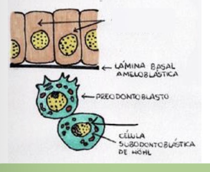
Young odontoblasts
• They develop adhesive bonding systems and communicate with each other.
• They undergo a change of polarity.
• As a result of such polarization the cell volume increases.
• The cell becomes cylindrical and the nucleus moves distally. At the proximal pole there is also a single, larger extension called the odontoblastic process

Secretory odontoblast
The young odontoblast increases in volume and adopts a more cylindrical morphology.
The odontoblastic process increases in length and is arranged perpendicular to the basal lamina.
Immediately the odontoblast begins its secretory activity and from that moment on it is called secretory odontoblast.
The secretory activity of this cell occurs towards the proximal pole, which secretes the predentin that occupies the space between the enamel organ and the odontoblasts.
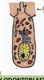
Secretory mature odontoblast
Once the predentin is formed, the odontoblast contributes to the first mineralization of the predentin and its transformation into calcified dentin matrix (formation of mantle dentin).
When the odontoblastic extension is found in the dentinal tubule of the newly formed dentin matrix, the odontoblast is called “mature”
In the process the odontoblast moves toward the center of the Pulp.

What contributes to the formation of dentin?
The proteins: DMP-1, DSPP, DSP and DPP synthesized by the mature
secretory odontoblasts.
What is the function of the Dentin matrix protein -1 (DMP-1)?
Inhibits mineralization, facilitating the formation of predentin and also stimulating it (DUal function)
It is capable of sequestering and stabilizing calcium phosphate nanoparticles that form in the matrix due to the accumulation of calcium from odontoblasts.
What is the function of the Dentin sialophosphoprotein (DSPP)?
It is elaborated by the odontoblast and splits into DSP and DPP . They are related to the mineralization process and to the beginning of the nucleation of the mineral component.
Which dentin is the first formed and what is its thickness?
Mantle dentin which located below the enamel and cementum with a thickness of 80-100 micrometer
Which protein is not present in mantle predentin ?
DPP
Process of mantle dentin formation
Secretion of Korff fibers: Large diameter type III collagen fibrils associated with fibronectin (0.1 to 0.2 μm in diameter). They extend from the odontoblasts towards the internal epithelium of the enamel.
Once the odontoblast process is formed, a series of small vesicles attached to the odontoblast membrane, known as matrix vesicles, are secreted. It is only after the presence of these vesicles that mineral crystals associated with the collagen fibers of the matrix are observed arranged so that their major axis is parallel to the fibers.
Non-collagenous matrix proteins produced by odontoblasts come into play to regulate mineral deposition (non-DPP). Coronal mantle dentin forms in a layer approximately 15 to 20 μm thick upon which circumpulpal dentin is added.
After the first deposition of mantle dentin, the ameloblasts phagocytize the basal lamina, leaving an interface of mixed dentin and enamel.


When does the formation of circumpumpal dentin occur?
When the mantle dentin calcifies, the mature odontoblasts continue to produce organic matrix to form the circumpulpal dentin.
What is the difference of the matrix of the circumpumpal dentin to the mantle dentin?
No matrix vesicles are formed and the mineralization follows a globular pattern (calchospherites) and then coalesce
What are the two steps of circumpumal dentin formation?
The formation of organic predentin on the side adjacent to the odontoblasts and matrix mineralization on the side adjacent to the preformed dentin.
During the mineralization of the predentin, changes in its composition occur, so that the predentin and the matrix of the already mineralized dentin have different components.
How does the mineralization of the circumpumpal predentin work?
odontoblastic processes, Ca²⁺, proteins, and a new series of proteoglycans are released.
At this mineralization front, DPP (only present here) can be detected, which initiates mineralization by binding to collagen fibers.
The molecular configuration of DPP facilitates the formation of hydroxyapatite crystals from the amorphous precursor, and its activity depends on the degree of phosphorylation.
When does the root dentin formation begin and how does it work?
It begins once the formation of the enamel is complete.
Root odontoblasts differentiate from ectomesenchymal cells in the periphery of the papilla under the induction of the inner epithelium of the enamel organ and the Hertwig sheath.
What is the difference of the root dentin formation to the coronary?
slower in the root than in the crown
The Pattern of mineralization is similar, but the calcospherites are smaller.
orientation and organization of the collagen fibers (collagen fibers of the cementum mixed with those of the dentin).
When is secondary dentin deposited?
Secondary dentin is deposited after root formation by the same odontoblasts that produced primary dentin, but at a much slower rate.
What happens with secondary dentin formation?
Dentinal tubules become less regularly organized and some areas even absent
continued secretory activity of odontoblasts leads to deposition of peritubular dentin, which progressively narrows the lumen of the tubules, reducing their permeability.
At the same time, the overall thickness of dentin increases, which gradually reduces the size of the pulp chamber.
As this process advances, odontoblasts become crowded and
some may eventually die (Continously in life)
When is tertiary dentin formed and what determines the deposition rate?
They are deposited at specific sites in response to injury, by damaged odontoblasts or pulp replacement cells.
Deposition rate will depend on the degree of injury: ↑↑ injury ↑↑↑ deposition rate.
Rapid deposition: cells often get trapped in the newly formed matrix (tubular pattern becomes very distorted)
Which molecules participate in Tertiary dentin formation?
↓↓ Collagen, DSP and DMP1.
↑↑ BSP (bones sialoprotein) and osteopontin.
What is reactionary and reparative dentinogenesis?
Reactionary dentinogenesis: Occurs in repsonse to a mild stimulus or slowly advancing carious lesion. It is compensated by the upregulation of surviving odontoblast activity and new tubular dentin formed
Reparative dentinogenesis: Occurs in Response to severe or aggressive stimulus or rapidly advancing carious lesion. The death of primary odontoblasts is compensated by replacement with odontoblast-like cells which form low quality atubular mineralised tissue
What are the types of dentin structural units?
Primary (basic)
Dentinal tubules
Matrix or intertubular dentin
Secondary
Incremental or growth lines
Interglobular dentin and Czermak spaces
Tomes granular zone
Schregers tooth lines or bans
Dentinoenamal and cemetnoenamel junction
What are dentinal tubules and what do they contain?
Cylindrical structures that extend through the entire thickness of the dentin.
From the pulp to the dentinoenamel (DEJ) and cementodentinal junction (CDJ).
Contain odontoblastic processes (many along their entire length) and the dentinal fluid that comes from the pulp

What is the function of dentinal tubules?
They give the dentin its vitality, nutrition and ability to respond to various
stimuli by conducting them to the pulp region.
What nutritions the dentin?
Dentinal fluid rich in Na+ and poor in K+ with albumins and globulins (1/5 less
than plasma).
What happens with dentinal tubules when there is caries?
They increase permeability facilitating carious process but also pulp response to dental restorative procedures.
The processes may disintegrate or retract leaving behind an empty tubule (dead zone).
Reparative dentin seals these dead tracts at their pulpal end, thus protecting the pulp from infection.
What is intertubular dentin and what are its characteristics?
• It is distributed between the walls of the dentinal tubules.
• All organic components of dentin can be detected.
• A network of type I collagen fibrils in which apatite crystals, non-
collagen proteins and some plasma proteins are deposited
What surrounds the dentinal tubules and what is the differenced to the intratubular dentin?
The tubules are surrounded by a highly calcified dentin called peritubular dentin. It is hypermineralized compared to the intertubular (higher amount of DSP and DMP1).
What is sclerotic dentin?
Sclerotic dentin appears when several dentinal tubules become clogged with calcified material.
Glassy appearance; dentin becomes translucent


A - dentin affected by caries
B – sclerotic dentin
C – dead tubules
D – secondary dentin
E - dentin
F - enamel
G – pulp


What are van ebner lines?
• Lines of less mineralization due to physiological pauses of the odontoblast (every 5 days).
• Their arrangement is perpendicular to the dentinal tubules

What are pathological or owens lines?
• Lines spaced further apart (neonatal line)
• They occur due to periods of inadequate malnutrition or febrile periods


Van ebner lines

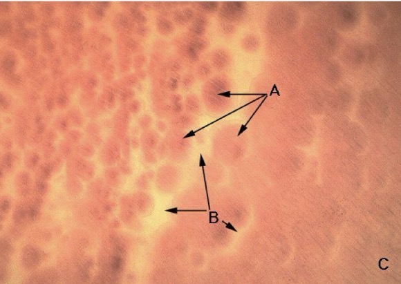
What are czermack spaces and what is their cause?
Areas of non-mineralized or hypomineralized dentin where globular areas of mineralization due to improper bonding in the calcospherites.
Vitamin D deficiency or exposure to high levels of fluoride at the time of dentin formation.
Greater presence in the circumpulpal dentin just below the mantle dentin.
(In the near of cementoenamel junction)


A = Calchospherides
C = Czermack spaces
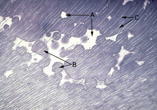
A = Czermack spaces
B = Calchospheride
C = interglobular dentin

What is tomes granular zone and where is it found?
It is observed as a thin dark stripe parallel to the cementodentinal junction.
It is located only at the periphery of the entire root dentin
Numerous cavities and small irregular spaces can be distinguished at higher magnification
At higher magnification, numerous cavities and a small irregular spaces can be observed

What forms tomes granular zone?
Due to the special arrangement of collagen and non-collagenous matrix proteins at the dentin- cementum interface

Schregers tooth lines or bands
Represent the more or less abrupt change of direction of the dentinal tubules during primary curvature
Homologous to the Hunter-schreger bands of enamel

DEJ and CDJ

What are the three theories of dentin sensitiivty?
Dentin contains nerve endings that respond when stimulates
Odontoblasts act as receptors and are coupled to the pulp nerves
Tubular nature of dentin allows fluid movement to occur within the tubule when a stimulus is applied. The movement would be detected by the detected by the pulpal free nerve endings near the odontoblasts

Why is pulp innervation important?
It plays an important role in pulp homeostasis and its defense mechanisms
Dentin contains nerve endings that respond when stimulated
Pro: Some nerves are arranged within certain tubules in the internal dentin.
Against: But dentin sensitivity does not depend solely on the stimulation of such nerve endings.

Odontoblasts serve as receptors and are occupied to the pulp nerves
In favor:
Odontoblast of neural origin: retains a capacity to transduce and propagate an impulse.
Against:
No synaptic relationship between odontoblast and pulpal nerves.
The membrane potential of odontoblasts measured in vitro is too low to allow transduction.
Local anesthetics and protein precipitants do not eliminate sensitivity

Hydrodynamic theory: Movement of the fluid within the tubule when a stimulus is applied.
Pressure changes that take place are transmitted to the intertubular free nerve endings affecting the subodontoblast nerve plexus
Movement of fluid through the tubule distorts the local pulp environment and is detected by free nerve endings in Raschkow´s plexus.
Movement of interstitial fluid which depends on the physiology of blood vessels will result in intravascular and extravascular pressure changes
