9. Angle Closure Glaucoma
 Call Kai
 Learn
 Practice Test
 Spaced Repetition
 Match
 Flashcards
1/142

There's no tags or description
Looks like no tags are added yet.
143 Terms
___________________ is contact between the iris and trabecular meshwork.
iridotrabecular contact (ITC)
what are the 2 types of ITC?
appositional closure (reversible)
peripheral anterior synechiae (PAS, chronic)
What is appositional closure?
Apposition/touch (not adhesion) between the iris and trabecular meshwork
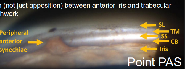
What is PAS?
Adhesion between the iris and trabecular meshwork

PAS =
chronic adhesion (not just apposition) btw anterior iris & trabecular meshwork

posterior synechiae =
adhesion btw posterior iris and anterior lens
ITC of ______________, is considered an occludable angle
180 degrees or more
ways to classify angle closure:
• Primary angle closure suspect (PACS)
• Primary angle closure (PAC)
• Primary angle closure glaucoma (PACG)
Primary angle closure suspect (PACS)
• At least 180˚ of iridotrabecular contact in the absence of peripheral anterior synechiae (PAS) plus normal IOP, disc, and visual field.
• This is an at-risk eye.
Primary angle closure (PAC)
• At least 180˚ of iridotrabecular contact with either elevated IOP and/or PAS plus normal disc and visual fields
• The angle is abnormal in structure (PAS) or function (elevated IOP)
Primary angle closure glaucoma (PACG)
• At least 180˚ of iridotrabecular contact plus elevated IOP plus optic nerve and visual field damage
• The angle is abnormal in structure and function, and there is glaucomatous optic neuropathy
_______________ are considered primary angle closure diseases (PACD)
Primary angle closure (PAC) & Primary angle closure glaucoma (PACG)
PACS, PAC, and PACG are all considered ____________________________.
Spectrum of Primary Angle Closure (sPAC)
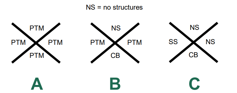
Assume normal IOP, disc, and visual field and no PAS. Which gonio findings below indicate an occludable angle (and therefore a primary angle closure suspect)?
C
What are the anatomical mechanisms of angle closure?
• Pupillary block
• Plateau iris
• Lens-related, including phacomorphic
_________________ can and often do occur with a single angle closure pt.
multiple anatomical mechanisms
• Sudden onset of IOP elevation resulting from total angle closure
• Accompanied by symptoms of severe, usually unilateral, ocular pain, red eye, blurred vision, haloes, headache, nausea, and vomiting.
acute angle closure
• An episode of sudden IOP elevation that is spontaneously aborted
• Condition may be recurrent but transient (“intermittent”)
• Symptoms may be mild or even absent (“subacute”)
• PAS may form and progress (“creeping”)
intermittent angle closure
• Chronic IOP elevation due to the presence of peripheral anterior synechiae (PAS) that permanently close the anterior chamber angle
• Symptoms are usually absent.
chronic angle closure
a pt may shift btw _______________ throughout their lifespan.
acute, intermittent, and chronic angle closure
Anything in the posterior chamber that ________ the angle closed
pushes
Anything in the anterior chamber that _____ the angle closed
pulls
PACG is _____ compared to the number of occludable angles.
rare
_____ of occludable angles progress to PACG.
<5%
Acute PACG is _________ compared to chronic PACG.
uncommon
1 acute PACG for every ___ chronic PACG
3
what group of people have the most PACG?
inuit women
________ of people have narrow angles in US
2-6%
_______ total glaucoma cases are PACG.
6%
3 biggest risk factors for PACG:
older age
female sex
inuit/asian ancestry
4 methods to evaluate angle closure:
van Herick
gonioscopy (clinical standard)
Ant Seg OCT
ultrasound biomicroscopy (UBM)
______________ is not a replacement for gonio.
van Herick
van Herick is a _____________ to determine who needs gonio during routine/comprehensive exams.
triage tool
van Herick CANNOT identify __________________.
occludable angles
van Herick is not an acceptable evaluation tool in an established __________________________ pt.
PACS, PAC, or PACG
gonio allows for ________________ view.
circumferential
there is no replacement for gonioscopy. Not even _____________.
AS-OCT
Why is gonioscopy necessary?
to identify TM pigmentation, Sampaolesi line, angle recession, and angle neovascularization
gonioscopy is the ONLY way to identify ______________________, and therefore differentiate btw normal, PACS, PAC, and PACG
an occludable angle
gonio remains indispensable for diagnosis but insufficient for _______________________.
risk stratification
when performing compression (or indentation) gonio, the contact area must be less then the _____________________. You also must use a 4-mirror lens.
corneal diameter
______________________ often cannot be visualized without compression.
PAS
ant seg OCT pros
non-contact method
biometric data
room lighting adjustable
less stray light vs gonioscopy
longitudinal changes
ant seg OCT cons
not circumferential
optical (reduced penetration vs ultrasound)
Central ACD (ant chamber depth) is a measurement tool on the AS-OCT. What is the normal ACD range?
2.5mm to 3.5mm
What does the angle opening distance measure from?
posterior scleral spur to iris surface
What is the normal angle opening distance?
250um to 750um
(0.25mm to 0.75mm)
_____________ is the perpendicular distance btw the anterior lens apex and the spur-to-spur reference line.
lens vault
lens vault values anterior to the spur-to-spur reference line are ____________________.
positive
lens vault values posterior to the spur-to-spur reference line are ____________________.
negative
UBM pros
deeper penetration then AS OCT
allows imaging of posterior chamber (especially ciliary body pathology)
some biometric data
UBM cons
not circumferential
decreased resolution vs AS-OCT
What do we use to predict PACS to PAC?
baseline IOP
anterior chamber depth (ACD)
angle opening distance 500, especially nasal
the risk for progression from PACS to PAC uses the IOP recommendation of:
risk increases for ever 1mmHG increase over 15mmHg
the risk for progression from PACS to PAC uses the ACD recommendation of:
risk increases for every 100um (or 0.1mm) shallower than 2.2mm
the risk for progression from PACS to PAC uses the angle opening distance 500 recommendation of:
risk increases for those with nasal AOD500 </= 90um
pupillary block has the highest prevalence from the __________________, but it is possible at any decade.
6th - 7th decade
(67 LETS GOOOOOO)
_________________ have a higher risk of pupillary block by 2-3x.
females
pupillary block has _________________ heredity.
mostly sporadic
what are the ocular risk factors for pupillary block?
hyperopia
shallow AC centrally and peripherally
short axial length
small corneal diameter
anterior position of the lens
pathophysiology of pupillary block
1. Lens-iris apposition
2. Pressure differential between posterior and anterior chambers
3. Anterior bowing of peripheral iris (iris bombe)
4. Appositional closure
PAS (may or may not be present)
5. Elevated IOP
6. Glaucomatous optic neuropathy
Elevated IOP and optic neuropathy can occur even in the absence of ___________.
PAS

What features of pupillary block can be seen at the slit lamp?
narrow angles
shallow central depth
iris bombe
a narrow angle on gonioscopy is characteristic of _________________.
pupillary block
_________________ can be used to help identify appositional closure.
corneal wedge
if there is appositional closure, the angle will ______________ with indentation/compression.
widen
if PAS is present, ______________ is necessary to visualize it.
indentation
how will pupillary block present on AS-OCT?
narrow angle depth
shallow central chamber depth
small corneal diameter
what can a UBM tell us about pupillary block?
rules out secondary causes of pupillary block
not routinely used in “run-of-the-mill” pupillary block
what even is pupillary block?
Iridolenticular contact sufficient to limit the normal flow of aqueous from posterior chamber to anterior chamber
pupillary block leads to a _________________ btw the 2 chambers.
pressure differential
When there is a pressure differential, USUALLY the pressure is _____________ in the posterior chamber than the anterior chamber (“normal” pupillary block)
greater
in what syndrome does REVERSE pupillary block occur?
pigment dispersion syndrome
what is REVERSE pupillary block?
the pressure is greater in the anterior chamber than in the posterior chamber, due to anatomical differences in iris insertion into the CB.
ways to manage pupillary block:
• Surveillance (Educate and document!)
• Laser peripheral iridotomy (LPI)
• Argon laser peripheral iridoplasty (ALPI)
• Cataract extraction
****NO PILOCARPINE******
LPI allows for __________________ the posterior and anterior chambers, allowing relief for pressure differentials.
communication btw

THE ANGLE WILL ______________ AFTER SUCCESSFUL LPI IN PTS WITH PUPILLARY BLOCK.
WIDEN
argon laser peripheral iridoplasty (ALPI) is not a common tx in _______________________.
pupillary block
iridioplasty (also called gonioplasty) applies ____________________ to the peripheral iris.
low-energy laser burns
the burns in ALPI are _______________________.
NOT full thickness
ALPI allows for ____________ of the peripheral iris away from the TM. It effectively widens the ACA.
contraction
What else can ALPI do?
break PAS
plateau iris occurs in ______ of PACG cases.
30%
plateau iris occurs in more in ______________.
females
plateau iris is less common in ____________.
hyperopes
pts with plateau iris are more likely to have _____________________.
(+)FHx of angle closure
plateau iris pathophysiology
1. Anomalous ciliary body position, size, and/or rotation creates plateau configuration
2. Bolstered peripheral iris crowds iridocorneal angle
3. Appositional closure
PAS (may or may not be present)
4. Elevated IOP
5. Glaucomatous optic neuropathy
plateau iris primary anomaly
ciliary body
plateau iris secondary anomaly
iridocorneal angle
how does plateau iris present on SLE?
narrow iridocorneal angles
deep central chamber
what is the characteristic sign found on gonioscopy with plateu iris syndrome 1?
double hump sign
the double hump sign associated with plateau iris 1 is only visible with ____________________.
compression gonioscopy
plateau iris can present with variable degrees of _________________________.
appositional closure and PAS
how will plateau iris present on AS-OCT?
• Improved appreciation of plateau configuration
• Narrow iridocorneal angles
• Often normal ACD and LV
• But cannot image ciliary body (primary anomaly in plateau iris)
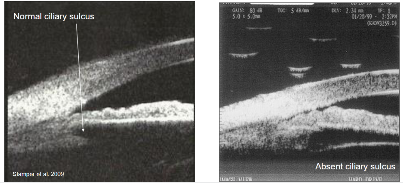
UBM reveals a pathognomonic sign of plateau iris: _______________________________.
absence of the ciliary sulcus
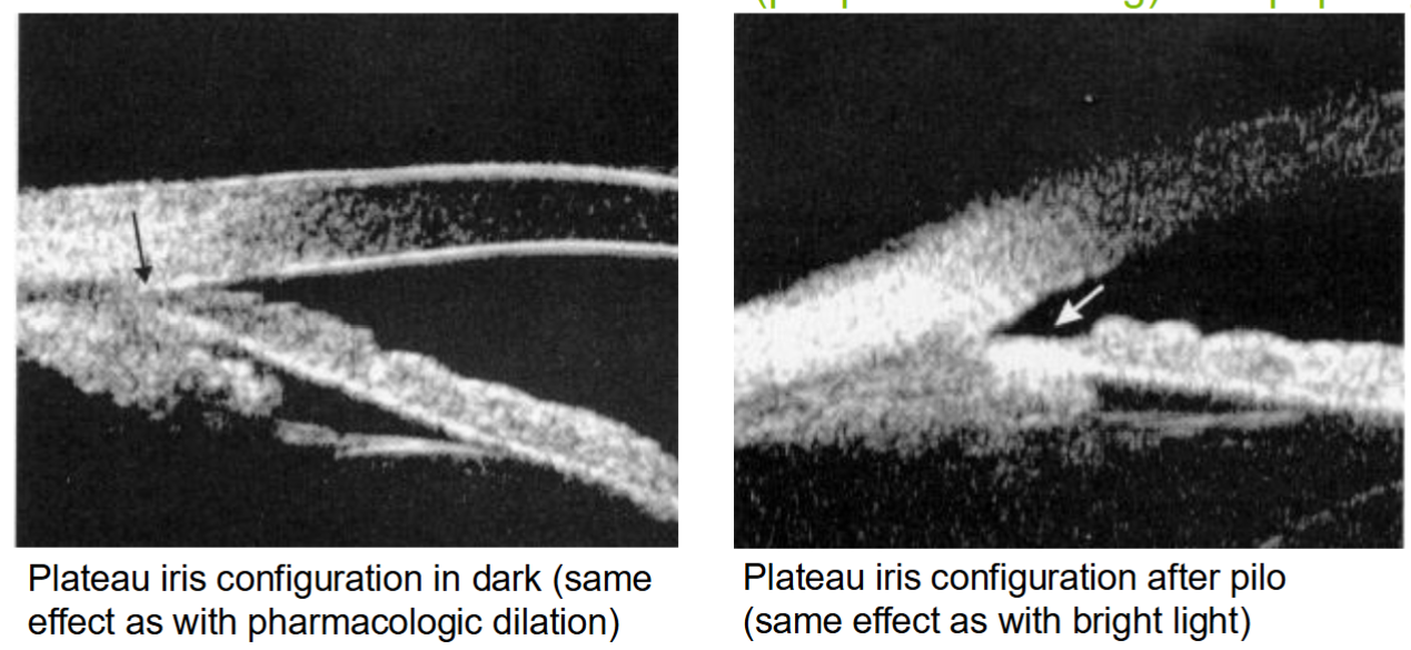
____________ can still close a plateau iris angle, but it is thru a different mechanism (peripheral crowding) than pupillary block.
dilation
What are the management strategies for plateau iris?
surveillance (educate & document)
LPI (NOT IN ISOLATION)
ALPI
cataract extraction (variable success)
What’s the deal with LPI and plateau iris?
• To relieve any pupillary block component
• LPI alone does NOT treat plateau iris
• A “failed” LPI means that the anterior chamber did not deepen centrally or peripherally following LPI.
• A “failed” LPI is strongly suggestive of plateau iris.
__________________ is most commonly used in plateau iris.
ALPI
Why has phacomorphic decreased in prevalence in developed countries?
cataract surgery