Primary Brain Cancer: Anatomy, Multimodality Management, and Radiation Therapy Techniques
 Call Kai
 Learn
 Practice Test
 Spaced Repetition
 Match
 Flashcards
 Knowt Play
1/33

Earn XP
Description and Tags
Comprehensive vocabulary flashcards covering the basic epidemiology, functional and cross-sectional neuroanatomy, imaging modalities, multimodality clinical management, and precision external beam radiation therapy planning principles for primary brain cancer.
34 Terms

Corpus Callosum Anatomical Segments
The major interhemispheric white matter tract comprising five anatomical segments from anterior to posterior: the rostrum, genu, body, isthmus, and splenium.

Major Cerebral Sulci and Fissures
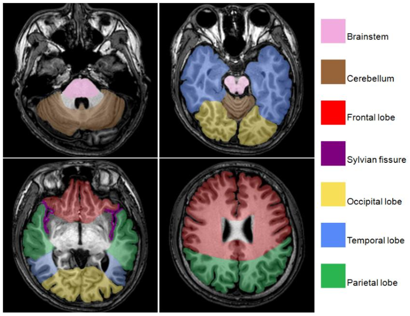
Key brain landmarks including the longitudinal fissure (divides the two cerebral hemispheres), the lateral (Sylvian) fissure (separates the temporal lobe from the frontal and parietal lobes), and the central sulcus (separates the frontal lobe from the parietal lobe).

Cranial Meninges
The three protective connective tissue envelopes surrounding the brain and spinal cord, arranged from superficial to deep as the dura mater, arachnoid mater, and pia mater.
Meningioma
The most prevalent primary non-malignant CNS tumor, originating from the arachnoid cells of the meninges and accounting for 54.5% of all non-malignant brain tumors.
Glioblastoma (GBM)
The most frequent and aggressive primary malignant CNS tumor, classified as WHO Grade IV, accounting for 48.6% of all malignant brain tumors, with a 5-year relative survival rate of 7.2% and a median survival of 8 months.
Radiologic Tumor Response Criteria
Standardized neuro-oncology imaging metrics: Complete Response (disappearance of all contrast enhancement), Partial Response (≥50% reduction in contrast enhancement), Progressive Disease (≥25% increase in tumor size), and Stable Disease (no significant change/no growth).
WHO CNS Tumor Classification Grading
A 4-tier grading scheme (Grades I through IV) indicating predicted biological aggressiveness rather than anatomical TNM stage; Grade I represents slow-growing/benign tumors, while Grade IV denotes highly malignant and diffusely infiltrative neoplasms like glioblastoma.
Acoustic Neuroma (Schwannoma)
A benign tumor originating on the myelin sheath of the vestibulocochlear nerve (cranial nerve VIII) that controls hearing and equilibrium, leading from the inner ear to the brainstem.
Turcot's Syndrome
A hereditary cancer predisposition syndrome characterized by the concurrent development of multiple adenomatous colorectal polyps and primary central nervous system neoplasms.
Craniotomy
A surgical procedure involving the temporary excision of a section of skull bone (bone flap) to permit direct visualization and resection or debulking of intracranial tumors.
AV-GBM-1
A personalized autologous cancer vaccine containing dendritic cells loaded with tumor neoantigens derived from self-renewing tumor-initiating cells harvested during surgical debulking, which achieved a median PFS of 10.4 months in Phase II testing.
Grade 2 Glioma Risk Stratification (RTOG 9802)
Prognostic categorization where 'High-Risk' is defined by patient age ≥40 years OR a subtotal resection (treated with radiation therapy plus chemotherapy), and 'Low-Risk' requires patient age <40 years AND a gross total resection (observation preferred).

Tumor Treating Fields (TTF / Optune)
A non-invasive, antimitotic regional treatment modality utilizing transducer arrays applied directly to the shaved scalp to deliver intermediate-frequency alternating electrical fields that disrupt dipolar protein alignment during spindle formation, preventing division and inducing cell death in glioblastoma.
Craniospinal Irradiation (CSI)
Radiation therapy delivering dose to the entire neuraxis (whole brain and spinal subarachnoid space down to the thecal sac) indicated for tumors prone to leptomeningeal CSF seeding, including medulloblastoma, PNETs, anaplastic ependymoma, pineoblastoma, germ cell tumors, choroid plexus carcinoma, and lymphoma.
Glioma Key Molecular Prognostic Biomarkers
Favorable molecular markers that correlate with enhanced treatment responsiveness and overall survival in glioma patients: IDH1 mutation, MGMT promoter methylation, and chromosome 1p/19q codeletion.
MRI Tissue Signal Characteristics (T1 vs. T2 vs. FLAIR)
Tissue signal intensity variations across pulse sequences: CSF is dark on T1, bright on T2, and suppressed/dark on FLAIR; inflammation/edema is dark on T1, bright on T2, and bright on FLAIR; white matter is light on T1 and dark gray on T2/FLAIR.

Glioblastoma Target Volumes (CTV1 and CTV2)
Target definitions for GBM: CTV1 encompasses the T2/FLAIR hyperintense edema plus surgical cavity and a 2 cm margin treated to 46 to 54 Gy; CTV2 encompasses the T1 gadolinium-enhancing residual abnormality plus surgical cavity and a 2 cm margin treated to a boost dose of 60 Gy.
Coplanar vs. Non-Coplanar Beams
Coplanar beams feature central axes entering along the single transverse plane defined by the central-axis cross section (couch angle 0^\tightcirc), whereas non-coplanar beams enter at angles outside this transverse plane through couch rotation.
Co-Axial Beams
Radiation beams that share an identical central path trajectory from opposite directions, such as parallel-opposed anterior-posterior (AP) and posterior-anterior (PA) field arrangements.
Wedge Angle Formula
The formula used to determine the necessary wedge filter angle based on the hinge angle (HA) between two intersecting radiation beam central axes: \text{Wedge Angle} = 90^\tightcirc - \frac{\text{HA}}{2}.
Vertex Beam Geometric Constraints
Non-coplanar beams entering through the crown of the head (couch at 90^\tightcirc or 270^\tightcirc); the gantry angle cannot be placed directly at 90^\tightcirc or 270^\tightcirc to prevent beam exit through the entire body axis and gonads, requiring a gantry offset of at least 20^\tightcirc.
Simultaneous Integrated Boost (SIB)
An IMRT or VMAT technique that simultaneously delivers differing prescribed fraction sizes and total doses to distinct risk volumes (e.g., 64 Gy to GTV, 60 Gy to CTV1, and 54 Gy to CTV2) within the identical treatment fraction and session.

Optic Structure Overlap Volume (PTVc)
A treatment planning volume defined as the exact spatial intersection/overlap between the planning target volume (PTV) and the optic pathway expansion (optic chiasm and optic nerves expanded symmetrically by 3 mm, termed Optic+3).
Radiation Falloff Gradient in Brain IMRT
The steep spatial dose falloff achievable with optimized IMRT treatment planning, capable of exceeding a dose gradient of 5% per mm outside the target while maintaining dose uniformity within the PTV.
Proton Beam Therapy in CNS Radiation
A particulate radiation modality possessing a Bragg peak with zero exit dose beyond the spread-out Bragg peak, facilitating high-dose escalation to ≥70 Gy adjacent to critical organs (brainstem, cochlea, optic pathway) and reducing acute and chronic toxicities in craniospinal irradiation.
Brainstem QUANTEC Dose Tolerance
Standard conventional fractionation constraints limiting the whole brainstem to ≤54 Gy and restricting maximum dose to small subvolumes (1 to 10 mL) to ≤59 Gy, with a single-fraction stereotactic maximum dose cap of 12.5 Gy.
Optic Nerves and Chiasm QUANTEC Dose Tolerance
Dose limits for anterior visual pathways requiring a maximum point dose of <55 Gy under conventional fractionation or <8 Gy in single-fraction radiosurgery to avoid radiation-induced optic neuropathy.
Cochlea QUANTEC Dose Tolerance
Radiation constraints to preserve sensorineural hearing, requiring a mean cochlear dose of ≤45 Gy (or conservatively ≤35 Gy) in conventional fractionation, and a single-fraction prescription dose of ≤14 Gy.
Ocular Dose Constraints (Lens, Whole Eye, Lacrimal Gland)
Organ-at-risk intracranial thresholds: Lens Dmax<200 cGy (up to 500 cGy), Whole Eye Dmax<45 Gy, and Lacrimal Gland Dmax<40 Gy (mean <20 Gy).
Dosimetric Predictors of Alopecia in VMAT
Scalp ROI metrics (hair-bearing tissue to outer skull table up to 5 mm) predicting hair loss: acute alopecia is best predicted by V16Gy<16.7 cc and V20Gy<5.2 cc, with a 50% incidence of acute alopecia at D0.1cc=33 Gy and a 50% risk of permanent alopecia at a follicle dose of 43 Gy.
3 Regions of the brain and their subdivisions
The three main regions of the brain are the cerebrum, cerebellum, and brainstem. Each region has specific subdivisions: the cerebrum includes the frontal, parietal, temporal, and occipital lobes; the cerebellum is divided into the anterior and posterior lobes; and the brainstem consists of the midbrain, pons, and medulla oblongata.