BIOL2217 9/21- 10/8
 Call Kai
 Learn
Learn Practice Test
 Spaced Repetition
 Match
Match Flashcards
 Knowt Play
1/139

There's no tags or description
Looks like no tags are added yet.
Name | Mastery | Learn | Test | Matching | Spaced | Call with Kai | Chat |
|---|
No analytics yet
Send a link to your students to track their progress
140 Terms
Functions of the skeletal system:
1.)
2.)
3.)
4.)
5.)
1.) support (supports the body)
2.) protection (protects internal organs)
3.) attachment for muscles (facilitates movement)
4.) energy and mineral reserves (stores and releases fat/stores and releases minerals)
5.) hemopoiesis (produces blood cells)
Bone is what kind of tissue?
Connective tissue
Cells of bone
Osteoblasts and osteoclasts
Osseous tissue (compared to cartilage, materials, nutrients?)
Denser and more durable than cartilage… organic and inorganic components (resistant to compression and tension)… highly vascular
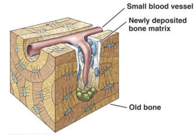
Osteoblasts
Secrete osteoid which mineralizes later… way to remember: osteoblasts=build

Osteocytes
Osteoblasts trapped in matrix… functions: maintain matrix, repair damage, sense mechanical stretch

Osteoclasts
Break down bone matrix… function: bone remodeling… way to remember: osteoclast=consume

Extracellular matrix
Part of the skeletal structure… made up of protein fibers (collagen) and ground substance (mineralized osteoid)… combination of organic and inorganic makes bones strong and slightly flexible
Composite material > organic and inorganic components
Organic: osteoid (collagen fibers, carbs, proteins), tensile strength
Inorganic: hydroxyapatite (calcium, phosphorus, oxygen), compressive strength
Rickets/osteomalacia
Disorder of skeletal matrix
Inadequate calcification of bone ECM (usually from vitamin D deficiency)
Bones are soft and easily deformed
Osteogenesis imperfecta
Disorder of skeletal matrix
Congenital defect of collagen
Bones are brittle and easily fractured
Cortical bone
Compact form of bone… the dense outer shell
Cancellous bone
Spongy form of bone… loosely organized interior bone

Cortical bone stucture
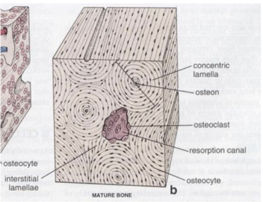
Organized into osteons… concentric lamellae (layers) of EMC… Central canal for passage of neurovasculature… oriented parallel to direction of compressive stress

Osteon stucture
Alternating collagen fiber orientation… multidirectional stability… resistance to torsion (twisting in opposite directions)

Cancellous bone structure
Bones arranged in lattice (strong but lightweight, no osteons)… spaces sometimes filled with red bone marrow… helps absorb and transmit forces

Periosteum
Layers of dense irregular connective tissue… periosteum is external covering… highly innervated… function in growth and repair (produce osteoblasts)

Endosteum
Layers of dense irregular connective tissue… endosteum is internal lining… highly innervated… function in growth and repair (produce osteoblasts)

Long bones
Longer than wide
Support and movement
Flat bone
Thin, often curved
Protection
Irregular bones
Complex shapes
Support and protection
Short bones
Cube-like shape
Stabililty and movement
Sesamoid bones
Form in tendons
Movement
Long bone stucture
Epiphyses- enlarged ends, which have cancellous bone internally and red bone marrow… articular cartilage externally
Diaphysis- shaft, which is cortical bone with medullary cavity (yellow bone marrow)

Flat/short/irregular bones stucture
Cancellous bone surrounds by cortical bone (no medullary cavity)
Protect underlying structures, shock absorption

Ossification
Bone formation… begins around embryonic week 8…intramembranous or endochondral
Intramembranous ossification step 1 and 2
Primary ossification center forms within fetal connective tissue… osteoblasts develop and begin to secrete osteoid… mineral crystals are incorporated so osteoblasts become osteocytes
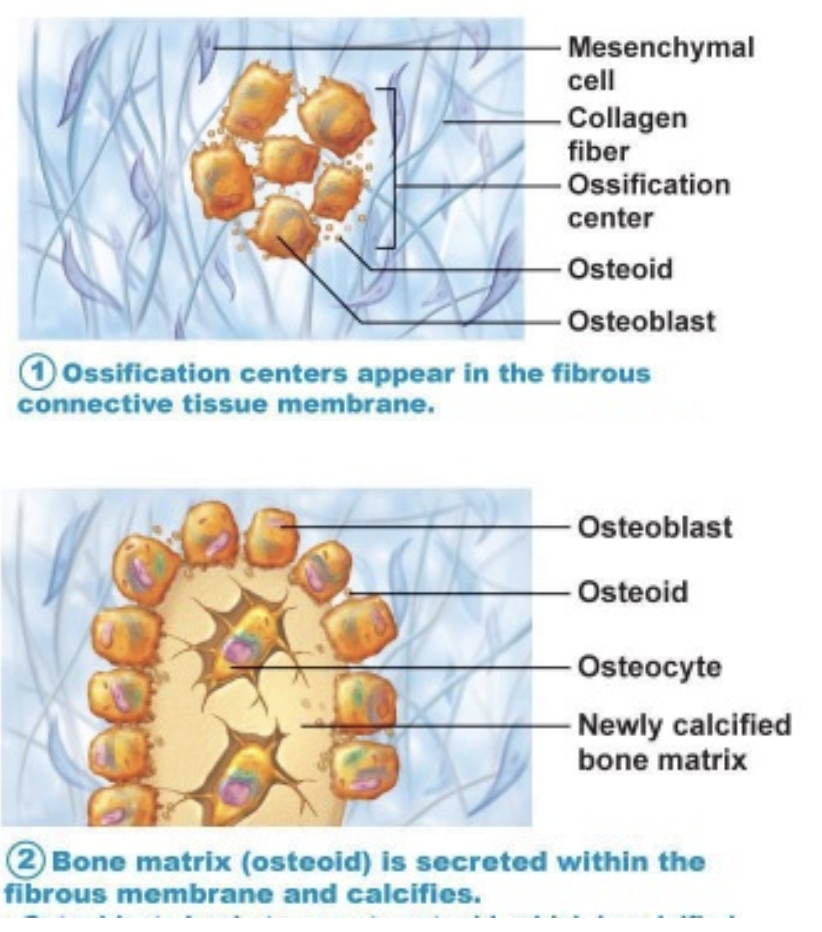
Intramembranous ossification step 3
Osteoblasts produce cancellous bone trabeculae… surrounding connective tissue becomes periosteum

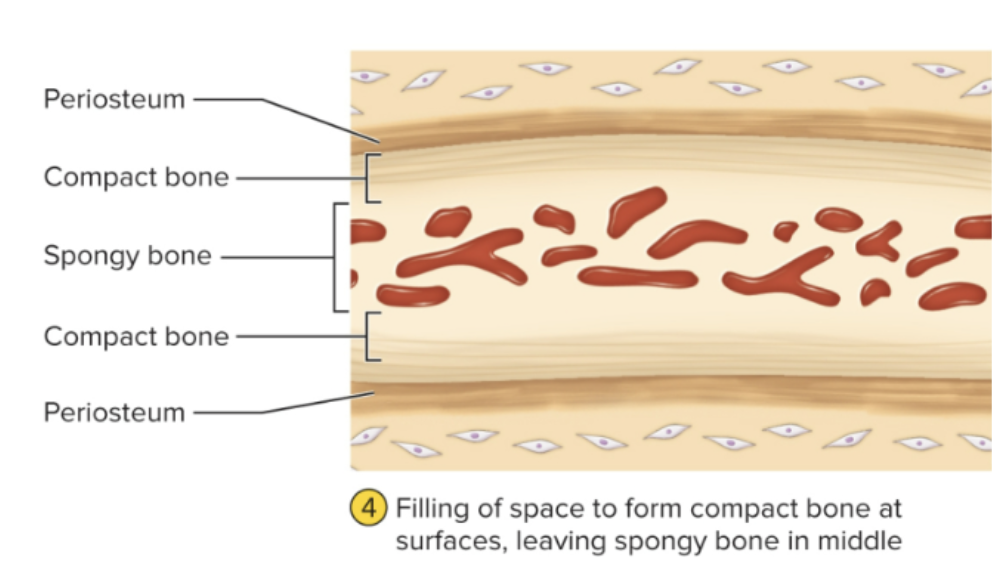
Intramembranous ossification step 4
Cortical bone forms on exterior surface… produces some bones of skull, clavicles
Endochondral ossification step 1
Beings with cartilaginous model covered by perichondrium… perichhondrium starts to produce osteoblasts = becomes periosteum

Endochondral ossification step 2
Bony collar forms around shaft (early cortical bone)… enlargment of chondrocytes and calcification of EMC at primary ossification center… calcification (deposition of minerals in matrix other than osteoid)… nutrients unable to diffuse through calcified ECM = chondrocytes die

Endochondral ossification step 3
Osteoclasts penetrate bony collar… break down calcified cartilage to form primary medullary cavity (only in long bones)… osteoblasts, blood vessels, and bone marrow enter… bony collar expands… secondary ossificetions center develop at epiphyses

Endochondral ossification step 4
Cartilaginous model enlarges… bony collar and primary medullary cavity expands… calcification of secondary ossification centers… osteoblasts secrete osteoid over calcified cartilage to form cancellous trabeculae… developmental stage of bone at birth… continues to develop postnatally

Endochondral ossification step 5
Bone ossification continues during early childhood… articular cartilage (hyaline cartilage retained on epiphyses)… epiphyseal plate (site of continued bone growth) which is present through early childhood and body growth

Endochondral ossification step 6
Epiphysial fusion marks end of skeletal growth… bone completely ossified… only articular cartilage remains (caps to form joints)

Note: only long bones have a medullary cavity
Note: only long bones have a medullary cavity
Primary bone
Immature… produced by osteoblasts very quickly…benefit: quick, good for initial skeletal element or bridging a fracture site… cons: haphazardly built, little inorganic (mineralization) matrix, irregularly arranged collagen fibers… eventually reabsorbed and replaced by secondary bone

Secondary bone
Mature bone… produced much slower than primary… fully formed osteons… parallel collagen orientation… higher percentage of inorganic material (mineralization) = stronger
Bone marrow
Type of tissue/organ… two different types (red and yellow)
Red bone marrow
Hematopoietic tissue… children have it in every bone, adults have it in the head and trunk…
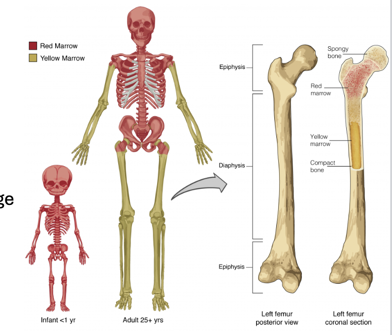
Yellow bone marrow
Fat storage… replaces red bone marrow of children as we age… adults have it in the limbs

Longitudinal bone growth
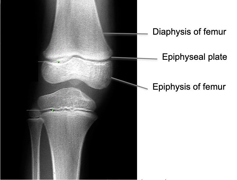
Occurs at epiphyseal plate condrocytes divide and mature (push epiphysis away from diaphysis)… cartilage matrix ossifies and is replaced with bone… rates of growth and ossification balance during childhood (continues as long as proliferative zone is active)
Zones: zone of reserve cartilage (inactive chondrocytes)… zone of proliferation (rapid cell division)… zone of hypertrophy (chondrocytes increase in size)… zone of calcification (dead chondrocytes, calcified matrix)… zone of ossification (osteoblasts ossify calcified cartilage)

Zones of longitudinal bone growth
Zone of reserve cartilage (inactive chondrocytes)… zone of proliferation (rapid cell division)… zone of hypertrophy (chondrocytes increase in size)… zone of calcification (dead chondrocytes, calcified matrix)… zone of ossification (osteoblasts ossify calcified cartilage)

Epiphyseal plate closure
Proliferation rate slows near end of adolescence… cartilaginous tissue overtaken by ossification… epiphyseal plate ossifies entirely = fusion/closure which is the end of longitudinal growth
Achondroplasia
Failure of cartilage growth in epiphyseal plate (results of spontaneous mutation)… long bones stop growing in childhood… disproportionately short limbs
Appositional growth
Occurs at periosteal surface… periosteal osteoblasts secrete new matrix (new cortical bone develops superficially)… osteoclasts erode medullary cavity (bone widens but remains proportional)… can continue after cessation of longitudinal growth (due to additional force/muscle activity or response to increase in body weight, in addition to other reasons)

Hormonal regulation of bone growth
Growth hormone increases chondrocyte cell division at epiphyseal plate, increases activity os osteogenic cells, and stimulates osteoblasts in periosteum… growth hormone is responsible for development in adolesense… testosterone increases appositional growth and increases chondrocyte cell divison… estorigen inhibits osteoclast activity and accelerates epiphyseal closure
Growth hormone impact on bone growth
Increases chondrocyte cell division at epiphyseal plate
Increases activity os osteogenic cells
Stimulates osteoblasts in periosteum
Growth hormone is responsible for development in adolescence
Testosterone impact on bone growth
Increases appositional growth
Increases chondrocyte cell divison
Estrogen impact on bone growth
Inhibits osteoclast activity
Accelerates epiphyseal closure
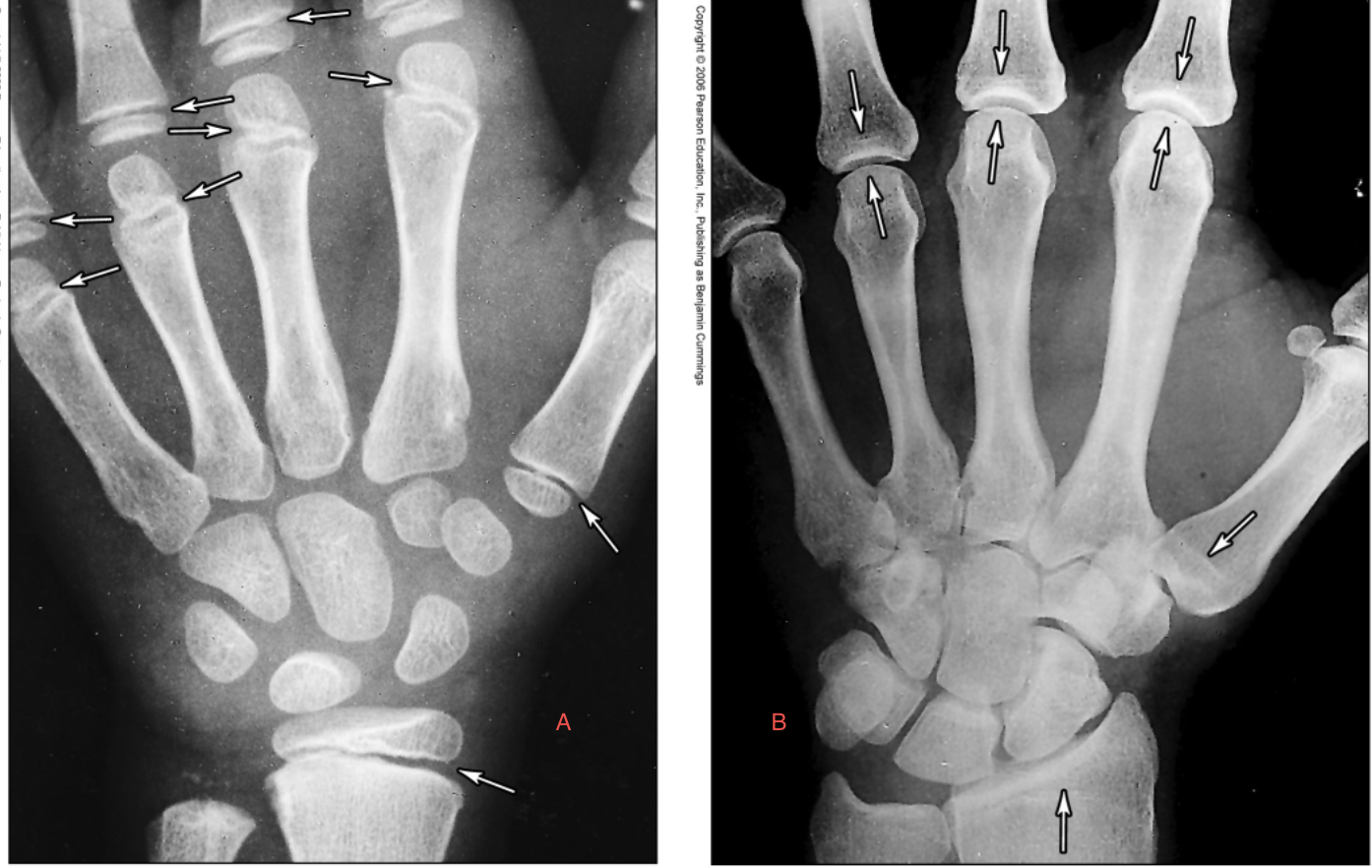
Which is an adult, a or b?
B

Skeletal remodeling: function
Reshape bone during growth
Reshape bone in response to use
Repair damage
Release minerals in blood
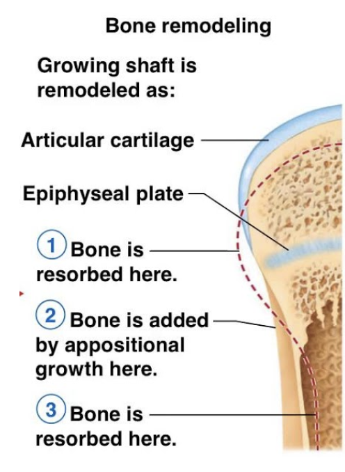
Skeletal remodeling occurs where
Occurs on periosteal and endosteal surfaces
Skeletal remodeling: deposition
Osteoblasts secrete osteoid (collagen fibers arranged in a spiral pattern along osteon), mineralization occurs (calcium and phosphate crystalize on collagen fibers as hydroxyapatite)
Skeletal remodeling: rebsorption
Osteoclasts dissolve bone and return minerals to blood… secrete H+ ions and enzymes
Regulation of remodeling: calcium homeostasis
Needed for neuron signaling, muscle contraction, blood clotting… balance between intake, excretion, and exchanges between skeleton and blood… regulated by calcitriol, calcitonin, and parathyroid hormone

Calcitriol
The active form of vitamin D… produced by actions of skin, liver, and kidney (initiated by UV radiation)… Raises blood calcium levels (which promotes calcium absorption in small intestine, increases calcium resorption for skeleton, reduces calcium excetion in kidneys)

Calcitonin
Secreted by thyroid in response to hypercalcemia… it inhibits osteoclasts and stimulates osteoblasts

Parathyroid hormone (PTH)
Secreted by parathyroid glands in response to hypocalcemia… stimulates osetoclast activity, inhibits osteoblast activity… reduces calcium excretion in kidneys… promotes calcitriol synthesis

Regulation of remodeling: mechanical stress
Stress: force/area
More bone deposition in areas under high stress (monitored by osteocytes)
Wolff’s law: bones remodel in response to habitual demands… compact bone is thickest in diaphysis… large projections occur where heavy, active muscles attach… exercise increases bone formation

Wolff’s law
Bones remodel in response to habitual demands… compact bone is thickest in diaphysis… large projections occur where heavy, active muscles attach… exercise increases bone formation

Orthodontic braces
Wires place tension on the teeth, alveolar bone is resorbed in response to increased pressure (allows teeth to shift)… additional bone deposited to fill in spaces
Fractures
Two types: stress fractures and pathological fractures
Stress: caused by abnormal trauma to a bone
Pathological: break in bone weakened by disease
Healing of fractures: step 1
Inflammatory response
Blood clot formation (clot invaded by blood vessels, fibroblasts, macrophages, osteoclasts, osteoblasts, chondrocytes) and those invasions produce granulation tissue

Healing of fractures: step 2
Soft callus formation
Fibroblasts secrete collagen, chondrocytes produce fibrocartilage

Healing of fractures: step 3
Hard callus formation
Osteoblasts produce bony collar around fracture… takes 4-6 weeks

Healing of fractures: step 4
Remodeling
Osteoclasts remove bon fragments, osteoblasts lay down cancellous bone, continued ossification produces cortical bone

Effects of aging
Children and adolescents: deposition>resorption (greatest period of bone growth, attainment of peak bone mass)
Young adults: deposition=resorption
Older adults: deposition<resorption (bone mass, mineralization, and healing ability decrease)
Osteoporosis
Excessive resorption leads to severe loss of bone density… cancellous bone of spine and femur are the most vulnerable (vertebral and hip fractures are common)… related to decline in estrogen production (postmenopausal women have greatest risk, occasionally in younger females with very low body fat)… Other risk factors = race, smoking, diabetes, nutritional deficit, activity level

Osteoporosis treatment and prevention
Treatments= estrogen replacement therapy (which increase risk of heart disease, stoke, breast cancer), medications, weight bearing exercise, nutritional supplements
Preventative behaviors= calcium-rich diet and weight bearing exercise increase peak bone mass… reduced consumption of alcohol and carbonated drinks
Axial skeleton
Skull, hyoid, vertebra, ribs/sternum

Appendicular skeleton
Clavicle, scupula, upper limb, os coxae, lower limb

Cranial bone function
Protect brain
Attachment sites for head/neck muscles

Facial bone function
Form the framework of face, hold the teeth, and anchor muscles of the face

Cranial bones (6)
Parietal, occipital, frontal, temporal, sphenoid, and ethmoid (others: occipital condyles and foramen magnum)

Sutures
Immobile joints, filled with dense connective tissue… wide and flexible during infancy (easier passage through birth canal and allows for brain growth)… interdigitate (get closer) as cranial bones grow… fuse over time
Major sutures (4)
Coronal, sagittal, squamous, and lambdoid

Vertebral column (numbers and sections)
26 total= 7 cervical, 12 thoracic, 5 lumbar, 5 sacral (fused into 1), 4 coccyx (fused into 1)
Vertebral column functions
Protect spinal cord, support body axis, attachment points for ribs and muscles of back and neck

Basic vertebral anatomy
Articular processes/facets (superior and inferior), vertebra arch, vertebral body, vertebral foramen, transverse process, spinous process
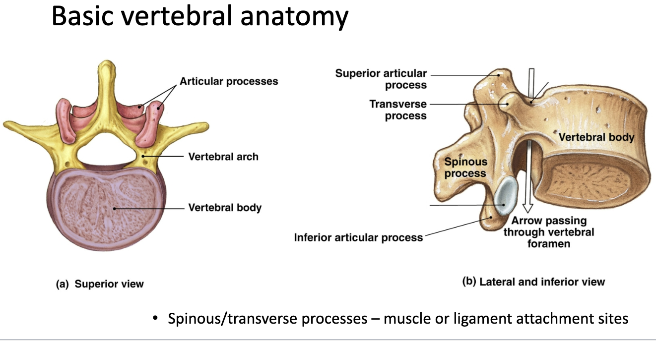
Spinous and transverse process function
Muscle and ligament attachment sites
Cervical vertebra
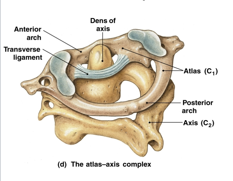
C1-C7, top two different (atlas which has no body and a large superior articular facet and axis which has a dens)
All cervical vertebrae are distinguishable because they have transverse foramina (which transmit vertebral arteries)

Atlas (C1)
No body, superior articular facet articulates with occipital condyle, atlano-occipital joint allows for head flextion and extension

Axis (C2)
Modified body (dens), atlas-axis joint allows heat L/R rotation
Thoracic vertebrae
T1-12, has costal facets (which articulate with ribs)

Lumbar vertebrae
L1-5, large bodies and lack of other structures
Intervertebral discs
Fibrocartilage with gel-like center, join vertebrae into column, allow for mobility, and cushion/absorb force

Ribs
12 pairs
1-7=true ribs that attach to sternum by costal cartilages
8-10=false ribs that attach to sternum via shared cartilage
11 and 12=floating ribs
All are attached to the thoracic cage

Thoracic cage
Protects underlying organs and attachment site for respiratory muscles
Rib structure
Body/shaft: main portion
Head and tubercles: articular surfaces
Angle: most lateral point
Costal groove: channel for neurovasculature

Clavicle
Provides muscle attachment, braces scapula and arms (only point of attachment between upper limb and axial skeleton), only intramembranous long bone

Pectoral girdle: scapula
Acromion articulates with clavicle, glenoid fossa articulates with humerus, other fossae: muscle attachment sites

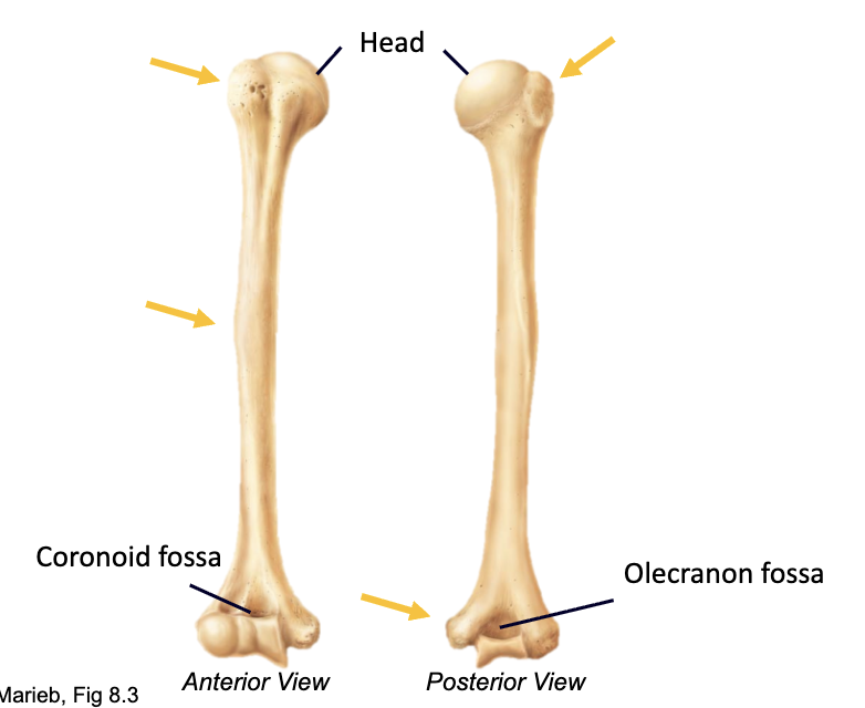
Humerus
Articulations: proximal (scapula) distal (radius, ulna)
Fossae: articulate with ulna
Tubercles/tuberosities: muscle attachment
Ulna and radius
Ulna: olecranon forms majority of elbow
Radius: head allows for rotation
Styloid processes: ligament attachment sites
Interosseous membrane: Dense connective tissues and allows rotation while maintaining spatial relationships

Os coxae
AKA innominate bones
Formed from fusion of ilium, ischium (ischial tuberosity: hold weight while sitting), pubis (pubic symphysis: anterior articulation via interpubic disc)
Acetabulum: articular site for femur and composed of all three bone

Pelvic sexual dimorphism
90° or less space in pelvic girdle=male
100° or more=female
Femur
Articulation: proximal (acetabulum) distal (tibia and patella)
Large trochanters and epicondyles=muscle attachments
Fractures most frquently at neck

Patella
Largest sesamoid bone
Cartilaginous at birth (ossifies at age 3-6)
Anchored to rectus femoris proximally and tibia distally

Tibia and fibula
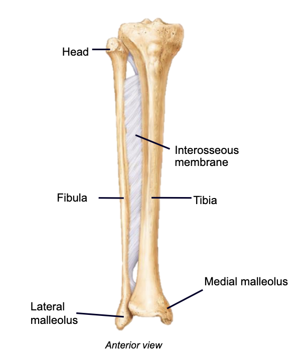
Tibia: receives and transmits body weight, flat anterior surface→shin, articulations (proximal: femur, patella, fibula… distal: talus, fibula)
Fibula: stabilizes ankle, articulations (proximal: tibia… distal: tibia, talus)
Interosseous membrane
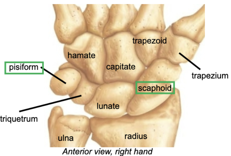
Carpals
Allow wrist to be very flexible and mobile
Pisiform: sesamoid bone, develops by age 12
Scaphoid most frequently fractured (FOOSH)
Tarsals
Calcaneus→heel (attachment for achilles tendon)
Talus: articulates with tibia and fibula
Calcaneus and talus bears most of weight
