Quiz 5 Chapter 6 vocab
 Call Kai
 Learn
Learn Practice Test
 Spaced Repetition
 Match
Match Flashcards
 Knowt Play
1/26

There's no tags or description
Looks like no tags are added yet.
Name | Mastery | Learn | Test | Matching | Spaced | Call with Kai | Chat |
|---|
No analytics yet
Send a link to your students to track their progress
27 Terms

Dentin Dysplasia ( Type I: Radicualr)
Autosomal Dominant
Type I: Radicular (the teeth have normal crowns and abnormal roots)
The teeth are generally exfoliated prematurely
Multiple periapical radiolucencies are associated with this condition (root defects)
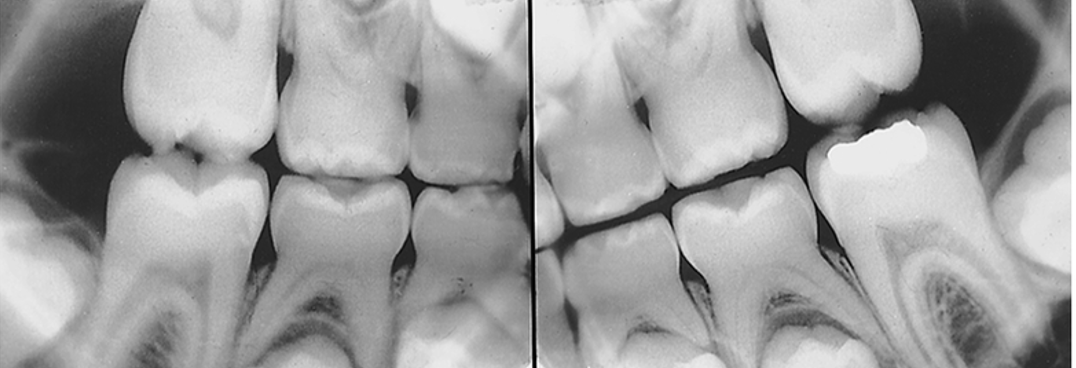
Dentin Dysplasia ( Type II: Coronal)
Type II coronal
Primary teeth are translucent with an amber color
Adult teeth appear normal
Permanet teeth may or may not have pulp stones
Radiograph: thistle-shaped pulp chambers in single-rooted teeth and a bow-tie appearance of the pulp champers of permanent molars

Ectodermal Dysplasia (hypohidrotic)
A genetic heterogeneity
Characterized by: Hypodontia (partial adontia), Hypotrichosis (decreased hair), Hypohidrosis (decresed sweating)
Although in the majority of the families it is inherited as an X-linked recessive trait, in some families it is inherited as an autosomal-recessive trait.
Female carriers of the X-linked form have minor clinical manifestations such as thin or sparse hair, cone-shaped teeth, hypodontia, and variable degrees of reduced sweating.

Amelogenisis Imperfecta
A group of inherited conditions affecting the enamel of the teeth with no assocated systemic defects
There are four types of amelogenesis imperfecta, as described by Witkop and Sauk (pitted teeth)
Amelogenisis Imperfecta (Type 1)
Hypoplastic amelogenisis imperfecta
the tooth enamel does not develop to normal thickness
autosomal dominant and autosomal recessive
teeth have random to pinhead and the pits are observed mostly on the labial and buccal surfaces of the permanent teeth

Amelogenisis Imperfecta (type II)
Hypocalcified amelogenisis imperfecta
normal thickness but poorly calcified
At eruption teeth appear yellow-orange enamel
Enamel is very soft leaving only dentin

Amelogenesis Imperfecta (type III)
Hypomaturation amelogenesis imperfecta
Enamel is softer than normal (Snowcapped)
Characterized: large amounts of enamel matrix; therefore the enamel is softer than normal
Amelogenesis Imperfecta (type IV)
Hypoplastic-hypomaturation amelogenesis imperfecta
Associated with taurodontic teeth
Yellow to brown pitted enamel
Hard to diagnose the exact type
•Radiographically:
Enamel has a radiodensity similar to dentin
Single-rooted teeth have large pulp chambers

Hypophosphatasia
Autosomal Recessive
A decresed in serum alkaline phosphates levels
Affects formation of bone and cementum
Teeth do not have cementum and are exfoliated prematurely
which shows the histologic section of a tooth from a patient with hypophosphatasia. The cementum is entirely lacking.

Peutz-Jeghers Syndrome
Autosomal Dominant
Characterized by:
Multiple melanotic macular pigmentations
Gastrointestinal polyposis—hamartomas
•The pigmentations occur around the eyes, nose, and mouth.
White Sponge Nevus (Familial White Folded Mucosal Dysplasia)
•An autosomal-dominant inheritance pattern with complete penetration
•Characterized by:
White, corrugated, soft, folding buccal oral mucosa
Thick layer of keratin that desquamates and leaves a raw mucosal surface
Free gingiva is not affected
•This is also called Cannon disease.

Maxillary Exostosis
An autosomal-dominant inheritance pattern
Occurs on the buccal aspect of the maxilla
May be single, multiple, unilateral, or bilateral
These are generally symptomless unless traumatized
Osteogenesis Imperfecta
Thirty Percent: an autosomal-dominant inheritance pattern with variable expression
Seventy percent: sporadic cases suggesting autosomal-recessive in heritance
Mutations occur that affect collagen, resulting in abnormally formed bones that fracture easily
In mildest cases, individuals may only show blue sclera (that portion of the eye that is usally white)

Osteogenesis Imperfecta (2/2)
Dentinogenesis imperfecta-like condition
Microdontia
Teeth appear opalescent or translucent but darken with age
Enamel is lost because of abnormal dentin
•Multiple spontaneous bone fractures are the main clinical complication of this syndrome.

Nevoid Basal Cell Carcinoma Syndrome (Gorlin Syndrome)
•An autosomal-dominant inheritance pattern with high penetrance and variable expressivity
•Characterized by:
Mild hypertelorism (increased distance between the eyes)
Mild prognathism
Frontal and parietal enlargement
A broad nasal root
•Oral lesions consist of multiple cysts of the jaws; histologically, they are odontogenic keratocysts (OKCs)
Nevoid Basal Cell Acarcinoma Syndrome (Gorlin Syndrome) 2/2
•Nevi are observed on the skin; typically, they are basal cell carcinomas
•Skeletal anomalies include:
Bifurcation of ribs
Shortening of metacarpals
Spina bifida occulta
Kyphoscoliosis
•Various neoplasms include:
Medulloblastoma
Calcified ovarian fibromas
Mesenteric cysts
•Cysts can develop as early as 5 to 6 years of age in some affected patients; these cysts interfere with normal development of the jawbones and teeth.
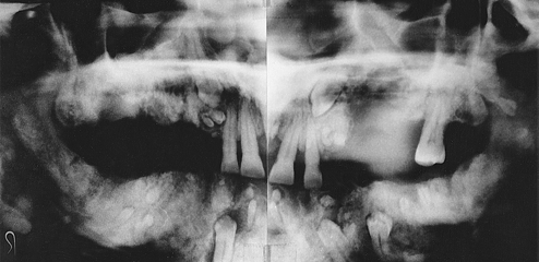
Gardner Syndrome (Familial Colorectal Polyposis)
•An autosomal-dominant inheritance pattern with variable expressivity and marked penetrance
•Characterized by osteomas in various bones
•Osteomas of the facial skeleton will obliterate the sinuses and cause facial asymmetry
•Multiple odontomas can occur in jawbones
•Intestinal polyps occur that will become malignant at age 30 or later
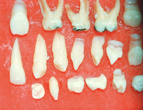
Cleidocranial Dysplasia
•Autosomal dominant, but about half of the cases are isolated examples caused by spontaneous mutation or a gene with poor penetrance
•The fontanelles remain open and the cranium develops a mushroom shape
•The neck is long and narrow as a result of unilateral or bilateral hypoplasia of clavicles
•The premaxilla is generally underdeveloped, resulting in pseudoprognathism
•Patients have many supernumerary teeth, which are crowded in the jaws and do not erupt
•Multiple cysts can develop in association with impacted teeth
Ehlers-Danlos Syndrome
a group of rare inherited connective tissue disorders caused by defects in the structure or processing of collagen
Characterized: unusally loose and weaken joints, skin hyperelasticity, patients more prone to joint issues (TMJ), severe brusing and bleeding
Oral: delicate mucosa, weakened gingiva tissue, BOP
able to touch their nose w/ tongue (gorlin sign)

Papillon-Lefevre Syndrome
•An autosomal-recessive inheritance pattern
•Peripheral blood neutrophils are depressed, and the theory is that chemotaxis is depressed
•Hyperkeratosis of the palms of the hands and soles of the feet
•The lesions on the hands and feet remain as reddish-white, scaly thick areas of hyperkeratinization.
•At about 1½ or 2 years of age, a gingivoperiodontal inflammatory process develops: Edema, bleeding, alveolar bone resorption, mobility of teeth
•Both primary and permanent dentition are lost prematurely
Klinefelter Syndrome
•Most are from nondisjunction of the X chromosome
•Male phenotype
•Not detected clinically until puberty
Taller than normal
Wide hips
Female pubic hair distribution
Development of female breasts
Intelligence levels may be lower than normal
Testes are smaller and harder
•The maxilla is slightly hypoplastic
May be XXXY or XXXXY

Trisomy 13
Multiple abnormalities in various organs
70% die within the first 7 months of life
Characteristic facial clinical findings include:
Bilateral cleft lip and palate
Microphthalmia (small eyes) or anophthalmia (no eyes), polydactyly (supernumerary digits)

Trisomy 21 (down syndrome)
•Nondisjunction
•Associated with late maternal age
Slanted eyes
Shorter stature
Heart abnormalities
Varied intelligence levels
Fissured tongue
Gingival and periodontal disease has been reported in 90% of affected individuals
Hypodontia (fewer teeth than normal)
Abnormally shaped teeth
Anomalies in eruption with malposition and crowding of teeth are common
Cyclic Neutropenia
•An autosomal-dominant condition
•Characterized by a cyclic decrease in the number of circulating neutrophils
•Systemic manifestations include fever, malaise, sore throat, and occasional cutaneous infections
Oral: severe ulcerative gingivits or gingivostomatitis, and ulcers
•Kostmann syndrome or chronic neutropenia: Autosomal-recessive condition
•The disorder is characterized by a cyclic decrease in the number of circulating neutrophilic leukocytes.
•A decrease in the number of circulating neutrophils is called neutropenia.

Cherubism
•Autosomal dominant with marked penetrance in males and variable expressivity and incomplete penetrance in females
•Clinical manifestation
Progressive bilateral facial swelling that first appears when the patient is 1½ to 4 years of age
Ocular hypertelorism (incresed distance between eyes)
Microscopically, bone lesions resemble central giant cell granuloma
Radiographs of the jaws reveal a characteristic “soap bubble” or multilocular appearance
Dentinogenesis Imperfecta
Multiple types
Associated with osteogenesis imperfecta
Hereditary opalescent dentin (Type II)
No pulp chambers or root canals are seen
Roots are short and thin with periapical radiolucencies

Hereditary Hemorrhagic Telangiectasia
•Autosomal dominant
•Characterized by:
Multiple capillary dilations of skin and mucous membranes called telangiectases
Lesions in mucosa of the nasal cavities may cause epistaxis (nose bleeds)
Risk of gingival hemorrhage