Comprehensive Respiratory Therapy and Pulmonary Medicine Review
 Call Kai
 Learn
 Practice Test
 Spaced Repetition
 Match
 Flashcards
 Knowt Play
1/65

Earn XP
Description and Tags
Comprehensive practice flashcards covering mechanical ventilation, pulmonary pathology, hemodynamics, pharmacology, arterial blood gases, pulmonary function testing, and cardiac dysrhythmias.
66 Terms
Pneumonia
An inflammation of the lung parenchyma that impairs gas exchange, progressing through four distinct stages: Stage 1 (congestion/edema within 24 hours), Stage 2 (red hepatization on days 2–3), Stage 3 (gray hepatization/consolidation on days 4–6), and Stage 4 (resolution on days 7–10).
CURB-65 Score
A simple bedside risk stratification tool used for community-acquired pneumonia where scores of 0–1 indicate outpatient care, 2 indicates hospital admission, and 3–5 mandate ICU admission.
Ventilator-Associated Pneumonia (VAP)
Pneumonia developing more than 48 to 72 hours after endotracheal intubation; categorized as early-onset (within 4 days, typically community pathogens) or late-onset (after 5 days, often multi-drug resistant organisms).
Lung Abscess
A localized area of suppurative necrosis within the lung tissue most commonly caused by aspiration pneumonia; typically located in the right lower lobe, producing foul-smelling, putrid sputum, and appearing on imaging as a cavity containing an air-fluid level.
Body Plethysmography
A sensitive pressure-transducer-based pulmonary function test that uses Boyle's law to measure total thoracic gas volume, residual volume (RV), total lung capacity (TLC), and airway resistance, successfully detecting all trapped gas.

Flow-Volume Loops (PFT Patterns)
Graphic representations of airflow versus lung volume: restrictive disorders present as narrowed, tall loops with normal contours; obstructive disorders display a scooped-out expiratory limb; fixed large airway obstructions demonstrate flattened inspiratory and expiratory limbs.
Diffusing Capacity of the Lung for Carbon Monoxide (DLCO)
A pulmonary diagnostic test measuring gas transfer across the alveolar-capillary membrane using a 10-second single-breath hold; normal values are 25 to 30 mL/min/mmHg or >75%.
Positive Bronchodilator Response
A post-bronchodilator spirometric improvement demonstrated by an increase in FEV1 or FVC of at least 12% and an absolute volume increase of at least 200 mL.
Premature Atrial Contraction (PAC)
A cardiac dysrhythmia originating from an irritable ectopic focus in the atria outside the SA node, characterized on ECG by an early abnormal P wave (which may be buried in the preceding T wave) followed by a compensatory pause.
Atrial Flutter
A rapid, organized atrial rhythm originating outside the SA node with an atrial rate near 300 bpm, producing characteristic regular saw-tooth or shark-fin flutter waves and an unmeasurable PR interval.
Atrial Fibrillation
A chaotic, irregularly irregular atrial dysrhythmia firing at 400 to 600 bpm across multiple reentry circuits, resulting in uncoordinated quivering of the atria, absent P waves replaced by fibrillatory (f) waves, loss of the atrial kick, and elevated risk of thromboembolic stroke.
Supraventricular Tachycardia (SVT)
A rapid, regular ectopic rhythm originating above the ventricles at a rate between 100 and 250 bpm, often treated acutely with vagal maneuvers or adenosine (6 mg rapid IV push, followed by 12 mg if unsuccessful).
Manual Resuscitator (BVM)
A portable hand-held bag-valve-mask device providing positive-pressure ventilation that does not require a compressed gas source to re-inflate; it must deliver at least 85% FiO2 at 15 L/min (preferred >95%) with a minimum tidal volume of 600 mL.
Fenestrated Tracheostomy Tube
An artificial airway with an opening on the posterior curve of the outer cannula that allows airflow past the vocal cords for speech when the inner cannula is removed and the cuff is completely deflated.
Passy-Muir Speaking Valve
A one-way valve attached to a tracheostomy tube that opens during inspiration and closes during expiration, directing exhaled gas through the vocal cords and upper airway; the cuff must always be deflated prior to application.
Endotracheal Tube Cuff Pressure
The safe pressure range maintained within an artificial airway cuff, strictly kept below 25 cmH2O (16 to 18 mmHg) to avoid mucosal ischemia, tracheal necrosis, and tracheal stenosis.
Light's Criteria
A diagnostic standard used to differentiate pleural effusions; an effusion is classified as an exudate if the ratio of pleural fluid protein to serum protein is greater than 0.5.
Transudative Pleural Effusion
A watery, protein-poor pleural fluid collection that develops when hydrostatic or oncotic pressure imbalances force fluid from pulmonary capillaries into the pleural space; most commonly caused by congestive heart failure.
Exudative Pleural Effusion
A protein-rich, cellular pleural fluid collection that develops when the pleura or adjacent lung parenchyma is directly involved in inflammation, infection, or malignancy; most commonly caused by bacterial pneumonia.
Pancoast Tumor
A superior sulcus apical lung tumor that typically invades surrounding nerves, presenting with severe shoulder/arm pain and Horner syndrome.
Sinus Arrhythmia
A variation in SA node discharge characterized by a normal heart rate (60 to 100 bpm) where the rhythm is irregular and cycles naturally in synchrony with the respiratory cycle.
Sinus Arrest
An episode of atrial standstill where the SA node fails to generate impulses for 3 or more consecutive beats, creating an unexpected pause where the subsequent beat does not return in cycle.
Kyphoscoliosis
A thoracic wall deformity combining posterior curvature (kyphosis) and lateral curvature (scoliosis) of the spine, resulting in a severe restrictive pulmonary defect, reduced chest wall compliance, and chronic alveolar hypoventilation.
Guillain-Barré Syndrome
An acute post-infectious autoimmune polyneuropathy characterized by progressive ascending symmetric flaccid paralysis and elevated CSF protein, presenting a risk of ventilatory failure if FVC<20 mL/kg or MIP<−30 cmH2O.
Myasthenia Gravis
An autoimmune disorder caused by circulating anti-acetylcholine (ACh) receptor antibodies at the neuromuscular junction, characterized by fluctuating, descending muscle weakness and fatigue, ptosis, and diplopia.
Cushing's Triad
A clinical triad indicating critically elevated intracranial pressure, consisting of systolic hypertension with widened pulse pressure, bradycardia, and irregular respirations.
Flail Chest
A mechanical chest wall injury caused by double fractures in at least three or more adjacent ribs, producing thoracic wall instability and paradoxical chest wall movement (inward during inspiration, outward during expiration).
Pendelluft
The inefficient movement of deadspace gas shifting back and forth between the injured and uninjured lung during breathing cycles in flail chest, leading to severe hypoventilation and hypoxemia.
Tension Pneumothorax
A life-threatening thoracic emergency in which air enters the pleural space during inspiration but cannot escape, building positive intrapleural pressure that collapses the ipsilateral lung, shifts the mediastinum to the contralateral side, and impairs cardiac return.

Chest Tube Indications
Clinical rationales for inserting intrapleural drainage tubes: removing air in pneumothoraces, performing emergent decompression in tension pneumothoraces, draining blood in hemothoraces, and evacuating pus/infection in empyemas.

Air-to-Oxygen Entrainment Ratios
The specific proportion of entrained room air to delivered pure oxygen required to obtain a specific target FiO2, such as 25:1 for 24%, 10:1 for 28%, 3:1 for 40%, and 1:1 for 60%.
Nonrebreathing Mask (NRB)
A high-concentration oxygen reservoir mask featuring one-way valves on the exhalation ports and between the bag and mask that prevent ambient air entrainment and rebreathing; the reservoir bag must remain at least one-third inflated.
Fractional Distillation
The most common and economical commercial process used to produce medical oxygen by cooling atmospheric air until liquefied and separating gases based on boiling points.
Heliox Therapy
A low-density gas mixture of helium and oxygen used to reduce turbulent airflow and decrease work of breathing in upper airway obstructions; calculated using correction factors of 1.8 for 80/20 and 1.6 for 70/30.
Inhaled Nitric Oxide (iNO)
A selective pulmonary vasodilator administered at 20 to 40 ppm to lower pulmonary vascular resistance and improve oxygenation without causing systemic hypotension; requires close monitoring for methemoglobinemia.
Dornase Alfa (Pulmozyme)
A recombinant human deoxyribonuclease administered via nebulizer at 2.5 mg daily to cleave extracellular neutrophil DNA in sputum, lowering mucus viscosity in cystic fibrosis patients.

Aerosolized Antimicrobial Medications
Nebulized anti-infectives targeting specific pulmonary pathogens, including Tobramycin and Aztreonam for gram-negative infections, Colistimethate to disrupt bacterial membranes, and Pentamidine to treat Pneumocystis jirovecii.
Respiratory Distress Syndrome (RDS)
A neonatal disease of lung immaturity and primary surfactant deficiency leading to increased surface tension, diffuse microatelectasis, grunting, retractions, and a ground-glass reticulogranular pattern on chest radiography.
Lecithin-to-Sphingomyelin (L:S) Ratio
An amniotic fluid test assessing fetal pulmonary surfactant maturity; an L:S ratio of 2:1 or greater indicates mature lungs, while an L:S<1:1 indicates a high risk of developing RDS.
Persistent Pulmonary Hypertension of the Newborn (PPHN)
A syndrome marked by sustained high pulmonary vascular resistance causing extrapulmonary right-to-left shunting across the patent ductus arteriosus or foramen ovale, confirmed by a pre- and post-ductal PaO2 gradient exceeding 20 mmHg.

Junctional Dysrhythmia Rate Ranges
The diagnostic intrinsic ventricular rate criteria for AV junctional rhythms: Junctional Escape operates at 40–60 bpm, Accelerated Junctional operates at 60–100 bpm, and Junctional Tachycardia operates at 100–200 bpm.
Incentive Spirometry (IS)
A hyperinflation lung expansion technique where a patient performs repeated sustained maximal inspirations (SMI) through a flow- or volume-metered device to mimic sighing and re-expand atelectatic alveoli.
Mechanical Insufflation-Exsufflation (MIE)
An airway clearance device (CoughAssist) that provides positive pressure inspiration followed immediately by a rapid shift to negative pressure exhalation to generate high expiratory flows and simulate a natural cough.
Static Compliance (Cstat)
A calculation reflecting the elastic recoil of the lungs and chest wall measured during an inspiratory hold without airflow: Cstat=Pplat−PEEPVT; normal values in mechanically ventilated patients are approximately 40 to 50 mL/cmH2O.

Peak Inspiratory Pressure vs. Plateau Pressure
Airway pressure components of mechanical ventilation where Peak Inspiratory Pressure (PIP) reflects the total pressure needed to overcome airway resistance and alveolar elastance, while Plateau Pressure (Pplat) during an inspiratory hold reflects true alveolar distending pressure.

Alveolar Overdistension ("Beaking")
An abnormal tapering appearance seen at the upper right corner of a dynamic pressure-volume loop signifying that lung compliance has fallen sharply at end-inspiration due to excessive tidal volume or high airway pressure.
First-Degree AV Block
A consistent delay in electrical conduction through the AV node characterized by a regular rhythm and a constant, prolonged PR interval exceeding 0.20 seconds (200 ms) without dropped beats.
Second-Degree AV Block Type I (Wenckebach)
An AV nodal conduction impairment characterized by progressive lengthening of the PR interval with each successive cycle until a sinus P wave fails completely to conduct to the ventricles, resulting in a dropped QRS complex.
Third-Degree AV Block
A complete cessation of AV nodal conduction where the atria and ventricles depolarize independently under separate pacemakers, causing complete AV dissociation with an atrial rate faster than the ventricular rate.
Pulsus Paradoxus
An abnormally large decrease in systolic blood pressure of greater than 10 mmHg during inspiration, frequently encountered during severe acute asthma exacerbations.
B-Type Natriuretic Peptide (BNP)
A cardiac neurohormone secreted by ventricular myocytes in response to increased cardiac wall stretch; levels <100 to 300 pg/mL are normal, while values >900 pg/mL indicate severe congestive heart failure.
Mean Arterial Pressure (MAP)
The average perfusion pressure driving systemic blood flow throughout the cardiac cycle, calculated by the formula MAP=3Systolic+(2×Diastolic), where values <60 mmHg indicate inadequate organ perfusion.
Total Arterial Oxygen Content (CaO2)
The total volume percentage of oxygen carried in arterial blood, calculated using the formula CaO2=(Hgb×1.34×SaO2)+(PaO2×0.003), with a normal value of approximately 20 vol%.
Alveolar Gas Equation (PAO2)
A mathematical formula estimating the partial pressure of oxygen inside the alveoli: PAO2=(PB−47)×FiO2−(PaCO2×1.25), where normal on room air is typically >100 mmHg.
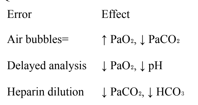
Blood Gas Pre-Analytical QC Errors
Documented sampling and handling errors that corrupt ABG measurements: air bubbles increase PaO2 and decrease PaCO2; delayed analysis decreases PaO2 and lowers pH; heparin dilution decreases PaCO2 and HCO3−.
Silhouette Sign
The loss of a normal radiographic anatomical contour or silhouette that occurs when an adjacent pulmonary infiltrate, mass, or fluid possesses the same radiodensity and touches that anatomical structure.
Reid Index
A histologic measurement of chronic bronchitis calculated as the ratio of mucous gland layer thickness to the total thickness of the bronchial wall; normal is <0.40, whereas a ratio >0.50 confirms glandular hypertrophy.

Noninvasive Ventilation (NIV) Interfaces
Patient interface options for positive-pressure NIV; nasal masks allow speech and coughing but allow mouth leaks, while oronasal and full-face masks control mouth leaks but carry higher risks of aspiration, claustrophobia, and gastric distention.
Sweat Chloride Test
The gold-standard quantitative diagnostic test for cystic fibrosis measuring sweat sodium and chloride collected via pilocarpine iontophoresis; sweat chloride levels >60 mmol/L on two occasions confirm CF.
Signet Ring Sign
A pathognomonic finding on high-resolution chest CT in bronchiectasis where the internal diameter of a dilated bronchus visibly exceeds the diameter of its adjacent accompanying pulmonary artery.
Virchow's Triad
The three broad pathological predispositions contributing to deep vein thrombosis and pulmonary embolism: venous stasis, hypercoagulability, and vascular endothelial vessel wall injury.
Pulmonary Capillary Wedge Pressure (PCWP)
The hemodynamic pressure measured by balloon flotation in a pulmonary capillary branch that estimates left atrial and left ventricular end-diastolic filling pressures; normal is 4 to 12 mmHg, with elevations above 12 mmHg signifying cardiogenic etiology.
Airway Pressure Release Ventilation (APRV)
A time-triggered, pressure-limited, and time-cycled CPAP mode alternating between a sustained high pressure (Phigh) and brief periodic releases to a lower pressure (Plow) allowing unrestricted spontaneous breathing throughout the cycle.
Kerley B Lines
Short, horizontal, linear radiopaque streaks found at the lung bases abutting the pleura that indicate fluid engorgement of the pulmonary interlobular septa, characteristic of cardiogenic pulmonary edema.
R-on-T Phenomenon
A cardiac electrophysiologic occurrence where an ectopic premature ventricular contraction falls directly on the vulnerable T wave repolarization period of the preceding beat, risking deterioration into ventricular tachycardia or ventricular fibrillation.
Pulseless Electrical Activity (PEA)
A clinical state characterized by the presence of organized electrical rhythm activity on the cardiac monitor in the complete absence of a palpable pulse or meaningful mechanical myocardial contraction.