Rest of the Objectives
 Call Kai
 Learn
Learn Practice Test
 Spaced Repetition
 Match
Match Flashcards
 Knowt Play
1/68

There's no tags or description
Looks like no tags are added yet.
Name | Mastery | Learn | Test | Matching | Spaced | Call with Kai | Chat |
|---|
No analytics yet
Send a link to your students to track their progress
69 Terms
What lab procedures can be used to work up Hemoglobinopathies?
CBC / RBC indicies
reticulocyte count
Peripheral blood smear
Hemoglobin solubility test
Hemoglobin electrophoresis
What is seen on a peripheral blood smear for HbSS disease?
Sickle cells
target cells
HJ bodies
pappenheimer bodies
basophilic stipplings
nucleated RBC
spherocyte
What is seen on a peripheral blood smear for HbC disease?
Target cells
microspherocytes
schistocytes
Hgb crystals “bar of gold”
What is seen on a peripheral blood smear for HbSC disease?
sickle cell
target cell
SC crystals
What is the Hgb Solubility Test?
blood is added to phosphate buffer and saponin
sample is deoxygenated
HbS becomes insoluble
produces a turbid suspension
positive = presence of HbS or other hemoglobinopathies
What does the Hgb Solubility Test NOT do?
tells you whether Pt has: HbSC, HbAS, HbS
positive result → follow with electrophoresis
What does hemoglobin electrophoresis do?
separates hemoglobins based on electrophoretic mobility
allows you to determine what hemoglobins are present and approx how much of each are present
What occurs when you have an alkaline pH for hemoglobin electrophoresis?
HbS, HbD, HbG migrate together
What occurs when you have an acidic pH for hemoglobin electrophoresis?
can separate HbS from HbD/HbG
What lab tests are used to diagnose thalassemias?
CBC
Blood smear
Hemoglobin electrophoresis
HPLC
Hemoglobin quantification
Routine Chemistry
What CBC results can indicate Thalassemia?
norm / ↑ RBC and MCHC
↓ MCV/ MCH
↓ HCT / Hgb
normal RDW
What blood smear results indicate homozygous B thalassemia?
extreme anisocytosis / poikilocytosis
target cells, ovalocytes, nRBCs
marker: hypo/micro
What blood smear results indicate heterozygous B thalassemia?
hypo/micro
mod/mild anisocytosis/poikilocytosis
target cells
basophilic stipplings
Why is hemoglobin electrophoresis important in diagnosing Thalassemias?
detect ↑ levels of abnormal/normal hemoglobins
Hgb H
Hgb Barts
Hgb Constant Spring
other abnormal hemoglobins
How does High-performance liquid chromatography (HPLC) help with diagnosing thalassemias?
replaces hemoglobin electophoresis in many labs
confirmatory method following abnormal hemoglobin electrophoresis results
separates molecules based on their movement through mobile/stationary phase
Why is hemoglobin quantification used for thalassemia diagnosis?
needed to establish true diagnosis
What can ↑ billirubin indicate?
thalassemia major / intermedia
1-6 mg/dL
higher in thalassemia intermedia than major
What can serum, iron, TIBC, and serum ferritin indicate?
differentiates thalassemia carrier from IDA
assessment of iron load in a patient with thalassemia major / intermedia
What is Hemoglobin H?
HbH = B4 ( 4 beta chains)
occurs when 3 of the 4 alpha genes are deleted
leaves 1 functional alpha gene
What is the hemoglobin concentration in Hemoglobin H disease?
5-40% HbH
rest is mostly HbA
small amounts: HbA2 and Hb Barts
What lab findings indicate Hbg H?
Hgb: 7-10 g/dL
retics: 3-10%
mod mico/hypo
target cells, heinz bodies
splenomegaly
BM: erythroid hyperplasia
What is Hemoglobin Barts?
y4 = consists of 4 gamma chains
occurs when all 4 alpha chains are deleted
no normal alpha chains are produced
What lab findings indicate Hemoglobin Barts disease?
approx 80% Hb barts
Hb barts can not carry O2 effectively
incompatible with life
death in utero or shortly after birth
Main findings of HbH.
chains: B4
alpha genes deleted: ¾
Timing: after birth
severity: mod/severe
Major clue: HbH inclusions
Major findings of Hb Barts.
chains: y4
alpha genes: 4/4
timing: fetal / newborn
severity: usually fatal
major clue: hydrops fetalis
What is the etiology of Gaucher’s Disease?
lack of beta-glucocerbrosidase enzyme
leads to - accumulation of glucocerebroside
autosomal recessive
What gene is involved in Gaucher’s Disease?
GBA 1
located on chromosome 1 at 1q21
What clinical triad indicates Gauchers Disease?
hepatomegaly
Gaucher’s cells in BM
↑ serum phosphatase
What is the etiology of Niemann-Pick Disease?
deficiency of sphingomyelinase
leads to accumulation of sphingomyelin and cholesterol
What can indicate Neimann-Pick Disease?
lipid laden giant foam cells in BM, tissues, and organs
cherry red spot in macula of eye
What is Tay Sachs Disease?
progressive neurodegenerative disorder
fatal by: 2-3 yrs
What is the etiology of Tay Sachs Disease?
lesions in cns caused by accumulation of gm2 ganglioside in neurocytes
deficiency of beta hexosaminidase enzyme
mutation in hexa gene
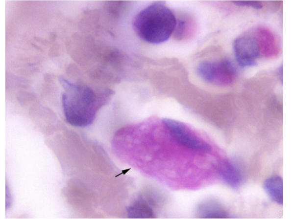
What stain is used for viewing Gaucher cells in Gaucher’s Disease?
Periodic Acid Schiff Stain
PAS +
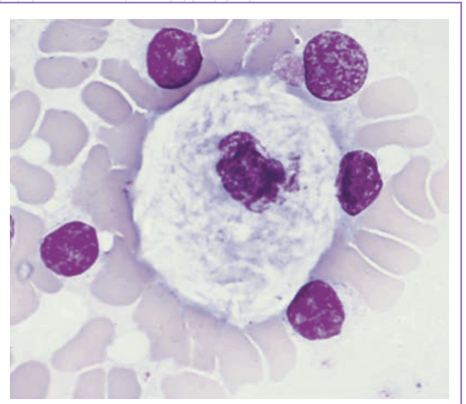
Describe the morphology of a Gaucher Cell.
macrophage like
20-80 micrometers
abundant blue-grey cytoplasm with wrinkled paper appearance
PAS +
cytoplasmic inclusions
aggregates of alucocerebroside
seen in: BM, liver, spleen

Describe the appearance of a Niemann Pick Cell
macrophage like
foamy cytoplasm with lysosomes and sphingomyelin
20-90 micrometers
eccentric nucleus
seen in BM / spleen
When is the clinical onset of Type 1 Gaucher’s Disease (Neuropathic)
childhood / adulthood
1/450 - 1/1,000
What do these lab findings indicate?
Hepatosplenomegaly +
Hematologic complications secondary to hypersplenism +
Skeletal deterioration (bone crisis/ fractures) +
Neurodegenerative course -
Life expectancy: 6-80+ yrs
Type 1 Gaucher’s Disease (adult form, neuropathic)
When is the clinical onset of Type 2 Gaucher’s Disease (Acute Neuropathic)
infant form
1/100,000
What do these findings indicate?
Hepatosplenomegaly +
Hematologic complications secondary to hypersplenism +
Skeletal deterioration (bone crisis/ fractures) -
Neurodegenerative course +++
Life expectancy: 2 yrs
Type 2, Acute Neuropathic Infant Form (Gaucher’s Disease)
What is the clinical onset of Type III Subacute Neuropathic Gaucher’s disease?
Childhood / juvenile
1/100,000
What do these findings indicate?
Hepatosplenomegaly +
Hematologic complications secondary to hypersplenism +
Skeletal deterioration (bone crisis/ fractures) +
Neurodegenerative course ++
Life expectancy: 20-40 yrs
Type III: Subacute Neuropathic Gaucher’s Disease
What causes type A Niemman-Pick Disease?
mutation: smpd 1 gene
most common
causes a deficiency in sphingomyelinase
What are characteristics of Type A Niemann-Pick Disease?
buildup of sphingomyelin in liver, kidney, lungs, brain
presence of niemann pick cells in BM
presents in infants
failure to thrive
hepatosplenomegaly
rapid neurodegeneration
fatal by 2 yrs old
What causes Type B Niemann-Pick Disease?
mutation: smpd 1 gene
deficiency in sphingomyelinase (exhibit 10-20% of normal enzyme activity)
buildup of sphingomyelin in lungs, liver, kidney
What findings can indicate Type B Niemann-Pick Disease?
presence of Niemann-pick cells in BM
presents later in life
does not live past adolescence
scant neurologic symptoms
progressive hepatosplenomegaly
heart disease
How does Type C Neumann Pick Disease occur?
mutations in npc1 or npc 2
cholesterol, biphosphate, and sphingolipid buildup in lysosomal storage organelles of macrophages
What findings indicate Type C Neumann Pick Disease?
systemic, neurologic and psychiatric symptoms
poor prognosis
affects children’s and adults - longer lifespan
What happens to a cell in Gaucher’s Disease?
macrophages swallow waste material
forms a lysosome containing waste material
no enzyme = majority of waste material remains in the lysosome
How do you interpret a scatterplot from a flow cytometry analysis?
flow cytometry scatterplot displays individual cells according to measured properties
each dot = individual cell or particle passing through instrument
there are two main light scatter measurements
forward scatter (FS/FSC)
Side Scatter (SS/SSC or RALS)
What does forward scatter do?
approximate cell sizes
What does side scatter do?
reflects cellular complexity / granularity
How do you read the axises on a FSC vs. SSC plot?
X axis: forward scatter (increasing from left →right)
Y axis: side scatter (increasing bottom → top)

What do Lymphocytes look like on a Flow Cytometry Plot?
relative FSC: low
Relative SSC: low
location: lower left

What do Monocytes look like on a Flow Cytometry Plot?
relative FSC: intermediate / high
Relative SSC: intermediate
typical location: middle
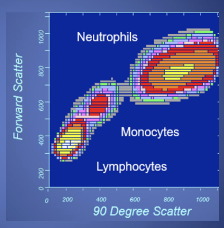
What do Granulocytes look like on a Flow Cytometry Plot?
relative FSC: intermediate to high
relative SSC: high
typical location: upper region often towards the right

What do the different quadrants mean on a plot?
Upper Left
Y marker +
X marker -
Lower Left
both markers -
Upper right
both markers +
Lower right
Y marker -
X marker +
What is Flow Cytometry?
flow - moving fluid
cyto- physical properties of cells
metry - measurement
differentiates cell counts / microparticles
10,000 cells per second
uses monoclonal Ab to differentiate cells
How does Flow Cytometry Work?
Fluids
transports particles / cells in single file through a laser
pressure and saline sheath fluid
optics
directs light to PTM using filters
electronics (photodetectors)
converts light → digital signals that are plotted as events on a scatterplot or contour plot
What occurs during Forward Scatter?
approximates cell size
larger cell = greater signal
What occurs during side or right angle scatter?
measures cell complexity / granularity
larger complexity = larger signal
Define gating.
separation of subpopulation from a heterogenous population to optimize analysis in multiparameter experiments
What is electron gating?
process of selecting a specific cell population for analysis based on its measured characteristics
What is a Gate?
region on a scatterplot of fluorescence plot
events inside region are included in selected population
events outside are excluded from particular analysis
Why is gating usfeul?
blood specimen contains many cell types
if you want to analyze a particular population, you need to separate it analytically from others
What is hydrodynamic focusing in flow cytometry?
process in which sheath fluid surrounds and narrows the sample stream so that cells pass through the laser in a controlled nearly single-file arrangement
Why is hydrodynamic focusing useful?
allows instrument to analyze cells individually and consistently
What CDs are expressed in hematopoietic stem cells?
CD34
What CDs are expressed in B cells?
CD34
CD19
CD10
CD21
CD22
What CDs are expressed in a proerthroblast?
CD33
CD72
GPA
CD36+