L3 - Hematopoiesis Diseases
 Call Kai
 Learn
 Practice Test
 Spaced Repetition
 Match
 Flashcards
 Knowt Play
1/46

There's no tags or description
Looks like no tags are added yet.
47 Terms
What are Hyperplasia, Hypoplasia, and Aplasia?
Hyperplasia:
Increased hematopoietic cellularity affecting one or more marrow lineages
Hypoplasia
Reduced hematopoietic cellularity affecting one or more marrow lineages
Aplasia
Near-complete or complete absence of hematopoietic cells in one or more marrow lineages
What are Pancytopenia and Myelophthisis?
Pancytopenia:
Concurrent anemia, neutropenia, and thrombocytopenia in peripheral blood
Myelophthisis:
Displacement or disruption of normal hematopoietic marrow by infiltrative tissue or fibrosis, impairing blood cell production
What are Myelodysplasia, Myelofibrosis, Myelonecrosis, and Myelitis?
Myelodysplasia:
Abnormal morphology and maturation of hematopoietic cells
Myelofibrosis:
Increased deposition of reticulin and/or collagen fibers in bone marrow
Myelonecrosis:
Necrosis of hematopoietic cells and supporting marrow tissue
Myelitis:
Inflammation of bone marrow
What is Ineffective Hematopoiesis?
Inadequate production of mature blood cells despite the presence of marrow precursors → due to defective maturation or premature precursor destruction
What are the Major Indications for Bone Marrow Evaluation?
Persistent, unexplained cytopenias
Diagnosis or staging of hematopoietic or metastatic neoplasia
Investigation of selected infectious diseases
Unexplained systemic abnormalities, including fever, hypercalcemia, weight loss, or hyperproteinemia
Suspected storage diseases
Which Cytopenias are the Primary Indications for Bone Marrow Sampling?
Key: Cytopenias should generally be persistent and unexplained
Anemia
Neutropenia
Thrombocytopenia
Any combination of these abnormalities
Why is Isolated Lymphopenia Not Typically an Indication for Bone Marrow Sampling?
Lymphocytes can proliferate in lymphoid tissues and circulate between blood and tissues
Therefore, lymphopenia does not necessarily indicate impaired bone marrow production.
Which Neoplastic Diseases May Require Bone Marrow Evaluation?
Lymphoproliferative diseases
Myeloproliferative diseases, including leukemia and MDS
Multiple myeloma
Histiocytic sarcoma
Mastocythemia
Metastatic neoplasia
Viral: FeLV, parvovirus, equine infectious anemia
Bacterial: Ehrlichia canis, Anaplasma phagocytophilum, mycobacteria
Fungal: Histoplasmosis, invasive mold infections
Protozoal: Leishmaniasis.
What is the Difference between Reduced and Ineffective Hematopoiesis?
Reduced hematopoiesis:
Too few precursors are present in the affected lineage
Ineffective hematopoiesis:
Precursors are present, sometimes increased, but defective maturation or destruction prevents adequate mature blood cell production
What Conditions Can Cause Reduced Hematopoiesis?
Hypoplasia
Aplasia
Myelofibrosis
Myelonecrosis
Infiltration or replacement of normal marrow
What Conditions Can Cause Ineffective Hematopoiesis?
Myelodysplastic syndrome (MDS)
Precursor immune-mediated anemia (PIMA)
Secondary dysplastic changes associated with infections or other marrow injuries
Why can Hypercellular Bone Marrow Still Have Hypoplasia of a Specific Lineage?
Overall marrow cellularity and individual lineage cellularity are different measurements
EX: granulocytic or megakaryocytic hyperplasia may increase total marrow cellularity while erythroid precursors remain decreased
What are the Major Categories of Bone Marrow Pathology?
Neoplastic
Inflammatory or infectious
Immune-mediated
Degenerative
Toxic
Idiopathic

What were the Major Clinical and Bone Marrow Findings in Elliot?
Clinical findings:
Persistent, progressive anemia
Bone Marrow Findings:
Hypercellular bone marrow
Trilineage dysplasia
Granulocytic left shift
Mild plasmacytosis
Dx: Myelodysplastic syndrome (MDS)

What Abnormal Granulocytic Morphology was identified in Elliot?
These findings support abnormal granulocytic development
Giant band neutrophils: Abnormally large band forms
Ring-shaped nuclei: Abnormal nuclear morphology in dogs
Myeloblasts: Immature myeloid cells identified in the marrow

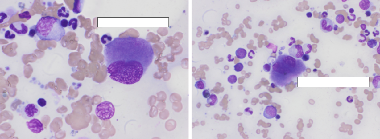
What Erythroid and Megakaryocytic Dysplastic Changes were identified in Elliot?
Erythroid: Nuclear-cytoplasmic maturation asynchrony
Megakaryocytic: Dwarf megakaryocytes, which are abnormally small megakaryocytes with relatively mature cytoplasm.

What is Trilineage Dysplasia?
Abnormal morphology or maturation affecting all three hematopoietic lineages
Erythroid
Granulocytic
Megakaryocytic
What is Myelodysplastic Syndrome (MDS)?
A PRIMARY clonal neoplastic myeloid disorder characterized by:
Ineffective hematopoiesis
Dysplastic hematopoietic cells
Abnormal maturation
Chronic or progressive peripheral cytopenias, and often hypercellular bone marrow

Why Does MDS Cause Peripheral Cytopenias Despite Hypercellular Bone Marrow?
Abnormal precursors proliferate but fail to mature appropriately and may die prematurely
Therefore, numerous marrow precursors produce too few functional mature blood cells

What Blast Percentage is Associated with MDS?
Less than 20% blasts
The lecture uses 20% as the threshold separating MDS from acute leukemia
What is the Main Takeaway from Elliot's Case?
A hypercellular marrow does not necessarily indicate effective hematopoiesis
Elliot demonstrates ineffective hematopoiesis caused by primary myelodysplastic syndrome

What were the Major Clinical and Bone Marrow Findings in Mama?
Clinical findings:
Persistent anemia
Neutropenia
Thrombocytopenia
Persistent fever
FIV-positive status
Bone marrow findings:
Marked hypercellularity
Granulocytic hyperplasia with marked left shift and mild dysplasia
Moderate erythroid hypoplasia
Mild megakaryocytic hyperplasia and dysplasia
What does a marked Granulocytic Left Shift Indicate in Bone Marrow?
An increased proportion of immature granulocytic cells relative to mature cells → This indicates abnormal maturation or increased immature cell production
What is the Difference between Primary Myelodysplasia and Secondary Dysmyelopoiesis?
Primary myelodysplasia (MDS):
Clonal neoplastic disease characterized by abnormal hematopoietic maturation
Secondary dysmyelopoiesis:
Abnormal marrow development associated with another disease or reactive process
What are Common Causes of Secondary Dysmyelopoiesis?
Immune-mediated: IMHA, ITP
Infectious: FeLV, FIV, Leishmania
Drugs/toxins: Chemotherapy, folate antagonists, estrogen, phenobarbital, heavy metals
Other marrow diseases: Lymphoma, myelofibrosis.
What is the Main Takeaway from Mama's Case?
Dysplasia does not automatically indicate MDS
Secondary diseases can cause dysplastic changes
Hypercellular marrow can contain hypoplasia of a specific lineage
FIV positivity alone does not prove the cause of every marrow abnormality
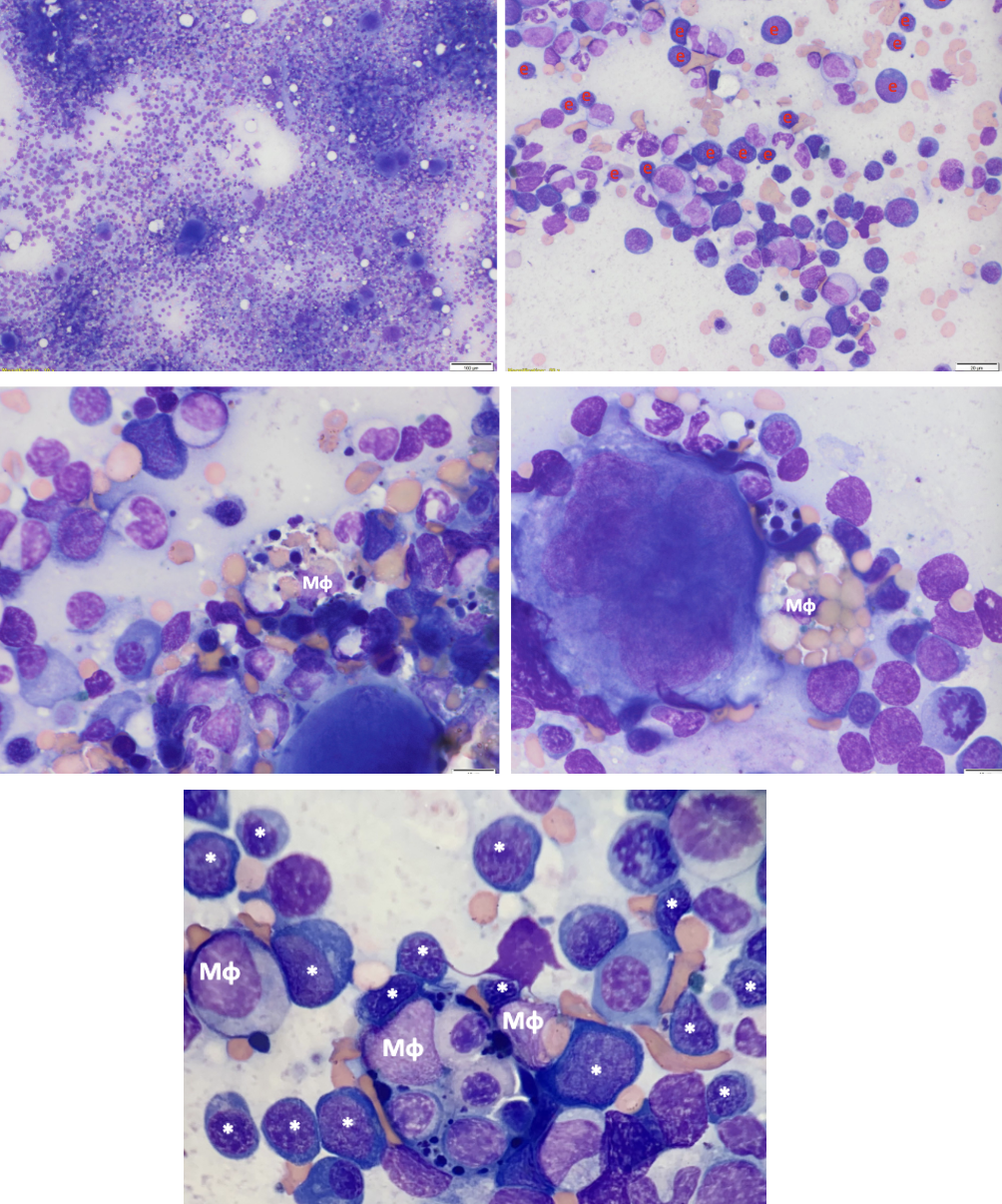
What were the Major Clinical and Bone Marrow Findings in Daisy?
Clinical:
Chronic, suspected immune-mediated anemia
Bone marrow:
Moderate to marked hypercellularity
Erythroid hyperplasia
Erythroid left shift
Erythrophagocytosis
Mild plasmacytosis
Megakaryocytic left shift
Mild dysplasia
What is Precursor Immune-Mediated Anemia (PIMA)?
An immune-mediated disorder in which the immune system targets developing erythroid precursors, typically late-stage precursors, within bone marrow → This prevents adequate production of mature circulating red blood cells
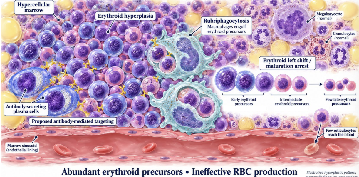
What are the Characteristic Findings of PIMA?
Non-regenerative anemia
Erythroid hyperplasia
Erythroid left shift or maturation arrest
Erythrophagocytosis may be observed

What is Erythrophagocytosis, and How is It Recognized in Bone Marrow?
Engulfment of erythrocytes or erythroid precursors by macrophages
Morphologic finding: Red blood cells or erythroid precursors visible within macrophage cytoplasm.

Why does PIMA Cause Erythroid Hyperplasia and a Left Shift?
1. Immune destruction of erythroid precursors contributes to anemia
2. Anemia causes tissue hypoxia
3. Kidneys increase EPO production
4. EPO stimulates erythroid precursor proliferation
5. Continued precursor destruction prevents normal maturation
Result: Erythroid hyperplasia with accumulation of immature cells
What is the Main Takeaway from Daisy's Case?
Peripheral anemia does not necessarily indicate reduced erythroid precursor production
Daisy demonstrates ineffective erythropoiesis caused by immune-mediated destruction of developing erythroid cells

What were the Major Clinical and Bone Marrow Findings in Bailey?
Clinical:
Severe anemia and thrombocytopenia
Bone marrow:
Myelofibrosis
Marked hematopoietic hypoplasia
Focal myelonecrosis and myelitis
Multifocal hemosiderosis
What is Myelofibrosis, and How Does It Cause Cytopenias?
Excessive deposition of reticulin and/or collagen fibers within bone marrow
Fibrous tissue replaces or disrupts normal hematopoietic tissue, reducing blood cell production and potentially causing marrow failure

How is Myelofibrosis Recognized in Bone Marrow Images?
Increased fibrous matrix replacing normal hematopoietic tissue
Reduced hematopoietic cellularity
Collagen appears pink in routine histologic sections
Special stains highlight collagen as blue fibrous material in the lecture images
Why is a Core Biopsy Preferred over an Aspirate for Evaluating Myelofibrosis?
Aspirate:
Often nondiagnostic because fibrotic marrow yields few cells
Core biopsy:
Preserves marrow architecture and reveals collagen deposition and loss of hematopoietic tissue
What is the Difference between Primary and Secondary Myelofibrosis?
Primary:
Neoplastic disorder associated with marrow fibrosis
Very rare in veterinary medicine
Secondary:
Reactive fibrosis associated with chronic inflammation, toxic injury, necrosis, immune-mediated disease, or other marrow injuries
What is the Main Takeaway from Bailey's Case?
Bailey demonstrates reduced hematopoiesis caused by structural damage to bone marrow
Unlike Elliot and Daisy, whose marrows contain abundant but ineffective precursors, Bailey's marrow has reduced hematopoietic tissue because of fibrosis
QUESTION 1: Why can hypercellular marrow coexist with anemia?
ANSWER → Precursors may fail to mature or be destroyed before release
Alternatively, overall hypercellularity may conceal hypoplasia of the erythroid lineage
QUESTION 2: Does dysplasia prove MDS?
ANSWER → No
Secondary infectious, immune, drug-related, toxic, and other marrow disorders can produce dysplasia
QUESTION 3: Which findings should you recognize in images?
ANSWER →
Dysplasia: Abnormal nuclear or cellular maturation
Erythrophagocytosis: Macrophages engulfing erythroid precursors
Fibrosis: Increased fibrous matrix with reduced hematopoietic tissue
Infection: Intracellular infectious organisms.
Bone Marrow Terminology SUMMARY
Term | Definition | Key Distinction |
Hyperplasia | Increased hematopoietic cellularity | More cells |
Hypoplasia | Decreased hematopoietic cellularity | Fewer cells |
Aplasia | Near-complete or complete absence of hematopoietic cells | Almost no cells |
Pancytopenia | Concurrent anemia, neutropenia, and thrombocytopenia | All three cell lines decreased in blood |
Myelophthisis | Replacement/disruption of marrow by infiltrative tissue or fibrosis | Normal marrow displaced |
Myelodysplasia | Abnormal morphology and maturation | Abnormal cell development |
Myelofibrosis | Excess reticulin/collagen deposition | Fibrous tissue replaces marrow |
Myelonecrosis | Death of marrow cells and supporting tissue | Tissue necrosis |
Myelitis | Inflammation of bone marrow | Marrow inflammation |
Reduced vs. Ineffective Hematopoiesis SUMMARY
Feature | Reduced Hematopoiesis | Ineffective Hematopoiesis |
Definition | Too few precursors are present | Precursors are present but fail to produce adequate mature cells |
Bone Marrow | Decreased precursors in affected lineage | Often increased precursors |
Main Problem | Insufficient cell production | Defective maturation or premature destruction |
Peripheral Blood | Cytopenias | Cytopenias |
Common Causes | Hypoplasia, aplasia, fibrosis, necrosis, infiltration | MDS, PIMA |
Clinical Example | Bailey (myelofibrosis) | Elliot (MDS), Daisy (PIMA) |
Key Concept | Not enough cells are made | Cells are made but do not mature or survive appropriately |
Important: Overall marrow hypercellularity does not exclude hypoplasia of a specific lineage, as demonstrated in Mama's case.
Clinical Case Comparison SUMMARY
Feature | Elliot | Mama | Daisy | Bailey |
Disease | MDS | FIV-positive, secondary dysplastic changes | PIMA | Myelofibrosis |
Mechanism | Neoplastic | Infectious/systemic disease association | Immune-mediated | Fibrosis |
Marrow Cellularity | Hypercellular | Hypercellular | Hypercellular | Hypoplastic |
Key Findings | Trilineage dysplasia, granulocytic left shift | Granulocytic hyperplasia, erythroid hypoplasia, dysplasia | Erythroid hyperplasia, left shift, erythrophagocytosis | Excess collagen, hypoplasia, necrosis |
Hematopoiesis | Ineffective | Mixed lineage responses | Ineffective | Reduced |
Main Takeaway | Abundant abnormal precursors fail to produce mature cells | Dysplasia does not always indicate MDS | Immune destruction of erythroid precursors | Fibrosis disrupts normal marrow production |
Remember:
Elliot: Neoplastic dysplasia.
Mama: Secondary dysplastic changes.
Daisy: Immune-mediated precursor destruction
Bailey: Fibrosis replacing hematopoietic tissue
Recognizing Bone Marrow Abnormalities SUMMARY
Abnormality | What to Look for in Images | Associated Disease/Case |
Dysplasia | Abnormal nuclear or cellular morphology, giant bands, ring-shaped nuclei, dwarf megakaryocytes | Elliot (MDS) |
Granulocytic left shift | Increased immature granulocytes relative to mature neutrophils | Elliot, Mama |
Erythroid left shift | Increased immature erythroid precursors | Daisy (PIMA) |
Erythrophagocytosis | Macrophages containing engulfed erythrocytes or erythroid precursors | Daisy (PIMA) |
Fibrosis | Excess fibrous matrix replacing normal hematopoietic tissue | Bailey (myelofibrosis) |
Infection | Infectious organisms within marrow cells, such as macrophages | Leishmaniasis |
Image recognition tips:
Dysplasia: Look for abnormal cell shapes and nuclear maturation
Erythrophagocytosis: Look for erythroid cells inside macrophages
Fibrosis: Look for collagen replacing normal marrow cells; collagen appears blue in the special-stain images
Infection: Look for intracellular organisms
Primary vs. Secondary Bone Marrow Disorders SUMMARY
Disorder | Primary | Secondary |
Myelodysplasia | MDS: Clonal neoplastic disease causing ineffective hematopoiesis | Dysmyelopoiesis associated with infection, immune disease, drugs, or toxins |
Myelofibrosis | Neoplastic marrow fibrosis; very rare in veterinary medicine | Reactive fibrosis associated with inflammation, toxic injury, necrosis, or immune-mediated disease |
Key Difference | Primary disease process | Develops in association with another underlying disorder |
Important distinction: Dysplasia alone does not prove MDS. Clinical history, peripheral blood findings, and other possible causes must be considered