CH 10 : The Central Vision System
 Call Kai
 Learn
 Practice Test
 Spaced Repetition
 Match
 Flashcards
 Knowt Play
1/31

Earn XP
Description and Tags
FNTB Exam 3
32 Terms
What are the other order cortical areas that vision travels to after the primary visual cortex
occipital, temporal, parietal
The retinofugal projection describes the movement of [what] to [where]?
retinal information to the LGN (then primary visual cortex, on…)
What are the steps of the retinofugal projection
APs enter optic nerve, partial decussitation in optic chiasm, through optic track to LGN, then projected to optic radiaiton, then primary visual cortex

How does the left and right visual hemifields get observed by the eye’s different zones of vision?
left visual hemishpield - observed by central left eye and peripheral right eye. L central crosses to R in optic chiasm, all left information is them together in right side
If there is blue color on the right side, where is the blue color sent/dispersed up to/including the pathway to the primary visual cortex?
Right side after chiasm

Cutting the L optic nerve results in a loss of _____
left eye vision, only left peripheral blindness
Cutting the L optic tract results in a loss of
Right sided information (right sided blindness)
Cutting the optic chiasm results in a loss of _____
Crossing fiber information - all peripheral blindness
What anatomical structures is the retinofugal projection close to that the optic tract can target in addition to the thalamus?
Hypothalamus - circadian rhythm
Superior colliculus - in midbrain, (around 10% ganglion cells) to have quick head and eye movements

Where is the LGN located?
Thalamus, dorsal

LGN has how many principle layers? numbered how?
6, increasing in number with ventral -→ dorsal
Which LGN layers receive input from ipsilateral axons? which from contralateral?
2,3,5 (ipsilateral) and 1,4,6 (contralateral)

Which LGN layers are magnocellular vs parvocellular, and how would these cells appear different? What about koinocellular - where are they and how do they look?
1&2 - magnocellular, larger cells
4-6 - parvocellular - smaller cells
koinocellular - between layers - very small

How does ganglion cell type compare to LGN cell type in terms of projection and visual field
Magoncellular receives projections form m type, parvo receivess from p type, and k receives from non-m-non-p type. The visual fields also match: Magnocellular is large fielded, quick to fade, insensitive to wavelength differences, parvo is small center-surround receptive field,sustained response, and can be color/opposing, koino is center surround and light/dark or color opponent
What area number is the primary visual cortex in
17 (or V1, striate cortex)

What is retinotopy?
neighboring retinal axons project to neighboring sections in the primary visual cortex → map of visual world
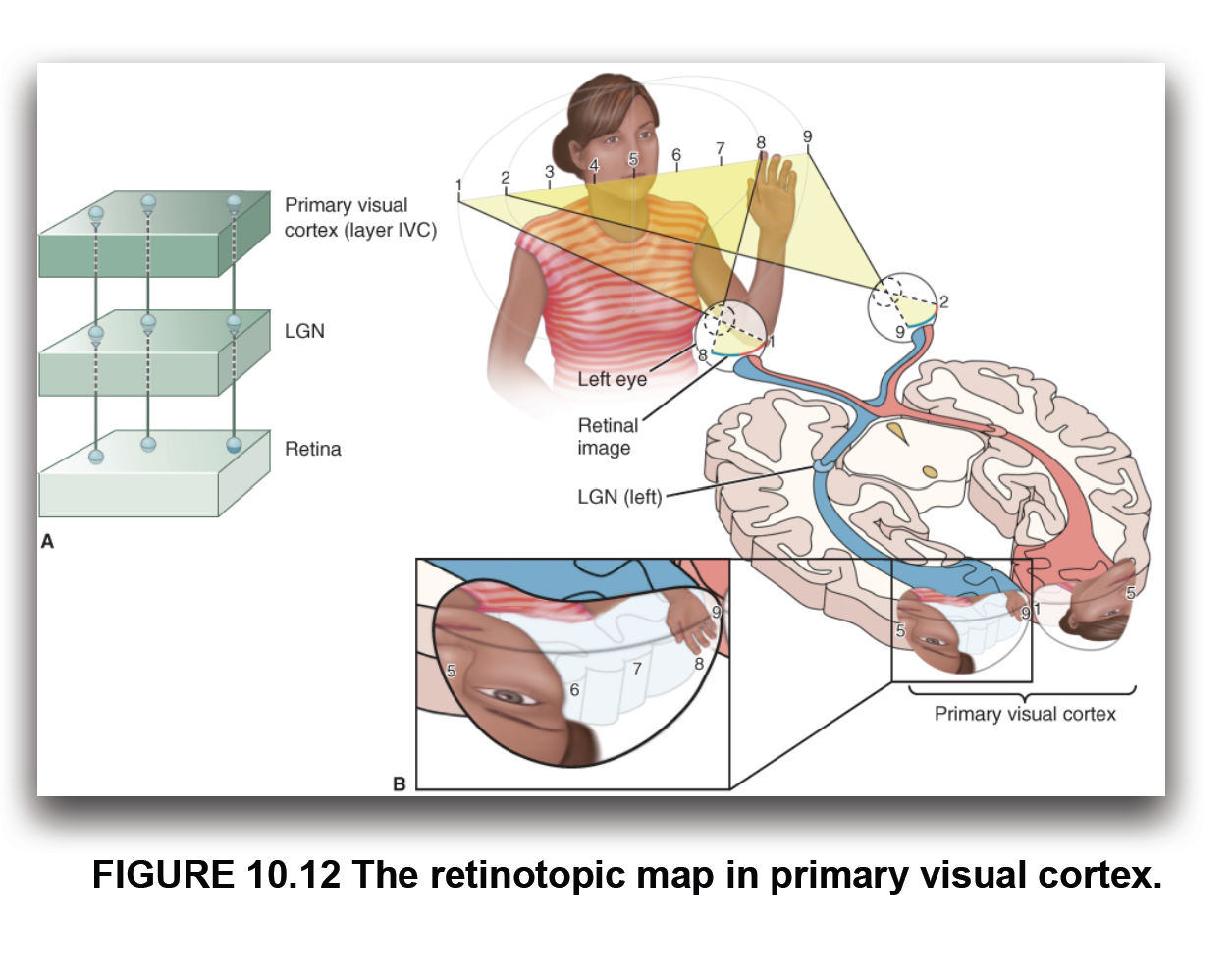
3 Points of retinotopy that influence organizaiton
distortion of areas based on density of receptors
one point of light appears as a gradient with a peak
not really a map, mor einformation to be interpreted
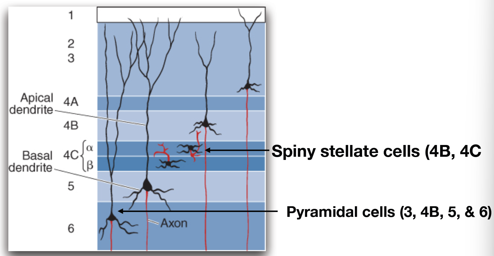
Layers of striate cortex, starting after white matter
VI, V, IV (IVA, IVB, IVC (alpha, beta)) III, II, I

What are the 2 different neuronal types in striate cortex, and what layers are they found in
spiny stallate - in IVC a, IVC b, and IVB
pyramidal (VI, V, IVB, III)
Which layers of the V1 receive input from koinocellular, magnocellular, and parvocellular
Koino - I and III
Magno - IVC a
Parvo - IVC b

How is eye specific information organize in layer IV
zebra stripes, some are left eye ocular dominant columns, some are right. some blend

Where do pyramidal cells in striate cortex layers output information
other cortical areas from 3/4B, pons and superior colliculus from 5, LGN from 6

How is cytochrome oxidase distributed in the striate cortex?
Blobs that span multiple layers (II - VI). They receive direct LGN input from koino, and parvo/magno input from IVC layer

Which primary visual cortex fields are bioncular and which are monocular
Binocular (having 2 receptive fields, one in ipsilateral and contralateral eye)- 4B - 1
monocular - 6-4C

What does orientation selectivity describe, and where are neurons like this found?
Response to light in a certain orientation - found in V1 neurons other than VI

What is direction selectivity
Response to motion in one direction but not the other

Simple cell receptive field vs complex
Simple - on off surrounding overlap - gives orientation specificity
Complex - orientation specific but not distinct on-off regions. Responses to both light on and off

What are the 3 parallel pathways
Magnocellular, koinocellular, parvocellular

What is a cortical module, what do they contain
2mm/2mm chunk that removes enough neurons to create a blind spot. contains ocular dominance columns, cytochrome oxidase blobs, and orientation columns

what are extrastriate streams for visual info to travel
Dorsal and ventral
Dorsal serves the analysis of visual motion and visual control of action (action stream)
Ventral is involved in perception of the visual world and recognition of objects (perception stream)

What cortical areas are associated with the dorsal stream, where does it feed
Extend to V2, V3, and V5 (MT) (processing of object motion). To posterior parietal cortex

What areas are involved with the ventral stream, where does it feed
Extend parallel with dorsal, progression from V1, V2, and V3 ventrally toward temporal lobes. V4 - receives blob/interblob inputs, important for shape and color perception. To Occititotemporal region
