NEUR 201 UNit 3
 Call Kai
 Learn
 Practice Test
 Spaced Repetition
 Match
 Flashcards
 Knowt Play
1/51

There's no tags or description
Looks like no tags are added yet.
52 Terms
proprioception
body’s ability to balance
dependent on info from the visual system, vestibular senses and touch senses
cardiac mucle cell
can contract rhythmically without innervation
regulated by autonomic NS (involuntary movement)
skeletal muscle cell
muscle fibers are individually innervated by axon branches from the CNS
smooth muscle cells
innervated by the autonomic nervous system (involuntary movement)
somatic motor system
moving different muscle groups produces a different type of movement in a different direction using flexion and extension of Synergistic and Antagonistic muscle groups
by knowing where our body is located in space + intention of movement- our brain can automatically determine what muscles to activate in order to achieve a movement
what is the pathway for extension?
biceps brachii (flexor)
brachialis (flexor)
triceps brachii (extensor)
anconeus (extensor)
what is the pathway for flexion?
anconeus (extensor)
triceps brachii (extensor)
brachialis (flexor)
biceps brachii (flexor)
what are characteristics of slow fibers (Type I, oxidative)
glycogen content: low
rate of fatigue: slow
ATPase activity: low
Fiber diameter: small
Motor unit size: small, slow
innervating by: small neurons
what are the two types of fast fibers?
fast oxidative (IIa) and fast glycolytic (IIb)
what are characteristics of fast oxidative (IIa) fibers
glycogen content: intermediate
rate of fatigue: intermediate
ATPase activity: high
Fiber diameter: intermediate
Motor unit size: intermediate, fast fatigue-resistant
innervating by: intermediate sized neurons
what are the characteristics of fast glycolytic fibers (IIb)?
glycogen content: high
rate of fatigue: fast
ATPase activity: high
Fiber diameter: large
Motor unit size: large, fast fatigue
innervating by: large neurons
what determines the amount of force that the muscle produces? how?
firing rate of the motor neuron
input to the (efferent) motor neuron is sent from upper motor neurons in the brain and sensory input from the muscles
how do motor neurons communicate with muscles?
neuromuscular junction
sarcolemma
excitable membrane around the muscle fiber
myofibrils
found within the muscle fiber
contract when an AP moves down the sarcolemma
what does the sarcoplasmic reticulum store?
calcium
how is the sarcoplasmic reticulum accessed by APs?
T-tubles within the sarcolemma that act like tunnels
how does the t-tubule carry APs?
when depolarized, a tetrad of voltage-sensitive calcium channels open allowing calcium to flow out of the sarcoplasmic reticulum leading to myofibril contraction
what happens when we want to contract our muscle?
excitation-contraction coupling
what are the steps of excitation
AP runs though a motor neuron and releases ACh at the NT junction
ACh binds to nicotinic receptors on the sarcolemma allowing Na+ to enter the cell causing depolarization called the end point potential (EPP)
Sarcolemma becomes depolarized moving into the T-tubules causing Ca2+ release from the sarcoplasmic reticulum
What are the steps of contraction?
Ca2+ binds to troponin (a protein that is covering attachment sites on actin within the thin filaments)
tropomyosin is shifted revealing binding sites on actin
myosin heads from the thick filament bind to actin and pivot, causing the filaments to slide along each other and the muscle to contract
ATP binds to each myosin head and detaches from actin
the cycle continues as long as Ca2+ and ATP are present
muscle spindles
type of stretch receptor that sends info to alpha motor neurons for sensory feedback
sensory info is about muscle stretch —> causes contraction producing reflex
intrafusal fibers
modified fibers within the muscle spindle
inntervated by gamma motor neurons instead of alpha motor neurons
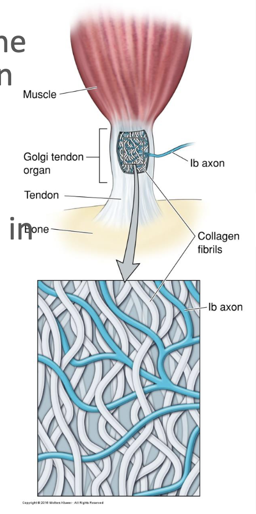
golgi tendon organ
provides info about stretch of the tendon attached to the muscle
uses Ib afferent axon instead of Ia like mucle spindle
as muscle contracts, firing rate of teh Ia axon is decreased, but the firing rate of the Ib axon is increased
what are the steps for the patellar reflex?
tap on patellar ligament excites nerve endings of muscle spindle in quadriceps femoris
stretch signals travel to spinal cord via afferent nerve fiber and posterior root
afferent neuron excites motor neuron in spinal cord
efferent signals in motor nerve fiber stimulate quadriceps to contract, producing knee jerk
at same time, a branch of the afferent nerve fiber stimulates inhibitory motor neuron in spinal cord
that neuron inhibits motor neuron that supplies hamstring muscles
hamstring contraction is inhibited so hamstrings (knee flexors) do not antagonize quadriceps (knee extensor)
spinal interneurons function
excitatory or inhibitory
allow for reflexes to coordinate proper muscle contraction or relaxation
homeostasis
maintenance of body’s internal environment with a narrow physiological range (temp, bp, etc)
Autonomic NS
function automaticcaly w/o conscious, voluntary control
Sympathetic division of the ANS
fight or flight —> prepare by dilating pupils, inc heart rate, dec digestion, etc
parasympathetic division of the ANS
rest + digest —> slow heart rate, inc intestinal and gland activity, relaxes spincter muscles in the GI tract
humoral response
hypothalamic neurons respond to sensory signals by stimulating or inhibiting the release of pituitary hormones in the bloodstream
visceromotor response
neurons in the hypothalamus respond to sensory signals by adjusting the balance of sympathetic and parasympathetic outputs of the ANS
somatic motor response
hypothalamic neurons (lateral hypothalamus) respond to sensory signals by inciting an appropriate somatic motor behavioral response
secretory hypothalamus
secretes chemicals into the bloodstream for long range impact on the brain and entire body
integrates somatic + visceral response depending on what the brain needs
recieves signals about the body and sends info to appropriate place to help maintain temp
neurosecretory cells
found in periventricular zone of the hypothalamus
extend axons to pituitary stalk —> release neurohormones into the blood (not normal NT)
which hormones are received by the posterior pituitary from what?
magnocellular neurosecretory cells: oxytocin + vasopressin/ADH
what is the function of teh anterior pituitary
actual gland that synthesizes and secretes many hormones to regulate other gland secretions in the endocrine system (thyroid, adrenal, mammary, etc)
controlled by the hypothalamus w/ parvocellular neurosecretory cells which secrete hypophysiotropic hormones into the blood
what is the function of oxytocin
social bonding + maternal instinct, contracts uterus to facilitiate delivery of baby + ejection of milk from mammary glands for feeding
what happens if too much oxytocin?
benigh prostatic hyperplasia
what happens if too little oxytocin?
prevent milk ejection + breast feeding
linked to autism + depressive disorder
what is the function of vasopression/ADH?
regulates BP, volume, salt concentration + water volume in tissues
acts on kidneys to retain water in the body
too much ADH?
caused by drug side effects + diseases of the lungs, chest wall, hypothalamus/pituitary
lung cancer can also lead to it
low levels ADH?
kidneys exrete too much water leading teh dehydration + fall in bp
indicate damage to hypothalmus/pituitary gland or excessive water drinking
diabetes insipidus
associated with inc thirst, urine production
cortisol function?
secreted from adrenal cortex of adrenal gland
mobilize energy reserves, supress immune system in a fight/flight situation stimulated by stress
CRH —> ACTH → adrenal cortex —> secrete cortisol
too much cortisol?
Cushing’s syndrome
rapid weight gain concentrated to the face, chest, abdomen, flushed round face, high BP, oseoporosis, immune suppression, sleeplessness, memory impairment/irritability
high levels in women —> dec sex drive + irregular/missed menses
too little cortisol?
Addison’s disease
fatigue, dizziness, weight loss, muscle weakness, mood changes, hyperpigmentation
life threatening condition
function of follicle stimulating hormone?
anterior pituitary —> targets gonads cause ovulation in women, spermatogenesis in men
too mcuh FSH?
hypergonadotrophic-hypogonadism
primary ovarian/testicular failure
Klinefelter’s syndrome (men), Turner syndrome (women)
rise naturally during menopause
too little FSH?
hypogonadotrophic-hypogonadism
incomplete development at puberty, ovarian failure
ovarian follicles don’t grow properly —> infertility
Kallman’s syndrome
lutenizing hormone function?
anterior pituitary —> act on gonads to regulate function and maturation of ovaries and sperm
Men: stimulates leydig cells —> prod testosterone
Women: easy in the cycle prod estradiol, later releases egg from ovary (ovulation), also required to prod progesterone