Anat 338 Lab 2: Spinal Cord | Vertebral Column | Trunk Wall Part I
 Call Kai
 Learn
Learn Practice Test
 Spaced Repetition
 Match
Match Flashcards
 Knowt Play
1/64

There's no tags or description
Looks like no tags are added yet.
Name | Mastery | Learn | Test | Matching | Spaced | Call with Kai | Chat |
|---|
No analytics yet
Send a link to your students to track their progress
65 Terms

Cervical region
7 vertebrae
neck portion of spine that supports the head

Thoracic region
12 vertebrae
middle part of trunk extending from neck to diaphragm
supported by rib cage and thoracic spine

Lumbar region
5 vertebrae (largest)
lower back segment and bear most body weight
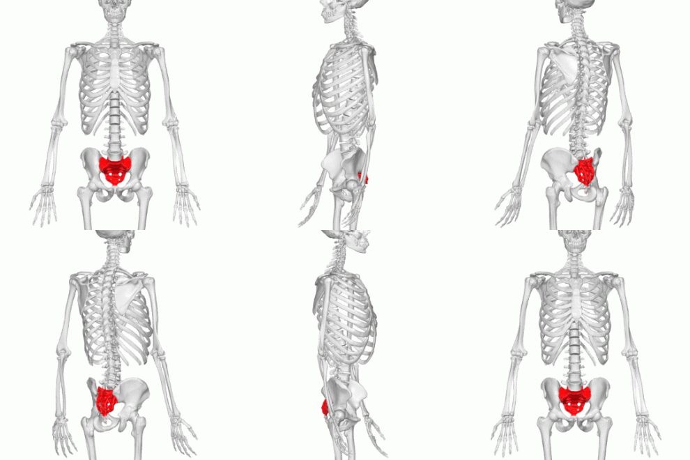
Sacral region
triangular shaped bone at base of spine between L5 and coccyx
formed by fusion of 5 sacral vertebrae during early adulthood

Cervical enlargement
Widened portion of the spinal cord spanning roughly C3–T2 (commonly C4–T1 or peaking at C5)
Extra motor and sensory neurons supply the upper limbs

Lumbar enlargement
Widened area of the spinal cord that supplies nerves to the lower limbs, located between the T11 and L2 vertebrae
Higher density of motor neurons in the ventral horns, which are responsible for innervating the lower limbs
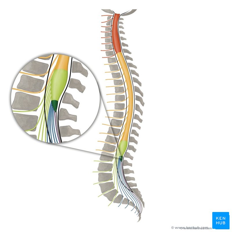
Conus medularis
tapered, end of spinal cord (L1-L2)
coordinates motor, sensory, autonomic functions of the lower body

Cauda equina
bundle of spinal nerves after conus medullaris essential for leg movement and bladder, bowel, and pelvic organ function


Meninges
3 protective membranes that cover and support the brain and spinal cord

Dura mater
outermost, thick, durable layer of meninges
attaches to inside of skull and vertebrae

Arachnoid mater
middle, thin, transparent layer of meninges
lies above pia mater
forms subarachnoid space which contains CSF and fine filaments that connect to pia mater

Pia mater
innermost layer, delicate membrane that closely adheres to surface of the brain and spinal cord
highly vascularized and allows blood vessels to pass through to supply CNS

Denticulate ligament
Horizontal anchors to provide support and stability to spinal cord
Triangular-shaped extensions of the spinal pia mater, extending sideways from the spinal cord.

Filum terminale
Anchors spinal cord to coccyx and stabilizes within vertebral column
Thread-like structure extending from the apex of the conus medullaris (the tapered end of the spinal cord) down to the coccyx

White matter
Connects different brain regions to transmit signals efficiently

Dorsal column
Location: Dorsal/posterior horns
Function: Sensory input
Receives fine touch, vibration, and proprioception of spinal cord

Ventral column
Location: ventral/anterior horns
Function: motor output
contains lower motor neurons → skeletal muscles
contains lateral neurons → visceral muscle control

Lateral column
Houses sympathetic preganglionic neurons
In white matter between anterior and posterior columns
Contains myelinated axons that transmit information up and down CNS so sensory and motor neurons can communicate

Gray matter
Processes information and controls thinking and movement

Posterior horn
processes somatosensory information i.e. fine touch, vibration, proprioception, pain, temperature
in gray matter - butterfly-like shape with 3 columns
contains second-order sensory neurons (receiving input from first-order neurons in dorsal root ganglia)

Lateral horn
projection of gray matter in thoracic spinal cord that contains preganglionic sympathetic neuron

Anterior horn
contains lower motor neurons that send axons via anterior nerve to innervate skeletal muscles

Central canal
Cerebrospinal fluid-filled space that runs through spinal cord
Located with gray commissure

Dorsal root ganglion
Cluster of sensory neuron cell bodies located near spinal cord that transmit sensory information from PNS → CNS
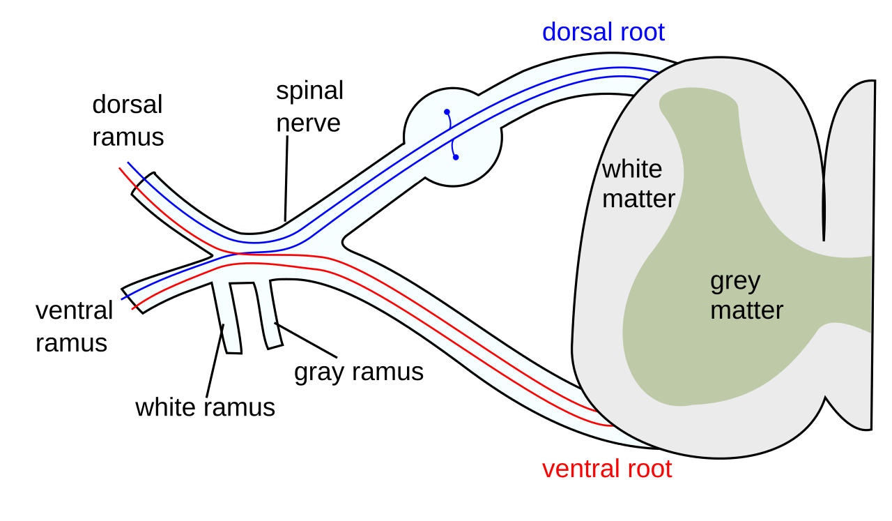
Dorsal root and rootlets
Dorsal rootlets are small nerve fibers that carry sensory info from body to spinal cord and converge to form dorsal root
Ventral root and rootlets
Ventral rootlets converge to form ventral root which carries motor (efferent) signals from spinal cord → muscles & glands
Spinal nerves
Mixed periphery nerves that connect spinal cord to body carrying sensory, motor, and autonomic signals

Dorsal rami
Posterior branch of spinal nerve
motor innervation → deep back muscles
sensory innervation → skin of posterior head, neck, back

Ventral rami
larger, anterior branch of spinal nerve
Sensory + motor fibers → front & sides of trunk & limbs
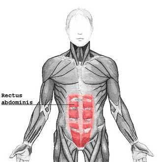
Rectus abdominis
6-pack muscle
Trunk flexion, stability, posture

Internal oblique
Deep to external obliques
Trunk rotation, lateral flexion

External oblique
largest, most superficial of the flat muscles
trunk rotation, lateral flexion

Tranversus abdominis
deepest abdominal muscle
stabilizes core, wrapped around abdomen like corset, keeps organs in
Erector spinae
Group of 3 main muscles in the back
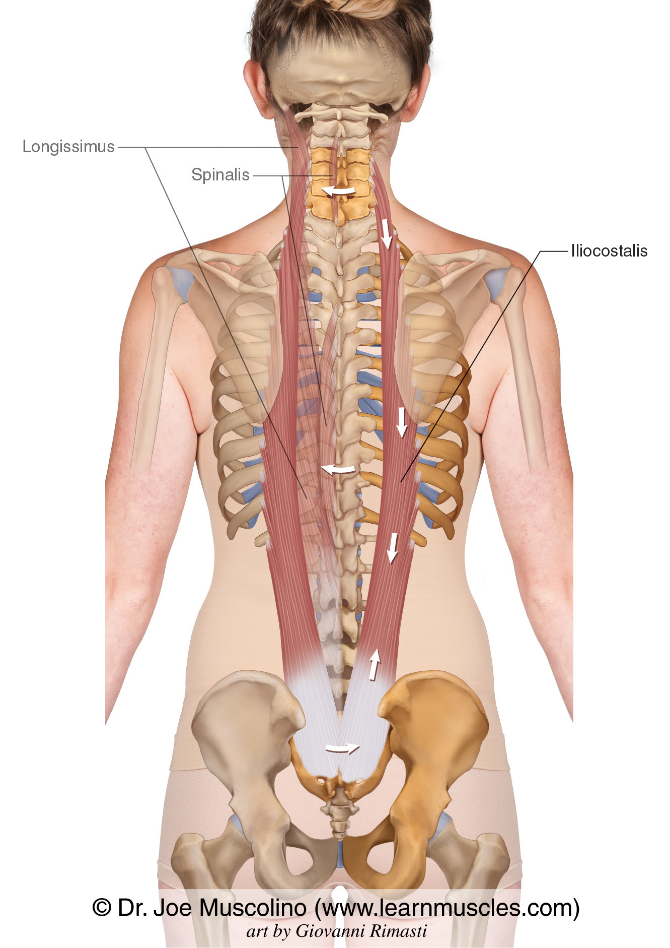
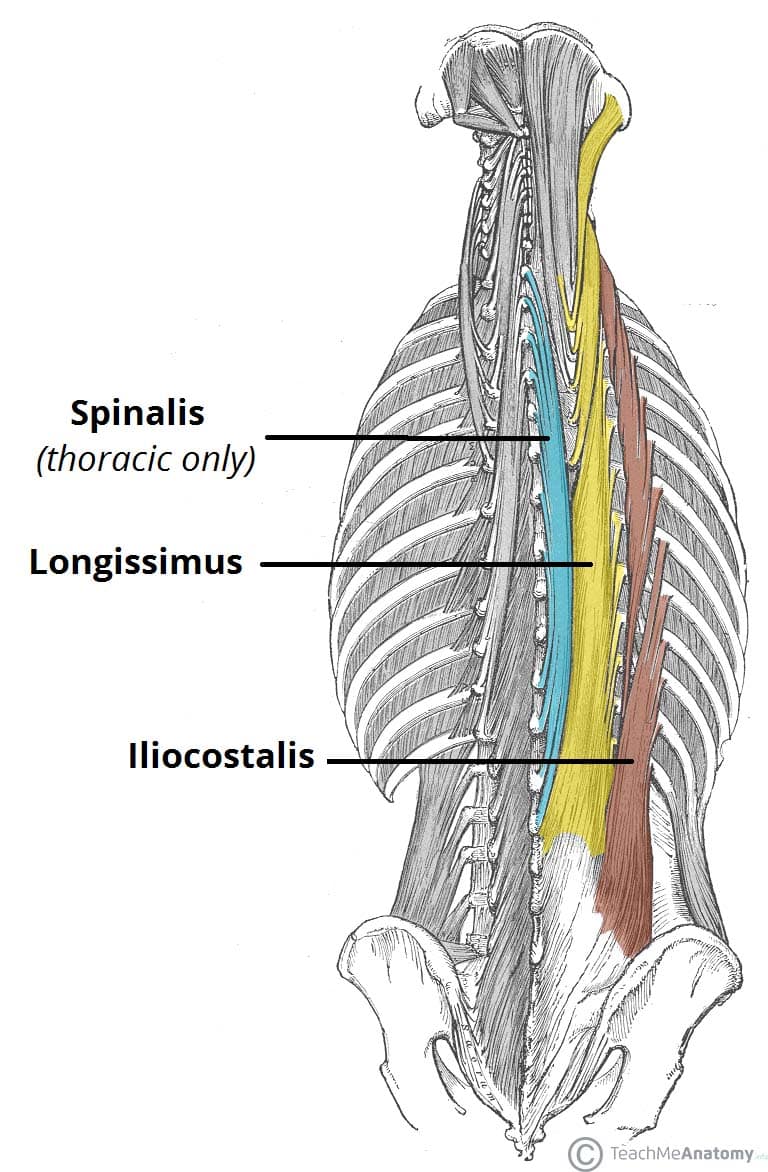
ilicostalis (I in I Love Sports)
most lateral of the erector spinae muscles. runs from the iliac crest of pelvis to the ribs
when contracted bilaterally: extend spine and help to maintain upright posture against gravity
when unilaterally contracted: lateral flexion

Longissimus (L in I Love Sports)
longest of the erector spinae muscles
when contracted bilaterally: extend spine and help to maintain upright posture against gravity
when unilaterally contracted: lateral flexion

Spinalis (S in I Love Sports)
thin and medial - closest to the spine
when contracted bilaterally: extend spine and help to maintain upright posture against gravity
when unilaterally contracted: lateral flexion

False ribs
don’t directly articulate with sternum (3 of them)

True ribs
directly articulate with sternum through costal cartilage (7 of them)
Floating ribs
only attach to the spine and not to the breast bone - they protect kidneys (2 of them) Found on the anterior (front) part of the chest wall, acting as an extension of the ribs.

Costal cartilage
Flexible band of hyaline cartilage that connects the front of the ribs to the breastbone, or sternum.

head of the ribs
wedge-shaped posterior end that connects the rib to the spine through costovertebral joints
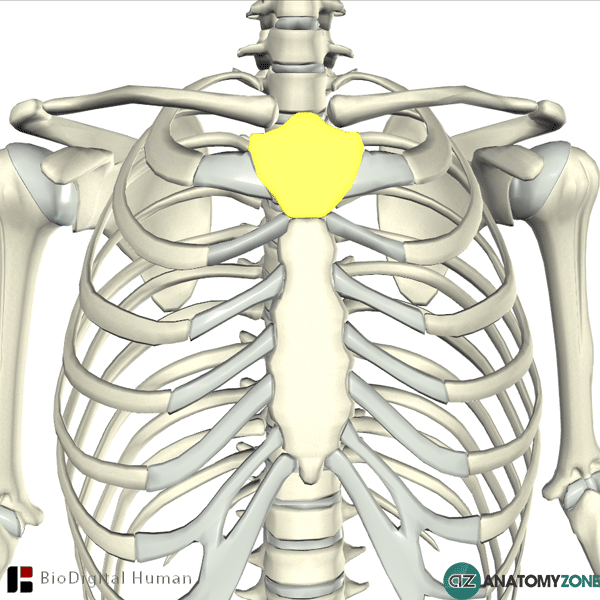
Manubrium - sternum
wide, triangular shaped, upper segment of the sternum (breastbone) located at the level of the T3 and T4 vertebrae
Body of the sternum
longest, flat middle portion of the sternum (breastbone) located in the center of the chest


Xiphoid process - sternum
the small, lowest pointed end of your sternum (breastbone) located in the center of your chest
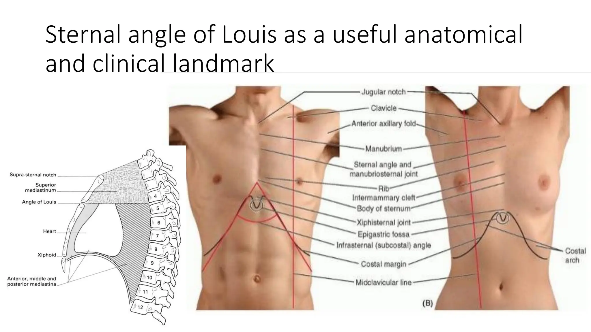
Sternal angle
the visible and palpable ridge on the chest where the manubrium meets the body (top meeds middle)

Suprasternal notch
the visible dip or hollow at the base of the neck, located right above the top of the breastbone (manubrium) and between the collarbones

Clavicular notch
small, oval depression located on the top corner of the manubrium (upper part of sternum)

Spinous process
the bony bump that sticks out from the back of each vertebra along your spine

Transverse process
small, bony projections pointing out from the left and right sides of each vertebra in the spine

Superior articular process
bony projection pointing upward on each side of a vertebra that locks with the vertebra above it to form a spinal joint

Superior articular facet
a smooth bone surface on the upper part of a vertebra that faces upward and connects with the inferior articular facet of the vertebra directly above it
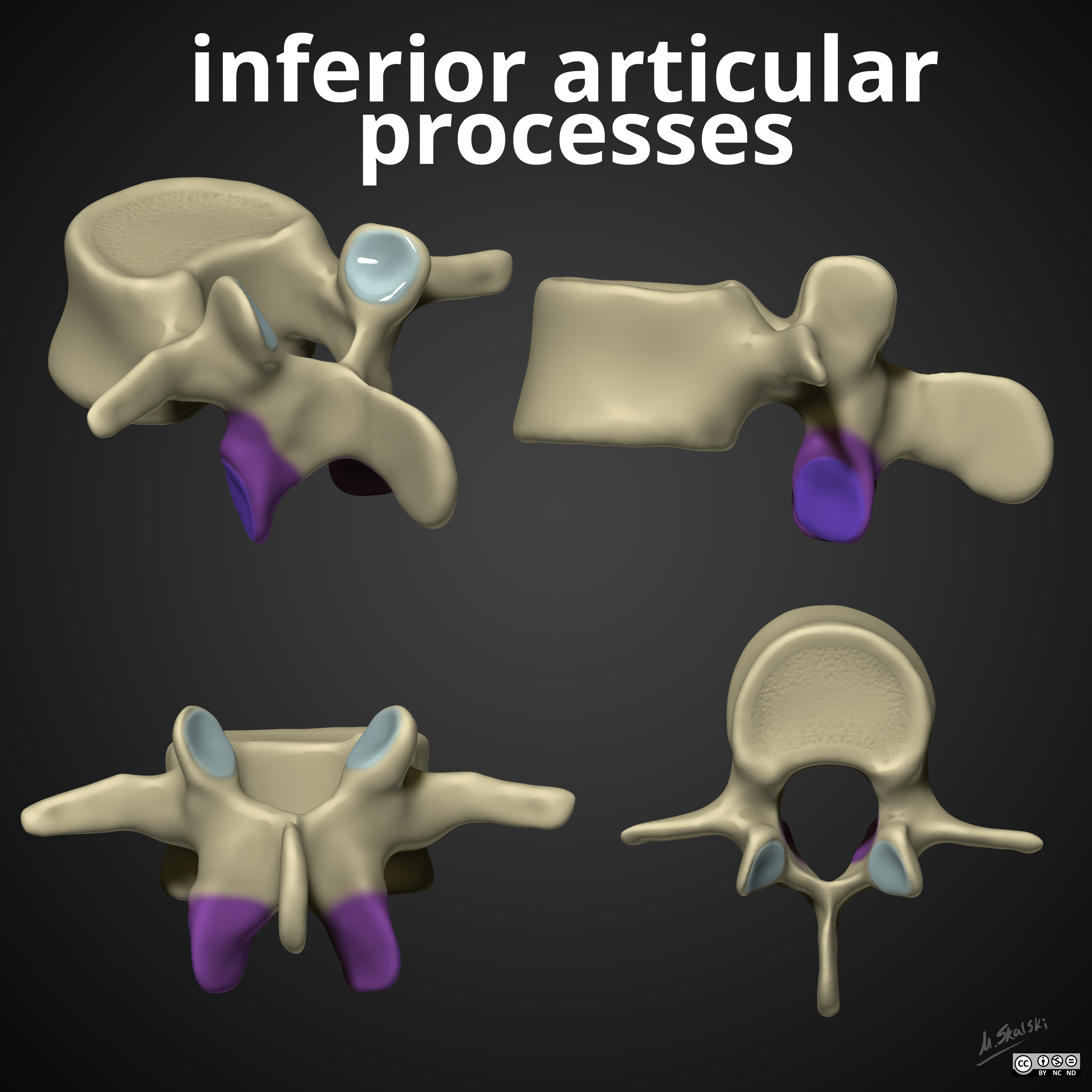
Inferior articular process
bony projection that extends downward from the articular process of a vertebra to lock with the vertebra directly below it
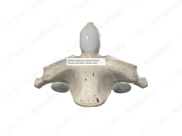
Inferior articular facet
smooth, bone-surfaced area on the bottom of a vertebra's lower process that links with the superior articular facet of the vertebra directly below it
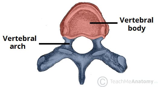
Body of vertebral column
thick, cylindrical front part of a spinal bone that bears weight and supports the upper body

Transverse foramen
opening found on each side of the transverse processes in the cervical (neck) vertebrae
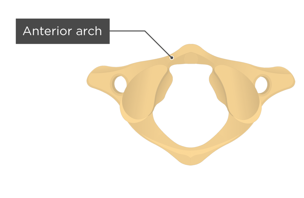
Atlas: anterior arch
C1 - supports skull and allows head to nod
anterior arch articulates with dens of axis

Atlas: posterior arch
C1 - larger than anterior arch - attachment site for muscles/ligaments and a groove for vertebral artery

Axis: dens
Dens/odontoid process - prominent projection of C2 that serves as pivot for head rotation & stabilized by ligaments connecting it to the atlas and occipital bone

Superior costal demifacet
the top half-facet depression surfaces on the sides of typical thoracic vertebral bodies (T2–T9) that join with adjacent parts to form complete sockets for rib heads

Inferior costal demifacet
the bottom half-facet depression surfaces on the sides of typical thoracic vertebral bodies (T2–T9) that join with adjacent parts to form complete sockets for rib heads

Costal facet of the transverse process
a smooth, oval spot on the transverse process of a thoracic vertebra that connects to a rib

Intervertabrael discs
Cushion of cartilage
Location: Between adjacent vertebrae in the spine
Function: Acts as a shock absorber and allows the back to bend and twist

Posterior longitudinal ligament
strong band of fibrous tissue that runs down the back of the vertebral bodies inside the spinal canal, stretching from the neck (axis) to the sacrum

Anterior longitudinal ligament
strong, fibrous band running down the front surface of the vertebral column from the base of the skull (occiput/atlas) to the sacrum