Cardiovascular Disease of Dogs and Cats
 Call Kai
 Learn
 Practice Test
 Spaced Repetition
 Match
 Flashcards
 Knowt Play
1/44

There's no tags or description
Looks like no tags are added yet.
45 Terms
What way do the arteries flow? What happens to the blood carried to the lungs
arteries flow away from the heart (Arteries= Away)
Blood carried to lungs to be oxygenated
What way do veins flow? what happens to the blood?
veins flow towards the heart
oxygenated blood throughout back to the heart to go out to the rest of the body
Filling of the ventricle
Diastole
Contraction of ventricle
systole
how many directions does blood flow
one

What is A
Cranial
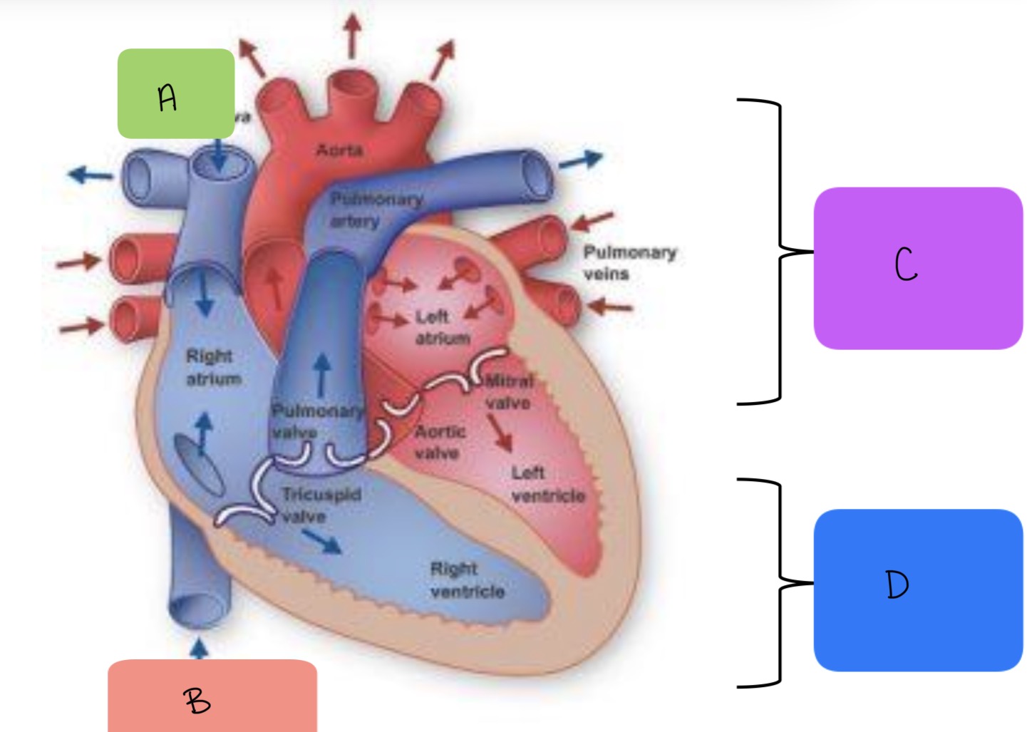
What is B
caudal

What is C
Base

What is D
Apex
What is a normal heart sound
“Lub-Dub”
What creates the sound of the heart? where is it best heard
The valves opening and closing within the heart. best heard at apical impulse which can be felt on the left side of the thorax (around 4th-6th intercostal space. always listen to both sides of the chest to fully assess the heart
What are the 3 major cardiac valves auscultated on the left
“PAM lives on the left”
P=pulmonic valve
A=aortic valve
M=mitral valve
What is the major cardiac valve auscultated on the right
tricuspid valve
What are the most common abnormal heart sounds, what do they indicate
heart murmurs is most common
indicates underlying heart disease
Are all heart murmurs bad? how can you tell the difference between ok and bad heart murmurs?
No, cats can have functional heart murmurs caused by stress
can be differentiated with pro-BNP test
What do heart murmurs mean and what sound does it make
Turbulent blood flow in which blood is moving multiple directions which the flow causes vibrations that are heard- make the “whoosh’ sound
How can heart murmurs be described and where are they best heard
based on location, timing, and loudness
best heard at point of maximal intensity (PMI)
What do heart murmurs grades describe
loudness of heart murmur, does not correlate with severity of disease
Mitral valve degeneration (MVD)
most common cardiac disease
degenerative disturbance of the connective tissue of the mitral valve leading to heart disease
leads to mitral valve regurgitation (heart murmur) resulting in cardiac volume overload (eventually heart will not be able to keep up with the high workload and results in congestive heart failure)
prevalence increases with age
breeds affected by mitral valve degeneration
Small breeds, Old dogs
cavalier King Charles spaniels
papillons
miniature poodles
yorkies
chihauhuas
dachshunds
Clinical signs of mitral valve degeneration
left sided apical systolic murmur
± tachypnea (increased respiratory rate) or dyspnea (difficult breathing)
Diagnosis of mitral valve degeneration
auscultation
radiographs
left atrial enlargement
cardiomegaly/ increased vertebral heart score (VHS)
echocardiogram
left atrial and ventricular size increases
treatment of mitral valve degeneration
not indicated for less severe disease
usually monitor with chest radiographs every 6 months
pimobendan (vetmedin)
positive ionotrope
helps heart pump more efficiently
reduces the rate of change the heart size will increase
monitor for signs of left sided congestive heart failure (CHF)
Dilated cardiomyopathy (DCM)
heart disease in which there is impaired, weakened systolic function of one or both ventricles
causes cardiac enlargement and possible CHF, arrhythmias or both
has been associated with certain diets
what diets has dilated cardiomyopathy been associated with
dogs-grain free
cats- taurine deficiency (before 1987)
who does dilated cardiomyopathy most impact
dogs but can also affect cats
adult onset disease:4-10 years old
males>females
breeds affected
dobermans
Irish wolfhound
great dane
typically bigger dogs
Clinical signs for dilated cardiomyopathy
may be without clinical signs
cardiac arrhythmias
syncope
temporary loss of consciousness due to low blood pressure (think fainting goats)
soft grade I-III systolic left and/or right apical murmur
± CHF signs
Diagnosis of dilated cardiomyopathy
suspected based on predisposed breeds with heart murmur or arrhythmias
radiographs
cardiomegaly
enlarged atriums
electrocardiogram (ECG)
sinus tachycardia or ventricular tachycardia
echocardiogram
ventricular and atrial dilation
reduced myocardial systolic function
treatment dilated cardiomyopathy
antiarrhythmics
digoxin
pimobendan (vetmedin)
ACE inhibitors
lower blood pressure
Hypertrophic cardiomyopathy (HCM)
Heart disease characterized by increased ventricular wall thickness
commonly left ventricle
results in altered contractions and relaxation as it pumps blood
common heart disease of older cats
What does hypertrophic cardiomyopathy often increase
often increases risk of thromboembolism
feline arteriothrombembolism (FATE)
saddle thrombus causing cats to be unable to use back legs
clinical signs of hypertrophic cardiomyopathy (HCM)
often none, incidentally found on physical exam
systolic murmur
arrhythmias
± CHF signs
Diagnosis hypertrophic cardiomyopathy
Pro-BNP
assess for heart muscle damage
echocardiogram
increased inter ventricular septum and/or left ventricular wall thickness
± left atrial enlargement
ECG
possible ventricular premature complexes (VPCs)
Radiographs
cardiomegaly
Treatment for hypertrophic cardiomyopathy (HCM)
ACE inhibitors
Clopidogrel
platelet aggregation inhibitor
decreases risk of clot formation
Don’t poke jugular
monitor for signs of CHF
patent ductus arteriosus (PDA)
arterial shunt between aorta and pulmonary artery that is present in fetuses but should constrict and close 24 hrs after birth
most common congenital heart defect of dogs

Who is patent ductus arteriosus (PDA) commonly seen with
affects young dogs
breeds:
maltese
pomeranian
shetland sheepdog
collie
irish setter
clinical signs patent ductus arteriosus
often found incidentally
continuous heart murmur
exercise intolerance
failure to thrive
±signs of CHF
Diagnosis of patent ductus arteriosus
radiographs
enlarged aorta
enlarged left side of the heart
echocardiogram
visualize patent ductus
patent ductus arteriosus treatment
surgery
PDA closure with ductal occluder
vessel ligation with thoracotomy
Congestive heart failure
sudden onset of clinical signs associated with pulmonary edema or cavitary effusions (fluid retention) due to heart disease
occurs because the pressure within the veins and capillaries draining into the diseased side of the heart increase to the point where the vessels leak
right-sided CHF
ascites(abnormal accumulation of excess fluid within the peritoneal cavity)
pleural effusion
left sided CHF
pulmonary edema
Congestive heart failure clinical signs
tachypnea
dyspnea
cough (esp dog)
pulmonary crackles
decreased breath sounds
exercise intolerance
restlessness
heart murmurs
diagnosis of congestive heart failure
radiographs
right sided
decreased serosal detail
pleural effusion
left sided
pulmonary edema in perihilar region
left atrial enlargement
treatment for congestive heart failure
furosemide
diuretic to treat fluid retention
oxygen therapy
ACE inhibitors