Lab Exam #1
 Call Kai
 Learn
Learn Practice Test
 Spaced Repetition
 Match
Match Flashcards
 Knowt Play
1/26

There's no tags or description
Looks like no tags are added yet.
Name | Mastery | Learn | Test | Matching | Spaced | Call with Kai | Chat |
|---|
No analytics yet
Send a link to your students to track their progress
27 Terms
Where should unused gloves, contaminated gloves, used slides and bacterial cultures be discarded?
Unused gloves → normal trash can
Contaminated gloves → Large red bucket
Used slides → sharps on table or smaller red bucket
bacterial cultures → large red bucket or designeated bucket

Where is the coarse knob, fine focus knob, slide-positioning knobs, objective lenses, ocular lenses, revolving nosepiece, and slide clip? What do they do?
Coarse knob - brings the slide closer to the objective lenses by a LOT
Fine focus knob - brings the slide closer to the objective lenses by a little
Slide-positioning knobs - move the slide duh
Objective lenses: changes the magnification
Ocular lenses: view the organism
Revolving nose piece: changes the objective lenses
Side clip: keeps slide in place
How do you calculate total magnification? How does immersion oil improve resolution and use it with the oil immersion objective?
Objective x Ocular lens = total magnification
Immersion oil gets rid of the air gap between the specimen and the objective lenses. The oil immersion objective prevents light from scattering and keeping the image crisp.
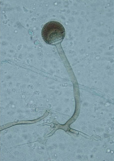
ID Eukaryotic
Rhizopus (tadpole looking)

ID Eukaryotic
Penicillum - branches with little flowers at the end

ID Eukaryotic
Candida Albicans - small ovals

ID Eukaryote
Paramecium Caudatum - long slipper cell with hella stuff in it

ID Eukaryote
Trypanosoma Cruzi - shrimp in blood

ID Eukaryote
Toxoplasma Gondii - small little crescents
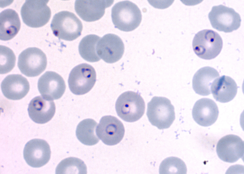
ID Eukaryote
Plasmodium falciparum - rings in blood cells with dark dots

ID Eukaryote
Trichomonas vaginalis - little leafs with dark circle inside
Bacterial cell shape and arrangements. What are the differences between the two?
Cocci: tetrad, sacrinae (3d cube), staph (irregular cluster)
Bacilli: palisades (clusters)
Simple and negative stains: how do they differ? name a dye for each and how dye charge plays a role in the staining.
Simple stains: Crystal Violet which are attracted to the negative bacterial surface
Negative stains: Nigrosin which is repelled by the negative bacterial surface and only stains the background
Gram Stain Procedure (4)
Primary Dye: Crystal Violet - stains the peptidoglycan layer
Mordant: Iodine fixes the violet in place to form a CVI complex
Decolonizer: Ethanol- rinses out CVI from Gram-negative cells
Counterstain: Safranin makes gram-negative cells pink
How do gram+ and gram- cell envelopes differ, and how do these explain their final colors? Distinguish the role of peptidoglycan from teichoic acids.
The gram+ cell has a thick cell wall while the gram- has two cell membrane and one thick cell wall. The thick cell wall and the alcohol traps the CVI complex from earlier. The thin cell wall can’t hold onto it and just rinses it out.
Peptidoglycan - stickys structural capsule
Teichoic: gives cell wall negative charge
Predict the colors of both groups if the decolorizer is omitted. What errors/culture-age effects can make gram+ cells pink?
no decolorizer → CVI still in cells, all purple
no mordant → no cvi, all pink
no counterstain → gram- are colorless
What color would human cells be after a Gram stain? Explain using cell structure and why they wouldn’t be gram negative.
They’d be red as the crystal violet would be washed out by the decolorizer and only the safranin would stick. It’s not gram-negative because there’s no peptidoglycan layer or cell wall.
Describe the acid-fast procedure in the lab including the primary stain, role of heat, two components of the decolorizer and the counterstain. What cell-wall substance allows stain retention?
Primary: Carbolfuchsin → colors AF b/c mycolic acid is sticky
Decolorizer: 95% ethyl and 3% HCl - washes out 1* dye from non-AF bacteria
Counterstain: Methylene blue → colors non-AF b/c it’s not lipophilic
Heat loosens the mycolic acid, making it waxier and stickier.
What colors indicate AF±? Why would safranin be a bad choice for a counterstain, as opposed to methylene blue?
positive is red/pink
negative is blue
safranin bad bc already red
What are the primary stain and counterstain in the endospore procedure? Explain the role of heat and how to recognize endospore/vegetative cells.
Primary: Malachite Green
Decolorizer: alcohol - washes alcohol off the cell, not the spore
Counterstain: Safranin
Heat softens the endospore coat so that malachite green could enter.
How do you recognize a capsule in a correctly prepared capsule stain? Why is heat-fixing avoided?
Capsules look like a clear halo around the nucleoid. Heat-fixing is avoided because heat can destroy the polysaccharide capsule.
How would you use growth on duplicate plates incubate with/without oxygen to classify an organism? Review obligate aerobes, obligate anaerobes, facultative anaerobes.
If growth is present on one but not the other, it would be an obligate. If growth is better on the oxygen one than the anaerobe jar, it’d be a facultative anaerobe.
What does the anaerobic jar indicator tell you about the conditions inside?
White → No oxygen
Pink → Oxygen breach
How do minimum, optimum, and maximum growth temperatures help classify bacteria? Compare psychrophiles, psychrotrophs, mesophiles, and thermophiles using growth patterns.
The different ranges help us differentiate between the different bacterial growths, such as psychrophiles having a max. temp that is the psychrotrophs’ optimum temp.
What makes MSA selective and differential? Name its’ pH indicator. Explain what growth means and what it tells you about mannitol fermentation.
Selective: high salt content
Differential: phenol red that shows yellow when mannitol is released and red when it isn’t.
Growth helps differentiate staphylcoccus species.
What makes MacConkey and EMB agar selective and differential? Name the two selective ingredients in MAC. Explain how colony appearance indicates lactose fermentation and what growth suggests about Gram reaction.
MAC:
selective - bile salts/ crystal violet
differential - neutral red shows red when lactose fermented and no color if not
EMB:
selective/differential: eosin Y/ methylene blue → select against gram+
measure lactose fermentation (green → red/pink) and colorless if not
Distinguish between alpha, beta, and gamma hemolysis on blood agar. Describe the appearance around the colonies and what it means for the RBC.
alpha = slight clearing/discoloration b/c oxidation causes by the partial hemolysis (dark line surrounded by fogginess)
beta= complete hemolysis and clearing of the bacteria (completely white line with fogginess)
gamma = no hemolysis (no fogginess)