ACS Pre Test JBL / AHA

0.0(0)
Studied by 0 people
call kaiCall Kai
learnLearn
examPractice Test
spaced repetitionSpaced Repetition
heart puzzleMatch
flashcardsFlashcards
GameKnowt Play
Card Sorting

1/19

encourage image

There's no tags or description

Looks like no tags are added yet.

20 Terms

1
New cards

Each of the following is a correct guideline for placing chest electrodes on a patient, EXCEPT

a. V4, 5 and 6 are in a straight line

b. the sternal angle is at the level of the second rib

c. V1 and V2 are parasternal

d. the interspaces are above the corresponding ribs

e. the interspaces are below the corresponding ribs

d. the interspaces are above the corresponding ribs

2
New cards

Each of the following is a contraindication (exclusion criterion) for fibrinolytic therapy, EXCEPT:

a. recurrent ventricular tachycardia

b. recent cervical disc surgery

c. suspected aortic dissection

d. active internal bleeding

e. recent stroke

a. recurrent ventricular tachycardia

3
New cards

Each of the following is a correct statement regarding the use of aspirin, EXCEPT:

a. prevents clot formation

b. promotes bleeding

c. lyses blood clots

d. decreases platelet stickiness

e. should be chewed for rapid effect

c. lyses blood clots

4
New cards

Each of the following bedside findings may be signs of congestive failure in the setting of an acute myocardial infarction, EXCEPT:

a. tachycardia

b. cool, diaphoretic skin

c. third heart sound

d. friction rub

e. crackles and wheezes

d. friction rub

5
New cards

Each of the following is a correct statement regarding nitroglycerin, EXCEPT:

a hypotension is a side effect

b. bleeding is a side effect

c. reduces cardiac work

d. improves coronary blood flow

e. headache is a side effect

b. bleeding is a side effect

6
New cards

The proper calibration for a 12-Lead ECG is 1 mV. This is equal to a height of

a. 1 small box

b. 5 small boxes

c. 10 small boxes

d. 15 small boxes

e. 20 small boxes

c. 10 small boxes

7
New cards

An acute ST-elevation myocardial infarction, regardless of the degree of coronary artery atherosclerosis, is mainly the result of

a. Coronary artery spasm

b. Ventricular fibrilation

c. Thrombotic occlusion

d. Ventricular aneurysm

e. Plaque formation

c. Thrombotic occlusion

8
New cards

The symptoms associated with an acute myocardial infarction usually last:

a. 5 to 15 minutes

b. more than 15 minutes

c. more than 60 minutes

d. more than 6 hours

e. more than 12 hours

b. more than 15 minutes

9
New cards

Morphine sulfate can cause all of the following, EXCEPT:

a. nausea and vomiting

b. hypotension

c. respiratory depression

d. reduced cardiac work

e. decreased platelet stickiness

e. decreased platelet stickiness

10
New cards

In the setting of AMI, all of the following define the high risk patient EXCEPT ONE:

a. heart rate of 100 BPM or more

b. pulmonary edema (crackles more than 1/2 way up)

c. atrial fibrillation

d. shock

e. hypotension

c. atrial fibrillation
11
New cards
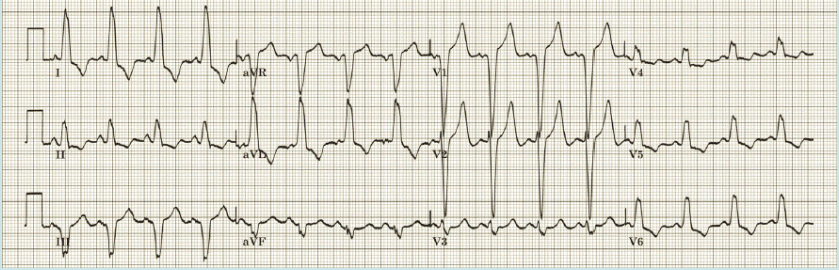
<p><span style="background-color: oklab(0.219511 0.00211 -0.007446); color: rgb(0, 0, 0);"><mark data-color="#ffffff" style="background-color: rgb(255, 255, 255); color: inherit;">What is your interpretation for the following 12-Lead ECG?</mark></span></p><p><span style="background-color: oklab(0.219511 0.00211 -0.007446); color: rgb(0, 0, 0);"><mark data-color="#ffffff" style="background-color: rgb(255, 255, 255); color: inherit;">a. Left bundle branch block</mark></span></p><p><span style="background-color: oklab(0.219511 0.00211 -0.007446); color: rgb(0, 0, 0);"><mark data-color="#ffffff" style="background-color: rgb(255, 255, 255); color: inherit;">b. Anterior wall subendocardial injury</mark></span></p><p><span style="background-color: oklab(0.219511 0.00211 -0.007446); color: rgb(0, 0, 0);"><mark data-color="#ffffff" style="background-color: rgb(255, 255, 255); color: inherit;">c. Acute inferior wall myocardial infarction</mark></span></p><p><span style="background-color: oklab(0.219511 0.00211 -0.007446); color: rgb(0, 0, 0);"><mark data-color="#ffffff" style="background-color: rgb(255, 255, 255); color: inherit;">d. Right bundle branch block</mark></span></p><p><span style="background-color: oklab(0.219511 0.00211 -0.007446); color: rgb(0, 0, 0);"><mark data-color="#ffffff" style="background-color: rgb(255, 255, 255); color: inherit;">e. Ventricular pacemaker</mark></span></p>

What is your interpretation for the following 12-Lead ECG?

a. Left bundle branch block

b. Anterior wall subendocardial injury

c. Acute inferior wall myocardial infarction

d. Right bundle branch block

e. Ventricular pacemaker

a. Left bundle branch block
12
New cards
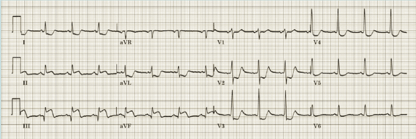
<p><span style="background-color: oklab(0.678923 0.00325415 -0.0111644 / 0.121569); color: rgb(0, 0, 0);"><mark data-color="#ffffff" style="background-color: rgb(255, 255, 255); color: inherit;">What is your interpretation for the following 12-Lead ECG?</mark></span></p><p><span style="background-color: oklab(0.678923 0.00325415 -0.0111644 / 0.121569); color: rgb(0, 0, 0);"><mark data-color="#fffcfc" style="background-color: rgb(255, 252, 252); color: inherit;">a. Acute anterolateral wall myocardial infarction </mark></span></p><p><span style="background-color: oklab(0.678923 0.00325415 -0.0111644 / 0.121569); color: rgb(0, 0, 0);"><mark data-color="#fffcfc" style="background-color: rgb(255, 252, 252); color: inherit;">b. Acute inferior wall myocardial infarction </mark></span></p><p><span style="background-color: oklab(0.678923 0.00325415 -0.0111644 / 0.121569); color: rgb(0, 0, 0);"><mark data-color="#fffcfc" style="background-color: rgb(255, 252, 252); color: inherit;">c. Left ventricular hypertrophy </mark></span></p><p><span style="background-color: oklab(0.678923 0.00325415 -0.0111644 / 0.121569); color: rgb(0, 0, 0);"><mark data-color="#fffcfc" style="background-color: rgb(255, 252, 252); color: inherit;">d. Ventricular pacemaker </mark></span></p><p><span style="background-color: oklab(0.678923 0.00325415 -0.0111644 / 0.121569); color: rgb(0, 0, 0);"><mark data-color="#fffcfc" style="background-color: rgb(255, 252, 252); color: inherit;">e. Acute pericarditis</mark></span></p>

What is your interpretation for the following 12-Lead ECG?

a. Acute anterolateral wall myocardial infarction

b. Acute inferior wall myocardial infarction

c. Left ventricular hypertrophy

d. Ventricular pacemaker

e. Acute pericarditis

b. Acute inferior wall myocardial infarction

13
New cards
<p><span style="background-color: oklab(0.678923 0.00325415 -0.0111644 / 0.121569); color: rgb(0, 0, 0);"><mark data-color="#ffffff" style="background-color: rgb(255, 255, 255); color: inherit;">What is your interpretation for the following 12-Lead ECG?</mark></span></p><p><span style="background-color: oklab(0.219511 0.00211037 -0.00744569); color: rgb(0, 0, 0);"><mark data-color="#ffffff" style="background-color: rgb(255, 255, 255); color: inherit;">a. Early repolarization </mark></span></p><p><span style="background-color: oklab(0.219511 0.00211037 -0.00744569); color: rgb(0, 0, 0);"><mark data-color="#ffffff" style="background-color: rgb(255, 255, 255); color: inherit;">b. Acute inferior wall myocardial infarction </mark></span></p><p><span style="background-color: oklab(0.219511 0.00211037 -0.00744569); color: rgb(0, 0, 0);"><mark data-color="#ffffff" style="background-color: rgb(255, 255, 255); color: inherit;">c. Acute anterior wall myocardial infarction </mark></span></p><p><span style="background-color: oklab(0.219511 0.00211037 -0.00744569); color: rgb(0, 0, 0);"><mark data-color="#ffffff" style="background-color: rgb(255, 255, 255); color: inherit;">d. Left bundle branch block </mark></span></p><p><span style="background-color: oklab(0.219511 0.00211037 -0.00744569); color: rgb(0, 0, 0);"><mark data-color="#ffffff" style="background-color: rgb(255, 255, 255); color: inherit;">e. Acute pericarditis</mark></span></p>

What is your interpretation for the following 12-Lead ECG?

a. Early repolarization

b. Acute inferior wall myocardial infarction

c. Acute anterior wall myocardial infarction

d. Left bundle branch block

e. Acute pericarditis

c. Acute anterior wall myocardial infarction

14
New cards
<p><span style="background-color: oklab(0.678923 0.00325415 -0.0111644 / 0.121569); color: rgb(0, 0, 0);"><mark data-color="#ffffff" style="background-color: rgb(255, 255, 255); color: inherit;">What is your interpretation for the following 12-Lead ECG?</mark></span></p><p><span style="background-color: oklab(0.678923 0.00325415 -0.0111644 / 0.121569); color: rgb(0, 0, 0);"><mark data-color="#ffffff" style="background-color: rgb(255, 255, 255); color: inherit;">a. Acute inferior wall myocardial infarction</mark></span></p><p><span style="background-color: oklab(0.678923 0.00325415 -0.0111644 / 0.121569); color: rgb(0, 0, 0);"><mark data-color="#ffffff" style="background-color: rgb(255, 255, 255); color: inherit;">b. Left bundle branch block </mark></span></p><p><span style="background-color: oklab(0.678923 0.00325415 -0.0111644 / 0.121569); color: rgb(0, 0, 0);"><mark data-color="#ffffff" style="background-color: rgb(255, 255, 255); color: inherit;">c. Acute inferorlateral wall myocardial infarction </mark></span></p><p><span style="background-color: oklab(0.678923 0.00325415 -0.0111644 / 0.121569); color: rgb(0, 0, 0);"><mark data-color="#ffffff" style="background-color: rgb(255, 255, 255); color: inherit;">d. Acute anterior wall myocardial infarction </mark></span></p><p><span style="background-color: oklab(0.678923 0.00325415 -0.0111644 / 0.121569); color: rgb(0, 0, 0);"><mark data-color="#ffffff" style="background-color: rgb(255, 255, 255); color: inherit;">e. Early repolarization</mark></span></p>

What is your interpretation for the following 12-Lead ECG?

a. Acute inferior wall myocardial infarction

b. Left bundle branch block

c. Acute inferorlateral wall myocardial infarction

d. Acute anterior wall myocardial infarction

e. Early repolarization

d. Acute anterior wall myocardial infarction

15
New cards
<p><span style="background-color: oklab(0.678923 0.00325415 -0.0111644 / 0.121569); color: rgb(0, 0, 0);"><mark data-color="#ffffff" style="background-color: rgb(255, 255, 255); color: inherit;">What is your interpretation for the following 12-Lead ECG?</mark></span></p><p><span>a. Left bundle branch block </span></p><p><span>b. Anterior wall subendocardial injury </span></p><p><span>c. Acute inferior wall myocardial infarction</span></p><p><span>d. Right bundle branch block</span></p><p><span>e. Ventricular pacemaker</span></p>

What is your interpretation for the following 12-Lead ECG?

a. Left bundle branch block

b. Anterior wall subendocardial injury

c. Acute inferior wall myocardial infarction

d. Right bundle branch block

e. Ventricular pacemaker

e. Ventricular pacemaker

16
New cards
<p><span style="background-color: oklab(0.678923 0.00325415 -0.0111644 / 0.121569); color: rgb(0, 0, 0);"><mark data-color="#ffffff" style="background-color: rgb(255, 255, 255); color: inherit;">What is your interpretation for the following 12-Lead ECG?</mark></span></p><p><span style="background-color: oklab(0.219511 0.00211037 -0.00744569); color: rgb(0, 0, 0);"><mark data-color="#ffffff" style="background-color: rgb(255, 255, 255); color: inherit;">a. Acute anterolateral wall myocardial infarction </mark></span></p><p><span style="background-color: oklab(0.219511 0.00211037 -0.00744569); color: rgb(0, 0, 0);"><mark data-color="#ffffff" style="background-color: rgb(255, 255, 255); color: inherit;">b. Anterior wall subendocardial injury</mark></span></p><p><span style="background-color: oklab(0.219511 0.00211037 -0.00744569); color: rgb(0, 0, 0);"><mark data-color="#ffffff" style="background-color: rgb(255, 255, 255); color: inherit;">c. Left bundle branch block</mark></span></p><p><span style="background-color: oklab(0.219511 0.00211037 -0.00744569); color: rgb(0, 0, 0);"><mark data-color="#ffffff" style="background-color: rgb(255, 255, 255); color: inherit;">d. Acute inferior wall myocardial infarction </mark></span></p><p><span style="background-color: oklab(0.219511 0.00211037 -0.00744569); color: rgb(0, 0, 0);"><mark data-color="#ffffff" style="background-color: rgb(255, 255, 255); color: inherit;">e. Old inferior wall myocardial infarction</mark></span></p>

What is your interpretation for the following 12-Lead ECG?

a. Acute anterolateral wall myocardial infarction

b. Anterior wall subendocardial injury

c. Left bundle branch block

d. Acute inferior wall myocardial infarction

e. Old inferior wall myocardial infarction

d. Acute inferior wall myocardial infarction

17
New cards
<p><span style="background-color: oklab(0.678923 0.00325415 -0.0111644 / 0.121569); color: rgb(0, 0, 0);"><mark data-color="#ffffff" style="background-color: rgb(255, 255, 255); color: inherit;">What is your interpretation for the following 12-Lead ECG (Case 1)?</mark></span></p><p><span style="background-color: oklab(0.678923 0.00325415 -0.0111644 / 0.121569); color: rgb(0, 0, 0);"><mark data-color="#ffffff" style="background-color: rgb(255, 255, 255); color: inherit;">a. Early repolarization </mark></span></p><p><span style="background-color: oklab(0.678923 0.00325415 -0.0111644 / 0.121569); color: rgb(0, 0, 0);"><mark data-color="#ffffff" style="background-color: rgb(255, 255, 255); color: inherit;">b. Acute inferior wall myocardial infarction </mark></span></p><p><span style="background-color: oklab(0.678923 0.00325415 -0.0111644 / 0.121569); color: rgb(0, 0, 0);"><mark data-color="#ffffff" style="background-color: rgb(255, 255, 255); color: inherit;">c. Acute anterior wall myocardial infarction </mark></span></p><p><span style="background-color: oklab(0.678923 0.00325415 -0.0111644 / 0.121569); color: rgb(0, 0, 0);"><mark data-color="#ffffff" style="background-color: rgb(255, 255, 255); color: inherit;">d. Acute pericarditis </mark></span></p><p><span style="background-color: oklab(0.678923 0.00325415 -0.0111644 / 0.121569); color: rgb(0, 0, 0);"><mark data-color="#ffffff" style="background-color: rgb(255, 255, 255); color: inherit;">e. Left bundle branch block</mark></span></p>

What is your interpretation for the following 12-Lead ECG (Case 1)?

a. Early repolarization

b. Acute inferior wall myocardial infarction

c. Acute anterior wall myocardial infarction

d. Acute pericarditis

e. Left bundle branch block

c. Acute anterior wall myocardial infarction

18
New cards
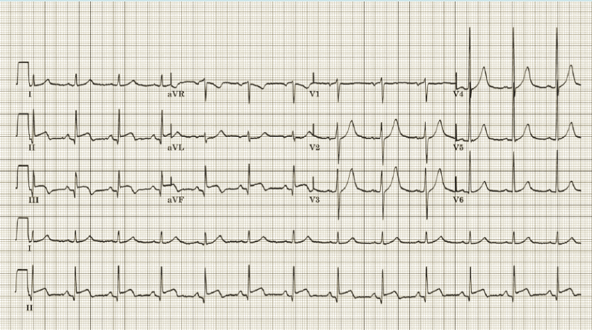
<p><span>What is your interpretation of this ECG (Case 2)?</span></p><p><span>a. Acute anterolateral wall infarction</span></p><p><span>b. Anterior wall subendocardial wall injury</span></p><p><span>c. Complete left bundle branch block</span></p><p><span>d. Acute inferior wall infarction</span></p><p><span>e. Early repolarization</span></p>

What is your interpretation of this ECG (Case 2)?

a. Acute anterolateral wall infarction

b. Anterior wall subendocardial wall injury

c. Complete left bundle branch block

d. Acute inferior wall infarction

e. Early repolarization

d. Acute inferior wall infarction
19
New cards
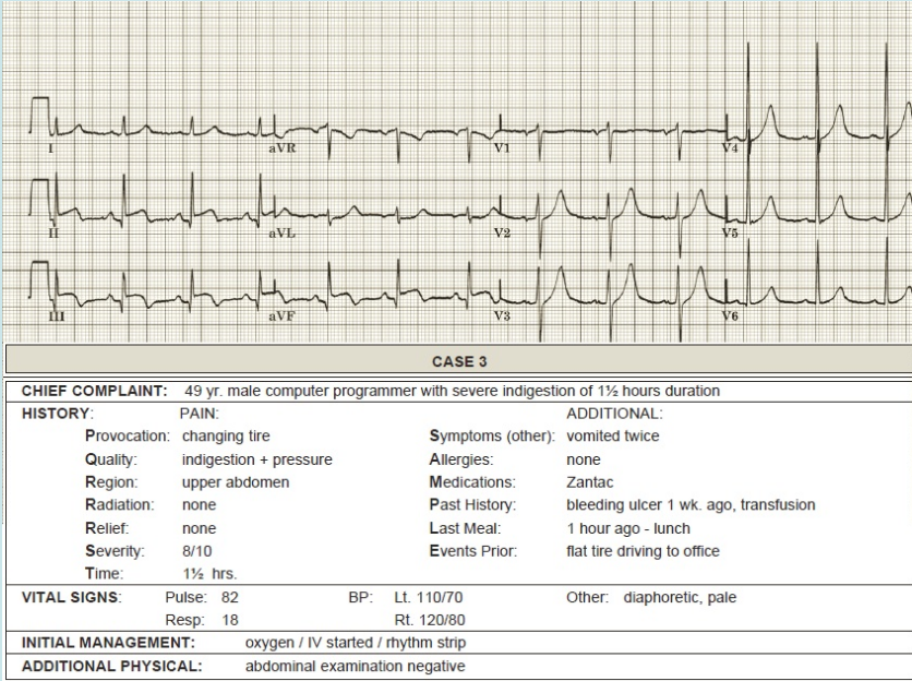
<p><span style="background-color: oklab(0.678923 0.00325415 -0.0111644 / 0.121569); color: rgb(0, 0, 0);"><mark data-color="#ffffff" style="background-color: rgb(255, 255, 255); color: inherit;">What is your interpretation of this 12-Lead ECG (case 3)?</mark></span></p><p><span style="background-color: oklab(0.678923 0.00325415 -0.0111644 / 0.121569); color: rgb(0, 0, 0);"><mark data-color="#ffffff" style="background-color: rgb(255, 255, 255); color: inherit;">a. Anterior wall subendocardial injury </mark></span></p><p><span style="background-color: oklab(0.678923 0.00325415 -0.0111644 / 0.121569); color: rgb(0, 0, 0);"><mark data-color="#ffffff" style="background-color: rgb(255, 255, 255); color: inherit;">b. Acute inferior wall infarction </mark></span></p><p><span style="background-color: oklab(0.678923 0.00325415 -0.0111644 / 0.121569); color: rgb(0, 0, 0);"><mark data-color="#ffffff" style="background-color: rgb(255, 255, 255); color: inherit;">c. Complete left ventricular hypertrophy </mark></span></p><p><span style="background-color: oklab(0.678923 0.00325415 -0.0111644 / 0.121569); color: rgb(0, 0, 0);"><mark data-color="#ffffff" style="background-color: rgb(255, 255, 255); color: inherit;">d. Ventricular pacemaker </mark></span></p><p><span style="background-color: oklab(0.678923 0.00325415 -0.0111644 / 0.121569); color: rgb(0, 0, 0);"><mark data-color="#ffffff" style="background-color: rgb(255, 255, 255); color: inherit;">e. Acute pericarditis</mark></span></p>

What is your interpretation of this 12-Lead ECG (case 3)?

a. Anterior wall subendocardial injury

b. Acute inferior wall infarction

c. Complete left ventricular hypertrophy

d. Ventricular pacemaker

e. Acute pericarditis

b. Acute inferior wall infarction
20
New cards
<p><span>What is your management for this patient (case 3)? (V4R result showing negative) </span></p><p><span>a. NTG, ASA and morphine </span></p><p><span>b. NTG and morphine </span></p><p><span>c. NTG, morphine and lasix </span></p><p><span>d. NG, morphine and fluids </span></p><p><span>e. ASA and morphine</span></p>

What is your management for this patient (case 3)? (V4R result showing negative)

a. NTG, ASA and morphine

b. NTG and morphine

c. NTG, morphine and lasix

d. NG, morphine and fluids

e. ASA and morphine

b. NTG and morphine



