Neuro - Blueprint
 Call Kai
 Learn
Learn Practice Test
 Spaced Repetition
 Match
Match Flashcards
 Knowt Play
1/99

There's no tags or description
Looks like no tags are added yet.
Name | Mastery | Learn | Test | Matching | Spaced | Call with Kai | Chat |
|---|
No analytics yet
Send a link to your students to track their progress
100 Terms
postconcussive syndrome
having difficulty thinking, HA/dizzy/vomiting, irritability
→ refer to neuro
Transient Ischemic Attack (TIA) causes
Cardioembolic sources: A-fib, subtherapeutic anticoagulation, thrombi
Other: Sickle cell, atherosclerosis, stenotic vessels, antiphospholipid syndrome, thrombotic endocarditis
TIA dx*
NONCONTRAST CT of head
more sensitive = MRI
CT angiography
low risk TIA vs. High risk TIA
low risk: ABCD2 score < 4 = treat w/ just ASA
high risk: ABCD2 score > 4 = dual antplt therapy w/ ASA and clopidogrel
clopidogrel MOA
irreversibly blocks plt aggregation
ABCD2 score
Age ≥ 60 yrs, BP (systolic ≥140 or diastolic ≥90), Clinical features (unilateral weakness, +/- speech impairment), Duration of TIA ≥60 mins or less, DM
bells palsy cause
HSV
herpes zoster
swelling of CN VII
unilateral facial nerve paralysis, hyperacusis, taste disturbance unable to raise eyebrow
bells palsy
bells palsy complications
ramsay hunt syndrome
(watch external ear canal and face)
tx of bell’s palsy**
prednisone (steroid and antivirals in 3 days)
valacyclovir

subarachnoid hemorrhage cause
ruptured aneurysm (saccular or berry)
worst HA of life

subarachnoid hemorrhage dx*
initial: noncontrast CT (white on CT)
if unremarkable → lumbar puncture, CTA, 4 CSF tubes
main complication of subarachnoid hemorrhag
vasospasm

subarachnoid hemorrhage - tx
nimodipine (CCB, relaxes vessels)
supportive
definitive = surgical clipping or endovasc embolization
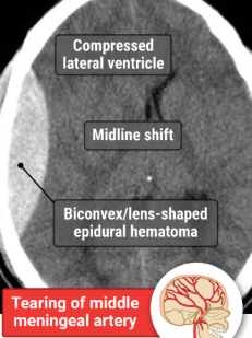
epidural hematoma
middle meningeal artery → rapid bleed
MC in adolescents = traffic collisions
convex shaped on CT
epidural and subdural hematoma tx*
hematoma evacuation
subdural hematoma risks
tearing of bridging veins slow bleed
RISKS: advancing age**, alcohol use**, traumatic brain injury, coagulopathy
crescent-shaped CONCAVE hematoma hypodensity = ??
subdural hematoma
hydrocephalus
Children → increased occipitofrontal circumference (macrocephaly)/ aberrant head shape, N/V, irritability, development delays
Urgent signs → bulging fontanels, papilledema, headache, altered mental state)
hydrocephalus dx
MRI/CT
hydrocephalus tx*
shunt to DEC CSF level
Cerebellum
balance and coordinatoin
best initial dx study for syncope*
EKG
CHESS (high risk criteria for syncope)
CHF
Hematocrit < 30%
ECG abnormal
SOB
Systolic BP < 90 mm Hg
Malignancy or inflammatory condition + sudden-onset SOB + syncope
pulmonary embolism
Young female adult + abdominal pain + syncope
ectopic pregnancy
Older male adult + abdominal or flank pain + syncope
AAA
tension HA tx*
abortive = nsaids
preventative = tca
cluster HA tx
acute = 100% oxygen and triptans
prophylaxis = verapamil (CCB) or steroid
sudden unilateral electric shock-like pains in gums, cheek, chin, temporal forehead
trigeminal neuralgia
trigeminal neuralgia tx*
carbamazepine
migraine HA tx
Abortive Rx: triptans, DHE, valproic acid, antiemetics, NSAIDs
Triptans, DHE: avoid in those with uncontrolled HTN or CV disease
Prophylaxis: TCAs, beta-blockers, anticonvulsants (valproic acid, topiramate), CCBs
Secondary (metastatic) brain tumors
MOST common cause of intracranial neoplasms
MC metastasize from lung cancer, breast, melanoma, kidney cancer
brain tumor dx
MRI with contrast
reversible causes of dementia
depression, B12 deficiency, syphilis, hypothyroidism, NPH, drug use, intracranial mass
dementia
amyloid plaques and neurofibrillary tangles
short term memory loss
when to hospitalize delirium
highly agitated
tx for delirium
haloperidol
avoid benzos
Neonatal Meningitis age groups
≤ 1 month → E.coli, Group B Strep, Listeria meningitidis, gram neg bacilli
1 month to 3 months → Group B Streptococcus & gram negative bacilli
3 months to 10 years → Streptococcus pneumoniae
≥ 10 years old → Neisseria meningitidis
neonatal meningitis tx
cefotaxime and ampicillin
neisseria would need 3rd gen ceph + vanco
late onset → vanco, aminoglycoside, 3rd ceph or merepenem
bacterial meningitis adult sx
headache, neck stiffness, photophobia, phonophobia, fever
brudzinski sign
kernig sign
cause of bacterial meningitis
strep pneumo
listeria if young/elderly and alcohol
meningitis dx
cerebrospinal fluid culture
LP
bacterial meningitis tx
18-50 yo: ceftriaxone + vanco
penicillin G is abx of choice
>50: ceftriaxone + vanco + AMPICILLIN
postexposure abx = rifampin
essential tremor tx
propranolol
Guillain-Barré Syndrome cause*
campyloacter jejuni
symmetric PROGRESSIVE ASCENDING muscle weakness with lack of deep tendon reflexes =
GBS
GBS dx
lumbar puncture with INC CSF protein and normal cell count
albuminocytologic dissociation
GBS tx
supportive
Multiple sclerosis
optic neuritis (monocular vision loss), sensory abnormalities, lhermittes sign, bilateral opthalmoplegia, afferent pupillary defect
multiple sclerosis dx
MRI (white matter lesions)
CSF = oligoclonal igG bands
multiple sclerosis tx*
acute: methylprednisone + plasma exchange
relapsing remitting → disease modifying agents (-umab)
myasthenia gravis is associated with
thymoma**
myasthenia gravis
Autoantibodies to nicotinic acetylcholine receptors resulting in impaired transmission at the neuromuscular junction → progressive muscle weakness
MG sx
ocular/proximal muscle weakness, ptosis diplopia
tensilon test, serologic testing
MG tx*
acetylcholinesterase inhibitors (TOC: pyridostigmine)
complex regional pain syndrome
history of previous extremity injury, fracture, or surgery
light touch causes extreme pain (pain out of proportion to exam
Absence Seizures
Brief < 15 seconds of sudden impaired consciousness
To others their attention will seem “absent”
absence seizure dx
EEG → 3 Hz spike and wave discharges
Hyperventilation for 3-5 minutes can precipitate the seizures
absence seizure tx*
ethosuximide
simple febrile seizure tx*
reassurance
antipyretics (tylenol/ibuproen)
complex febrile seizure tx*
benzos
monitor if weakness, cognitive deficit
Simple partial epilepsy (focal aware seizures)
seizure sx without loss of consciousness
EEG shows focal rhythmic discharge
TX IS THE MIGRAINE PROPHYLAXIS MEDS
focal seizure with NO impairment of consciousness
Simple partial seizures (IF IMPAIREMENT = COMPLEX)
Tonic clonic (aka grand mal) seizures TX
carbamazepine, phenobarbital, levetiracetam, phenytoin
Status Epilepticus
PE will show ≥ 5 minutes of continuous seizure activity OR more than one seizure without recovery
status epilepticus tx*
benzos (IV lorazepam diazepam) , if not available IM midazolam
2nd = Keppra
3rd = pentobarbital, propofol, midazolam
Turner Syndrome (45,X)
short, webbed neck, dwelayed 2ndary sex characteristics
karotype analysis = dx (45,x)
assoc with premature ovarian fialure, CoA, CV malforation
Most commonly caused by mutation in the fibrillin-1 (FBN1) gene
marfan syndrome (autosomal dominant)

marfan syndrome
pectic carinatum, ectopia lentis, scoliosis
monitor with echo
assoc with aorti canerusym and disection
anterior cerebral artery stroke
frontal lobe - bad insight and impaired judgement
LE more effected than UE
CT scan w/o contrast
middle cerebral artery stroke
significant motor and sensory loss on the opposite side of the lesion
more UE affected than LE
possible aphasia
posterior cerebral artery stroke
visual changes, nyastagmus, N/V
vertebral artery stroke
dizziness and diplopia
difficulty sitting up, nyastagmus, ipsilateral loss of pain and temp on face and contra in body
trigeminal nerualgia tx
cause by compression of cerebellar artery
→ craniotomy to decompress aberrant vascular structure

canadian CT head rule
if no signs of basilar skull fracture or dangerous mechanism → observe
if >65 → neuro intervention
when suspecting brain TUMOR, what dx
MRI with contrast
CT of the head w/o contrast is done for
skull fractures
trauma
most common sources of brain metastateses
lung cancer
melanoma
kidney cell cancer
breast cancer
colorectal cancer
migraine HA tx
abortive: acetaminophen, triptans
prophylaxis: BB, TCA, Va, anticonvulsants, ergot derivatives, tizanidine, occipital nerve blocks, botulinum toxin injection, and calcium channel blockers
A 28-year-old woman reports 3 months of worsening vision, noting blurred and diplopic abnormalities. She also has left leg and right arm weakness. Examination reveals extraocular palsies, poor visual acuity, nystagmus, left leg and right arm strength deficits, and a positive Romberg sign. A central inflammatory demyelinating disease process is suspected. Which of the following diagnostic studies and results would help to confirm a diagnosis?
MS → noncontrast MRI findings are multiple T2 hyperintensities found most commonly in the periventricular white matter
lesion of the left optic tract
dysfunction of the left nasal and right temporal visual fields = right homonymous hemianopia
ankylosing spondylitis tx*
#1: exercise and pt
#2: NSAIDS (celebrix or naproxen)
#2: TNF alpha inhibitor (infliximab)
myoclonic seizures
muscle contractions without losing consciousness
EEG
anticonvulsants
tonic seizure
udden muscle stiffening and are typically associated with impaired consciousness during the episode

huntington
Irritability, personality changes, and antisocial behaviors
dystonic posturing and choreiform movements
PE: difficulty holding sustained postures, decreased muscle tone, hyperreflexia, and impaired volitional saccadic eye movements.
genetic testing
caudate atrophy
huntington management tx
tetrabenazine
right lacunar stroke →
LEFT HEMIPARESIS
left hemineglect is seen in
right middle cerebral artery stroke
finding seen in right cerebral ehmisphere strokes
right gaze deviation
Correction of the systemic acidosis found in status epilepticus is…
not necessary since the acidosis is thought to have anticonvulsant effects (so if someone is seizing and acidotic, wait until seizing stops)
focal onset seizures
aura, automatisms, and dystonic posturing (smacking lips, tilts head, no memory)
retained or impaired consciousness
dx = electroencephalography
when suspecting dementia, what labs to get?
vitamin b12
TSH and T4
dementia tx
cholinesterase inhibitors such as donepezil, rivastigmine, and galantamine
stop driving
workup for polyneuropathy
electrodiagnostics FIRST
lab testing
you suspect ur pt has huntingtons and their dad had similar short-term memory sx. do you do a MRI or refer for genetic testing?
genetic testing
(imaging is not needed for a huntington diagnosis)
CN V
corneal blink reflex
weakness on the right side when he opens his jaw against resistance
lightly touching face
(sensory and motor to facial muscles)
What is the genetic basis of Huntington disease?
expansion of cytosine-adenine-guanine trinucleotide repeats in the HTT gene
what gene is mutated in marfans?
FBN1 gene
what is the mutation in factor v leiden?
f5 gene