anatomy - skeletal system: bone development and clinical application
 Call Kai
 Learn
Learn Practice Test
 Spaced Repetition
 Match
Match Flashcards
 Knowt Play
1/49

There's no tags or description
Looks like no tags are added yet.
Name | Mastery | Learn | Test | Matching | Spaced | Call with Kai | Chat |
|---|
No analytics yet
Send a link to your students to track their progress
50 Terms
osteoprogenitor cell
stem cell
osteoblast
matrix-synthesizing cell; responsible for bone growth
osteocyte
mature bone cell; monitors and maintains the mineralized bone matrix
osteoclast
bone-resorbing cell
formation of the skeleton
all skeletal features start as either fibrous connective tissue or hyaline cartilage(begins around week 8 in utero, endochondral ossification, intramembranous ossification)
intramembranous ossification
flat bones of the skull, parts of clavicle and mandible
step 1 of intramembranous ossification
mesenchymal cells cluster around a vessel and become osteoblasts, osteoblasts secrete osteoid and an ossification center forms in fibrous connective tissue

step 2 of intramembranous ossification
osteoblasts grow and continue to secrete osteoid, trapped osteoblasts now become osteocytes
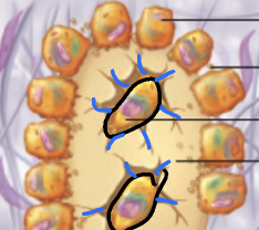
step 3 of intramembranous ossification
osteoid continues to form around vessels around vessels forming an immature spongy bone, mesenchyme adheres to the external surface and becomes the periosteum

step 4 of intramembranous ossification
immature spongy bone near the periosteum is remodeled and replaced with compact bone, central immature spongy bone matures

endochrondral ossification
process in which a bone develops from a preexisting model composed of hyaline cartilage
step 1 of endochondral ossification
mesenchymal cells specialize into osteoblasts; perichondrium becomes periosteum; osteoblasts secrete osteoid, creating a bone collar; chondrocytes left within hypertrophy and become primary ossification center

step 2 of endochondral ossification
chrondrocytes from the primary ossification center ossify surrounding cartilage; chrondrocytes die and matrix begins to dissolve; cavities are now formed within the model; remaining cartilage(ends) are healthy, growing rapidly to elongate the model

step 3 of endochondral ossification
cavities are invaded by the periosteal bud(nutrient a/v/n, red marrow elements, osteoprogenitor cells, osteoclasts); osteoclasts partially erode the cartilage matrix; osteoprogenitor cells become osteoblasts(spongy bone formation)

step 4 of endochondral ossification
osteoclasts break down the newly formed spongy bone and open the cavity; the epiphyses stay as cartilage ends through (growing rapidly); after birth, secondary ossification centers develop at epiphyses(short bones only have primary, irregular bones have multiple ossification centers)

step 5 of endochondral ossification
secondary ossification mimics that of primary except the spongy bone remains and no cavity is formed; the space between the diaphysis and epiphyseal plate and is hyaline cartilage

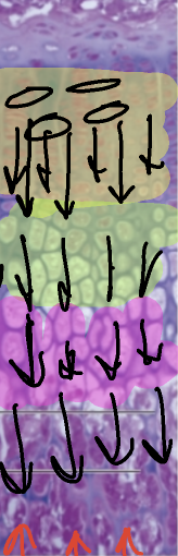
postnatal growth - length
proliferation(growth zone, chondrocytes begin rapidly dividing)
hypertrophic(increase in size, older chondrocytes enlarge/outgrow lacunae; erode leaving empty space)
calcification(surrounding matrix calcifies, chondrocytes die)
ossification(bone marrow elements come up and invade bringing osteoblasts(build new bone) and osteroclasts(old calcified cartilage)
postnatal growth - width
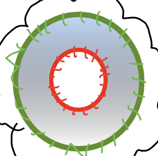
completed through appositional growth; osteoblasts in the periosteum secrete bone matrix to the external bone surface; osteoclasts on the endosteal surface remove bone; more osteoblast activity compared to osteoclast
remodeling bone
hormones determine if and when bones are remodeled; mechanical stress determines where bones are remodeled; takes 10 years to turn over
calcitonin
promotes mineralization and lowers blood Ca2+ concentration in children but usually has little effects in adults; may prevent bone loss in pregnent and lactating women
calcitriol
promotes intestinal absorption of Ca2+ and phosphate; reduces urinary excretion of both; promotes both resorption and mineralization; stimulates osteoclast activity
parathyroid hormone
indirectly activates osteoclasts, which resorb bone and raise blood Ca2+ concentration; inhibits urinary Ca2+ excretion; promotes calcitriol synthesis
hormonal influence
role of calcium(homeostasis for maintaining resting membrane potentials); 99% of the bodys calcium is stored in bone matrix(hormones will maintain a level of 9-11 mg/dL of blood); parathyroid hormone, calcitriol, and calcitonin(more important in children)
hormonal influence on calcium in hypocalcemia
blood Ca2+ deficiency—> parathyroid hormone secretion(signaled when calcium is low) leads to both increased osteoclast activity—more bone resorption and reduced osteoblast activity—> less bone deposition both lead to blood Ca2+ returns to normal
hormonal influence on calcium hypercalcemia
increased blood Ca2+ excess —> calcitonin secretion leads to both reduced osteoclast activity —> less bone resorption and increased osteoblast activity —> more bone deposition both lead to decreased blood Ca2+ returns to normal
parathyroid hormone
secreted by the parathyroid glands; stimulated the osteoclasts to promote bone resorption; promotes calcium reabsorption by the kidneys; promotes final step of calcitriol synthesis in the kidneys; inhibits collagen synthesis by osteoblasts
calcitriol and calcitonin
digestive tract

estrogen
sex hormones; increase osteoblast activity
testosterone
sex hormone; increase osteoblast activity
growth hormone
promotes growth at epiphyseal plates, but influences all aspects of bone metabolism; increases urinary excretion of calcium; increases digestive absorption of calcium; general influence on many tissues
mechanical stress influence
determines where the bone is remodeled; wolff’s law
wolff’s law
the body adapts to the forces placed upon it
non-displaced fracture
two ends are still touching
discplaced fracture
two ends are no longer touching
incomplete fracture
isnt all the way through
complete fracture
all the way through the width
closed fracture
bone is still inside the body
open fracture
bone is outside the body
transverse fracture
complete, straight line bone break that occurs horizontally or perpendicular to the long axis of the bone

oblique fracture
complete or partial bone break that runs diagonally across the shaft of a long bone

spiral fracture
complete bone break where a corkscrew shaped fracture line twists around a long bone

comminuated fracture
bone shatters or breaks into 3 or more pieces

avulsion fracture
tendon or ligament pulls a small piece of bone away from the main bone

impacted fracture
broken ends of a bone are driven or jammed into each other by a strong vertical force or high energy impact

fissure fracture
incomplete bone crack or hairline with no separation or displacement of the bone fragments

greenstick fracture
incomplete break where a supple bone bends and cracks on only one side

4 stages for traumatic fracture healing
hematoma formation(hematoma is converted to granulation tissue by invasion of cells and blood capillaries)
soft callus formation (deposition of collagen and fibrocartilage converts granulation tissue to soft callus)
hard callus formation (osteoblasts deposit a temporary bony collar around the fracture to unite the broken pieces while ossification occurs)
bone remodeling (small bone fragments are removed by osteoclasts, while osteoblasts deposit spongy bone and then convert it to compact bone

osteoporosis
group of diseases in which bone resorption outpaces bone deposit; 1:3 women > 50 will have fracture due to osteoporosis; risk factors: low calcium, decreased activity, PTH disorder, low estrogen, genetic

osteomalacia
group of diseases in which bones are poorly mineralized; results in weak and soft bones(calcium salts not sufficient); childhood version is rickets(bowed legs, deformities to the skull, pelvis, and ribs)

osteitis deformans
haphazard bone deposit and resorption; abnormally high ratio of spongy bone; commonly at the: spine, pelvis, femur, skull; 3% of elderly americans(rare before 40)