Cardio Block 4: Excitation + ECG
 Call Kai
 Learn
Learn Practice Test
 Spaced Repetition
 Match
Match Flashcards
 Knowt Play
1/43

There's no tags or description
Looks like no tags are added yet.
Name | Mastery | Learn | Test | Matching | Spaced | Call with Kai | Chat |
|---|
No analytics yet
Send a link to your students to track their progress
44 Terms
Give the name of the specific pore which essentially links two cells of the myocardium together?
Connexin 45 or CX45 pore
Why can arrhythmias occur in myocardium
One cardiac myocyte might have many different connections to allow it to transmit that signal throughout the myocardium.
a single cell could potentially fire a spontaneous action potential that could spread throughout the myocardium = this is the basis of many arrhythmias
What mechanism stops arrhythmias from happening?
cells of the myocardium have a refractory period where it is much more difficult to restimulate a cell once the depolarisation spreads through them
therefore, refractoriness makes the spread of excitation UNIdirectional in normal physiological conditions
What channel ensures the refractory peroid occurs?
voltage-gated sodium channels
these channels are basically inactive within 2 milliseconds of the action potential initiating and they won’t recover again until the end of the action potential
Name the 2 refractory periods of myocardial cells:
Absolute Refractory Period
Relative Refractory Period
What is the difference between the ARP and the RRP?
the ARP is where another action potential CANNOT be stimulated because ALL of the sodium channels are inactive
the RRP is where another action potential could potentially be stimulated because SOME of the sodium channels have recovered from inactivation
What happens to the sodium channels to inactive them?
a ball peptide on the end terminus of the channel moves into the pore and physically blocks it
Once that’s plugged in place, it is not coming out again until the membrane potential repolarises in phase 3.
What does the Electrocardiogram plot?
voltage changes of the heart tissue
against time
What heart problems can an ECG detect?
arrhythmias; tachycardia + bradycardia
atrial fibrillation
rate changes
rhythm disturbances
ischaemia (reduced blood flow to heart)
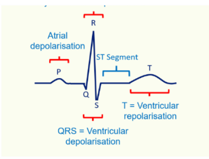
Draw and label a typical ECG graph:
What does the P wave represent?
atrial depolarisation
What does the QRS complex represent?
ventricular depolarisation
What does the R wave represent?
atrial repolarisation occurs here but is hidden by ventricular depolarisation
What does the T wave represent?
ventricular repolarisation
What is the ST segment?
represents the period when the ventricles are fully depolarised and contracting, just before repolarisation begins
What deflection do you see on an ECG when a wave is moving towards an electrode?
positive deflection (up stroke)
What deflection do you see on an ECG when a wave is moving away from an electrode?
small negative deflection (down stroke_
When there is no NET movement of charge, what can be seen?
an isoelectric flat line
How many ECG leads are there?
12
How many electrodes are there?
10
What are the two lead types for an ECG?
limb leads
chest leads
Where do the 6 chest leads lie for a 12 lead/unipolar ECG?
on the perpendicular transverse plane
from fourth intercostal space to sixth intercostal space

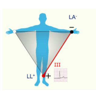
Where are the standard limb leads placed on bipolar ECG?
Lead I = right arm → left arm
Lead II = right arm → left leg
Lead III = left arm → left leg
How does Bipolar lead arrangement measure heart activity?
the leads record the electrical potential difference between 2 active electrodes
forms Einthoven triangle
What is Wilson’s central terminal?
a common virtual electrode
an average of the measurements from RA, LA and LL to give an ‘average potential of the body’
How does unipolar lead arrangement measure the activity of the heart?
uses on positive (exploring) electrode
uses one indifferent/reference electrode
List the Leads in a unipolar ECG arrangement:
chest precordial leads V1-V6
limb leads: RA, LA, LL, RL
Augmented limb leads aVR, aVL, aVF
What are augmented/virtual limb leads?
virtual leads use a reference created by combining signals from multiple electrodes to estimate the electrical activity from a specific direction.
What does the aVF measure?
the difference between positive LL and the mean of the negative RA and LA
What does the aVL measure?
the difference between positive LA and the mean of the negative RA and LL
What does the aVL measure?
the difference between the positive RA and the mean of the negative LA and LL
How can limb leads also be placed on the chest?
the leads are arranged in a wheel

Draw a diagram of the 3 augmented unipolar leads, coupled with the 3 standard bipolar leads:

Which segment of the ECG normally shows a myocardial infarction?
ST segment
instead of flat it will have elevation
What view of the heart do Leads II, III and aVF give?
inferior diaphragmatic surface of the heart
What view does LI. aVL, V5 and V6 give?
lateral wall of left ventricle
What view of the heart do leads V1 and V2 give?
septal surface (interventricular septum)
What view of the heart do leads V3 and V4 give?
anterior wall of right and left ventricles - sternocostal surface of the heart
How do you calculate the heart rate from an ECG?
count the number of large squares between an R and an R
do 300 divided by that amount to get rate
What is ventricular APD?
ventricular action potential duration
Faster heart rate, requires a ___ APD?
shorter
longer QT period =
slower heart rate
shorter QT period =
faster heart rate
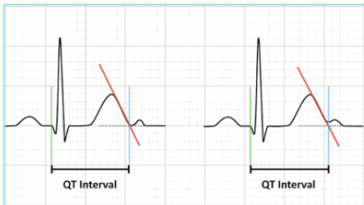
How is the QT period measured?
intersection of T wave maximum slope with the isoelectric line