22: Nucleic Acid Techniques 1
 Call Kai
 Learn
 Practice Test
 Spaced Repetition
 Match
 Flashcards
 Knowt Play
1/88

There's no tags or description
Looks like no tags are added yet.
89 Terms
What is restriction mapping?
A technique that uses restriction enzymes (from bacteria) to cut DNA and analyze the resulting fragments

What determines where a restriction enzyme cuts DNA?
The specific DNA recognition sequence recognized by the bacterial enzyme.
What information can restriction maps help locate?
Base sequences and genes on small genomes or chromosomes.
How can restriction mapping help identify inherited genetic defects?
Mutations can create or eliminate restriction sites, changing the resulting DNA fragment patterns.
It can also obtain other genetic information
How can restriction analysis be used in forensic science?
By comparing DNA fragment patterns to identify genetic differences between biological samples.
What is the natural biological function of restriction enzymes in bacteria?
To protect bacteria by recognizing and cutting foreign DNA
What do restriction enzymes recognize?
Specific nucleotide sequences within DNA
What does it mean for a restriction enzyme to be a four-cutter?
It recognizes a specific DNA sequence containing four bases.
What does it mean for a restriction enzyme to be a six-cutter?
It recognizes a specific DNA sequence containing six bases
Which restriction enzyme types does the slide identify as cleaving DNA at selected sites?
Type II and Type III restriction enzymes
What are nucleases?
Enzymes that hydrolyze nucleic acids by breaking phosphodiester bonds.
What is the difference between endonucleases and exonucleases?
Endonucleases cut internally; exonucleases remove nucleotides from the ends

What is a DNase?
A nuclease that acts on DNA
What is an RNase?
A nuclease that acts on RNA.
What are restriction endonucleases?
Enzymes that cut double-stranded DNA at internal locations.
Where are restriction endonucleases primarily isolated from?
Bacteria
What are the three types of restriction enzymes?
Type I, Type II, and Type III
What are the characteristics of Type I restriction enzymes?
They require ATP and perform nonspecific cleavage.
What are the characteristics of Type II restriction enzymes?
They do not require ATP and perform specific cleavage.
What are the characteristics of Type III restriction enzymes?
They require ATP and perform specific cleavage.
Which restriction enzyme type is particularly useful for predictable DNA cutting? Why?
Type II, because it cuts at specific locations without requiring ATP.
How many bases can restriction enzymes recognize?
Restriction enzymes can recognize 4, 6, or more bases in selecting sites
What determines whether a restriction enzyme produces sticky or blunt ends?
The relative positions at which it cuts the two DNA strands
How are sticky ends produced?
By staggered cleavage of the two complementary DNA strands.
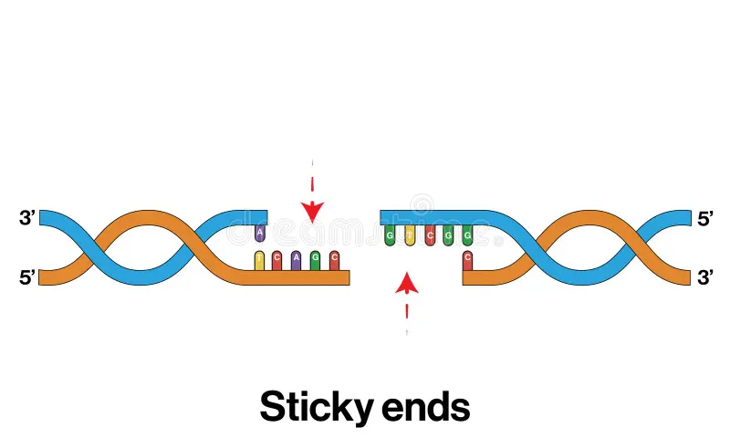
What is a DNA overhang?
An exposed, unpaired single-stranded region at the end of a DNA fragment.

Why are sticky ends useful in recombinant DNA technology?
Complementary overhangs can base-pair, helping DNA fragments join together
Why they’re called “sticky”

How are blunt ends produced?
When both DNA strands are cut at the same position.

Can blunt-ended DNA fragments still be joined together?
Yes. DNA ligase can join blunt-ended DNA fragments.
Why is DNA considered a polyanion?
Its sugar-phosphate backbone contains multiple negatively charged phosphate groups
Which direction does DNA migrate during gel electrophoresis?
Toward the positively charged anode
Why does DNA move toward the anode?
DNA has a negatively charged phosphate backbone and is attracted to the positive electrode.
What is the function of the gel matrix in electrophoresis?
It acts as a molecular sieve that separates DNA fragments based on size.
Which two gel matrices are used in gel electrophoresis?
Agarose
Popular due to the size of nucleic acids
Polyacrylamide
Used for small amino acids
How does DNA fragment size affect migration through a gel?
Smaller fragments migrate faster and farther than larger fragments.
Why is gel electrophoresis useful in restriction mapping?
It separates DNA fragments so their lengths can be determined and used to map restriction sites
What are the two major stabilizing interactions in DNA?
Hydrogen bonding between base pairs and van der Waals interactions between stacked bases.
DNA’s structure as a ladder:
IDK, it was just on the slides :/

What is the distance between adjacent DNA base pairs?
Approximately 0.34 nm.
What is the pitch length of the DNA helix?
Approximately 3.4 nm per complete turn.
What three factors impact to the stability of the DNA double helix?
Hydrogen bonding
Electrostatic interactions
Base-pair stacking.
Where do hydrogen bonds form in double-stranded DNA?
Between complementary nitrogenous bases on opposite strands.
How does do electrostatic interactions impact the stability of DNA’s double helix?
It decreases DNA stability because the negatively charged phosphate groups on the two strands repel each other.
This makes them more stable on the exterior of the helix
What are intercalating agents?
Molecules that insert themselves between stacked base pairs in DNA.

Why can intercalating agents enter the DNA double helix?
DNA is a flexible, dynamic structure that can accommodate certain molecules between its base pairs.
What structural characteristics of intercalating agents allow them to fit between DNA bases?
They are flat, hydrophobic, large cyclic (macrocyclic), aromatic molecules with fused heterocyclic rings.

What happens to DNA when an intercalating agent inserts between base pairs?
The bases are forced apart, distorting the normal double-helix structure
The bases are force apart to accommodate these intercalating agents
Three examples of intercalating agents:
Ethidium bromide
Acridine orange
Actinomycin D.
What is ethidium bromide used for?
Visualizing DNA fragments (DNA bands) separated through gel electrophoresis.
It inserts into DNA and fluoresces under appropriate illumination.

What is acridine orange used for?
Staining nucleic acids and helping determine cell-cycle stages as nucleic acid content and distribution change

What is actinomycin D?
An antibiotic with anticancer effects that binds DNA at the transcription initiation complex and prevents elongation.
How do ethidium bromide, acridine orange, and actinomycin D differ in their primary applications?
Ethidium bromide visualizes DNA, acridine orange stains nucleic acids, and actinomycin D inhibits transcription.
What is DNA denaturation?
The separation of the two complementary DNA strands due to disruption of interactions maintaining the double helix.
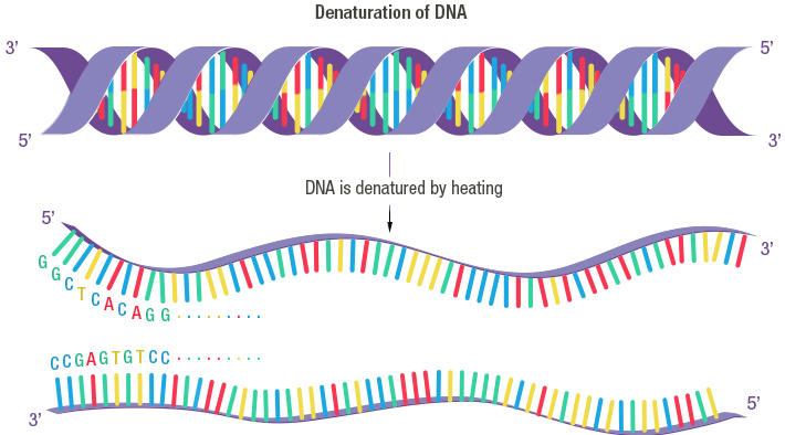
What causes DNA denaturation?
Heating the DNA

Does DNA denaturation normally break the phosphodiester bonds within each strand?
No. It disrupts the interactions between strands, leaving the individual strands intact.
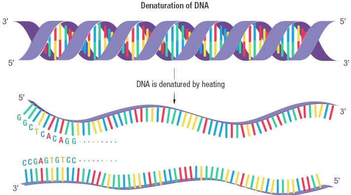
What happens to nitrogenous bases during DNA denaturation?
They become more exposed as the complementary strands separate.

What is the hyperchromic effect?
The increase in DNA absorbance at approximately 260 nm during denaturation.
Why does DNA absorbance increase during denaturation?
Separation of the strands exposes the bases, increasing their ultraviolet absorbance.
At approximately what wavelength is DNA absorbance measured to observe the hyperchromic effect?
260 nm
What is the melting temperature (Tm) of DNA?
The temperature at which 50% of the DNA is denatured
What happens to DNA absorbance as temperature increases during denaturation?
Absorbance at 260 nm increases as more DNA becomes denatured.
What is DNA renaturation?
The cooling, thus reassociation of complementary DNA strands to reform the double helix.
Denaturation conditions must be removed (i.e. heat)

What happens to the melting temperature as G+C content increases?
The melting temperature generally increases
G–C base pairs have three hydrogen bonds, compared with two in A–T pairs, and G+C-rich DNA generally has greater stability.
What is reannealing?
The reassociation of complementary DNA strands to reform a double helix.

What is the difference between DNA renaturation and reannealing?
Renaturation is the restoration of double-stranded DNA after denaturation. Reannealing is the process of single-stranded DNA molecules binding together through complementary base pairing.
The terms are often used interchangeably.
What must happen before DNA strands can renature?
The complementary strands must realign so their bases are correctly positioned opposite one another
What is nucleation during DNA renaturation?
The initial association of complementary regions between two separated DNA strands.
Why is nucleation considered the slow step of renaturation?
The two strands must encounter each other and establish correct initial base pairing.

What is zippering during DNA renaturation?
The rapid formation of base pairs after the strands have initially aligned.

Why is zippering faster than nucleation?
Once the strands are aligned, neighboring complementary bases can pair progressively.
What is the purpose of restriction mapping?
To determine the physical locations of restriction enzyme cutting sites along DNA
What two techniques are used to generate a physical map of genomes, genes, or other segments of DNA?
Restriction endonucleases (restriction enzyme digestion) and gel electrophoresis.
What are the three major steps of restriction mapping?
Cut DNA with restriction enzymes
Separate and analyze fragments using gel electrophoresis
Determine/map the locations of restriction sites
How many times does HindIII cut the DNA molecule?
Once
How many DNA fragments are produced when HindIII cuts a linear DNA molecule?
Two fragments
Why is the initial orientation of the restriction map arbitrary?
Gel electrophoresis reveals fragment lengths but does not identify which original DNA end each fragment came from.
How many times does BamHI cut a molecule?
Twice
How many fragments does BamHI produce when it cuts this linear DNA molecule?
Three fragments
What is the most important principle for interpreting a double digest?
If an original fragment disappears and smaller fragments replace it, the added enzyme cut within that original fragment.
What are restriction-fragment length polymorphisms (RFLPs)?
Differences in the lengths of DNA fragments produced when restriction enzymes cut DNA from individuals with different DNA sequences.
How can a mutation produce an RFLP?
A mutation can create or eliminate a restriction enzyme recognition site, changing where the enzyme cuts DNA and therefore changing the lengths of the resulting fragments.
How can a mutation eliminate a restriction site?
A mutation changes the nucleotide sequence recognized by a restriction enzyme, preventing the enzyme from binding and cutting at that location.
How can a mutation create a new restriction site?
A mutation changes a DNA sequence so that it now matches the recognition sequence of a restriction enzyme, allowing the enzyme to cut at that location.
Why are RFLPs useful for studying inherited genetic defects?
RFLPs allow researchers to identify differences in DNA fragment patterns that can be associated with inherited genetic variations or defects.
After how many base pairs do human chromosomes differ?
Human chromosomes differ in sequence every 200 to 500 bp, creating or eliminating restriction sites
How can RFLPs be used to study inheritance?
Researchers compare restriction-fragment patterns from parents and offspring to determine which DNA fragments were inherited from each parent.
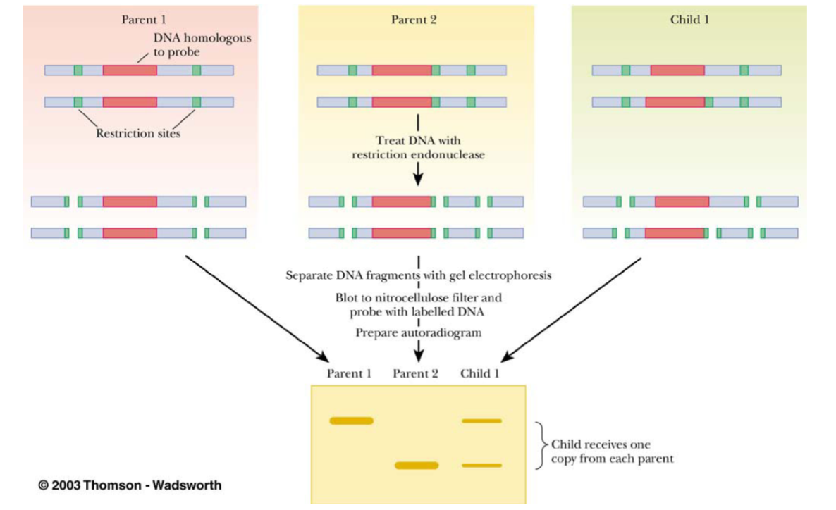
What is a DNA probe?
A labeled, single-stranded DNA sequence that binds to a complementary DNA sequence, allowing researchers to identify specific DNA fragments.
Why is a DNA probe used after gel electrophoresis?
The probe identifies specific DNA fragments containing a complementary sequence, allowing researchers to detect a particular DNA region.
Why does a child's DNA band pattern reflect both parents?
The child inherits one copy of each relevant DNA region from each parent, so the child's DNA pattern can contain fragments inherited from both.
