Ch 19: Lower Resp Disorders #2
 Call Kai
 Learn
Learn Practice Test
 Spaced Repetition
 Match
Match Flashcards
 Knowt Play
1/72

There's no tags or description
Looks like no tags are added yet.
Name | Mastery | Learn | Test | Matching | Spaced | Call with Kai | Chat |
|---|
No analytics yet
Send a link to your students to track their progress
73 Terms
Pleurisy (Pleuritis)
what is it
what kind of pain
pain exacerbates when? diminishes when ?
ascultation
often after ? 4
Inflammation of both layers of the pleura (parietal and visceral),
which then rub together during breathingClassic findings: sharp, 'knife-like' pain that intensifies with inspiration, coughing, or sneezing — pain diminishes when the patient holds their breath
Auscultation may reveal a pleural friction rub
Often secondary to pneumonia, viral URI, tuberculosis, or chest
trauma
managing pleurisy
treat ?
meds
positioning
application of
if severe
if untreated?
Management: treat the underlying cause:
NSAIDs,
positioning on affected side (acts as splinting), splinting to reduce pain during
breathing/coughing,application of heat or cold,
if severe – intercostal nerve block
if untreated or progressive, fluid can accumulate between the
pleural layers = effusion
Pleural Effusion
what is it
usually secondary to ? 5
what can cause reoccurring plueral effusion
• Abnormal collection of fluid in the pleural space
• Usually secondary to another disease process:
• Heart Failure - increased pressure = leaky capillaries
• Pneumonia - irritates pleura
• Tuberculosis
• Connective tissue disorders
• Malignancy - blocks blood flow → increases pressure (causes reoccurring plueral effusion)
s/s of plueral effusion
progressive ?
type of cough
what lessens as fluid accumulates
percussion ?
assessment findings?
Severity of symptom is determined by the size of effusion, the
spread of its formation and the underlying lung disease
• Progressive dyspnea
• Dry cough
• Pleuritic chest pain (may lessen as fluid accumulates)
• Dullness to percussion
• Diminished/absent breath sounds and ↓ fremitus over the effusion
dx for pleural effusion
• Chest X-R & Lateral decubitus X-R (pt lies on affected side)
• Thoracentesis - diagnostic & therapeutic
• Chest CT scan
transudate vs exudate fluid
transudate - thin, not a lot of proteins
exudate - thicker
guides underlying workup
tx of plueral effusion
major goal ?
what can we give
procedures ?
• Major goals: Discovering and treating underlying cause, Prevent
re-accumulation of fluid & Relieve discomfort
• Supplemental O2
• Thoracentesis
• Chest tube insertion - educate pt on drain management
• Pleurodesis - powder used to keep plueral space together
Empyema
complication of ?
• Accumulation of thick, purulent fluid (pus) in the pleural space
• Complication of pneumonia, thoracic surgery, or chest trauma
s/s of empyema
fever
night sweats
pleuritic chest pain
dyspnea
cough
anorexia, weight loss - cough uses a lot of ATP and lowers appetite
tx of empyema
goal?
medical tx?
nursing: what is required? monitor for? support ?
after discharge ?
Goal of treatment is to drain the pleural cavity and fully re-
expand the lungMedical: antibiotic therapy (4-6 weeks),
chest tube drainage required
monitor for sepsis
nutrition support
frequently discharged with chest tube & PICC line in place for IV antibiotics
fluid — chest tube drainage is frequently require
Blunt Chest Trauma
sudden compression or positive pressure into the chest wall
MVA, falls, bicycle crashes, hit in chest, compressed
penetrating Chest trauma
foreign object penetrates the chest wall
knife wound, gun shot
chest trauma results in
hypoxemia
lung tissue damage,
bleeding (hemorrhage),
collapsed lung (pneumothorax)
hypovolemia from fluid loss and cardiac failure
Primary vs Secondary survey
ABCDE
AMPLE
Primary - Emergency Assessment:
• find and fix what will kill the patient first, then look for everything elseABCDE: Assess airway, breathing, circulation, Disability (neurologic status), and expose pt for hidden injuries
Secondary Assessment
• Perform a systematic head-to-toe examination after stabilization
• Obtain AMPLE history: allergy, medications, past Hx, last meal, event that led to situation
sternal fractures
sings
common in motor vehicle crashes-direct blow
anterior chest pain, tenderness, ecchymosis, Bony
crepitus (bones rubbing together), swelling, possible chest wall deformity
rib fractures
what ribs are most frequently involved
if first 3 ribs are injured
if lower ribs are injured
if an older adult has broken ribs
most common chest trauma with blunt injury
Ribs 4–10 most frequently involved
First three ribs: high mortality due to subclavian artery/vein laceration
Lower ribs: spleen/liver injury
Older adults with three or more rib fractures:
• Fivefold increased mortality, fourfold increased pneumonia
s/s of rib fractures
severe pain,
point tenderness,
muscle spasm,
bruising,
Pain aggravated by coughing, deep breathing & movement,
Crepitus and swelling
consequences of rib and sternum fractures
diminished ventilation due to shorter breaths
atelectasis
pneumonia
hypoxia
respiratory insufficiency and failure
managing sternum and rib fractures
when do most rib fractures heal
control the pain
breathing exercises with mild sedation & Abdominal binder.
Most rib fracture heal in 3 to 6 weeks
flail chest
causes ?
occurs when ?
results in ?
consequences?
• Complication of blunt chest trauma
• Causes: steering wheel injury, motor vehicle crash, fall, assault
• Occurs when three or more adjacent ribs fractured at two or more
sites
• Results in free-floating rib segments
• Chest wall loses stability → causes respiratory impairment, severe distress
managing a flail Chest
what may be needed
what can control pain
procedure ?
tests?
encourage ? what do we not want to develop
WATCH FLAIL CHEST VIDEO IN PPT
ventilator at times and suctioning/oral care as needed
Pain control! - intercostal nerve blocks, splinting, binder
Chest wall stabilization (CWS) -plating system
Chest x-rays, ABGs , pulse oximetry, and bedside
pulmonary function monitoringencoruage deep breathing, cough and lung
expansion and secretion management - Don’t want pneumonia to develop

Penetrating Trauma
what organs can be injured
what are the main complications
chest wall, lung, pleura, tracheobronchial system, esophagus, diaphragm,
major thoracic blood vessels, heart, mediastinal structurespneumothorax and cardiac tamponade
pneumothorax
how does it happen
what is it
4 types
Air enters the pleural space, destroying the negative pressure thatkeeps the lung inflated
the lung partially or completely collapses
• Simple /spontaneous
• Traumatic
• Tensionopen
Spontaneous/Simple pneumothorax
how does it happen
often in?
occurs after ?
• Occurs without trauma
• Often in tall thin young apparently healthy males or in patients with underlying lung disease/ heavy smokers
• Occurs after a ruptured bleb (air bubble) or a bronchopleural fistula
Traumatic Pneumothorax
occurs when ?
often seen with?
how can this happen
Occurs when air escapes from a laceration in the lung and enters the pleural space or when air enters the pleural space through a wound in the chest wall
• Often accompanied with hemothorax(blood pooling in pleural space)
• Results from: Penetrating and/or Blunt chest trauma
• Pressure related trauma or Complication of procedure
Tension Pneumothorax
how does it happen
what are the signs
emergent tx?
s/s?
Air enters the pleural space but cannot escape → Progressively increasing pressure as pt takes each breath
Pushes the mediastinum toward the unaffected side
Compressing the heart and great vessels & tracheal deviation towards the
unaffected sideLife threatening! - in the field EMT will perform needle decompression to relive pressure
severe respiratory distress, hypotension, distended neck veins
• Life-threatening emergency
Open Pneumothorax
how does it happen
aka ?
what is the sign?
what does is effect ? vitals?
An opening in the chest wall allows air to move freely in and out of the pleural space with breathing
aka ‘Sucking chest wound’
Lung collapses and heart and great vessels shifting or swinging to
the unaffected side with each inspiration and back with expirationAffects cardiac output and circulation (BP and CO drops, HR goes up)
life threatening emergency
general s/s of pneumothorax
sudden ?
breathing ?
on affected side ? (2)
percussion ?
HR? O2?
• Sudden sharp chest pain
• Dyspnea
• Decreased/absent breath sounds on the affected side
• Hyperresonance to percussion (trapped air)
• Decreased chest expansion on affected side
• Tachycardia
• Low oxygen saturation (hypoxemia)
Management - Pneumothorax
main goal
small and simple?
large symptomatic?
tension?
open ?
Goal: evacuate blood or air from pleural space, restores the negative
intrathoracic pressureSmall, stable spontaneous - observation and supplemental oxygen
Larger/symptomatic pneumothorax: chest tube insertion - need to restore negative pressure!
Tension pneumothorax: needle decompression & chest tube placement
Open pneumothorax: cover open wound with sterile gauze & tape on three sides (not 4 b/c you can cause tension pneumothorax by trapping air)
chest tubes
purpose
indications
placement for what reasons?
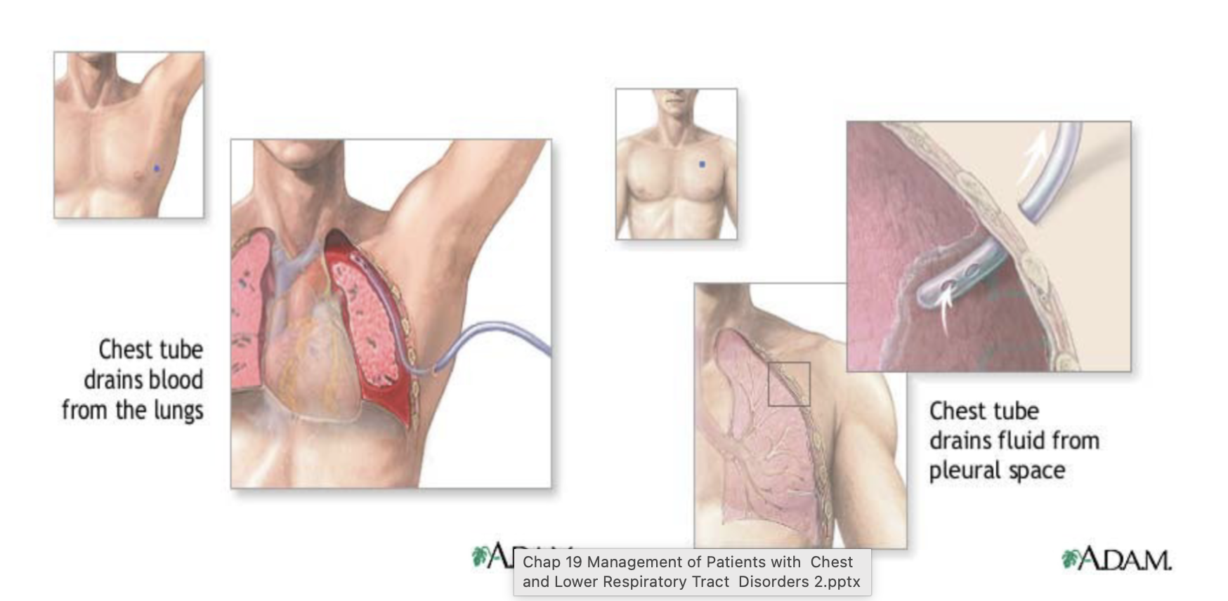
Purpose: remove air and/or fluid from the pleural space and restore
negative intrapleural pressure, allowing the lung to re-expandIndications: pneumothorax, hemothorax, pleural effusion, empyema, and
after thoracic surgery to restore pressurehigh placement if air in the lungs
low placement if fluid/blood is in the lungs
nursing management of chest tubes
what do we want in the water seal chamber? what are we looking for
where is the drainage system
never ?
what doe we monitor
@ bedside ?
Monitor for tidaling - we want in water seal chamber - shows inhalation and exhalation
Monitor for continuous bubbling in the water-seal chamber - indicates leak
Keep the drainage system below the level of the chest at all times
Never clamp a chest tube without a specific order - may cause hemothorax
Monitor and document drainage amount, color, and consistency & look at dressing
Keep two enclosed hemostats, a sterile occlusive dressing at the bedside
at all times
Subcutaneous Emphysema
how does it happen
often occurs when
can occur with?
manifestations
• Air trapped under the skin, usually in the chest or neck
• Often occurs when air leaks from the lung or pleural space
• Can occur with pneumothorax or chest tube complications
• Manifestation: Swelling and a crackling/crunchy sensation (crepitus)
managing Subcutaneous Emphysema
emergent ?
otherwise ?
if emergent (threatens airway patency) - tracheostomy STAT - can happen when its in the neck
otherwise: Treat the underlying air leak, maintain chest-tube patency,
supplemental o2 prn, monitor respiratory status and swellingusually dissipates on its own
Acute Respiratory Failure
occurs when ?
results in?
can result from ? 5
Occurs when the respiratory system can no longer maintain adequate gas exchange
results in inadequate oxygenation, inadequate ventilation (CO2 removal) or both
Hypoxemia, hypercapnia (too much CO2)
Can result from severe pneumonia, massive atelectasis,
pneumothorax, Pulmonary embolism or neuromuscular failure
who is at risk for ARF
cant drive breathing
cant move air
cant exchange gas
cant drive breathing - Neuromuscular/CNS
cant move air - Chest Wall / Mechanical
cant exchange gas - Airway/Lung Disease
what includes cant drive breathing - Neuromuscular/CNS
• Opioid/sedative overdose, anesthesia
• Stroke (esp. brainstem), High cervical spinal cord injury
• Myasthenia gravis, GBS, ALS, TBI, increased ICP
what includes cant move air - Chest Wall / Mechanical
• Flail chest, rib fractures, Kyphoscoliosis, morbid obesity
• Post-op abdominal/thoracic surgery (not breathing deep enough , pain)
what includes cant exchange gas - Airway/Lung Disease
• Sepsis / SIRS, Aspiration risk (↓LOC, dysphagia)
• Advanced age, smoking history
• Immunocompromised, immobility, Chest trauma, anaphylaxis
Early manifestations of ARF
restlessness, anxiety, tachypnea, and mild confusion — before dramatic
vital sign changes appear
late manifestations of ARF
severe hypoxemia, hypercapnia (high CO2), use of accessory muscles, cyanosis,
altered level of consciousness
Management of ARF
nursing priority
• supplemental oxygen, noninvasive positive pressure ventilation
(BiPAP/CPAP), or invasive mechanical ventilation for severe cases
• Nursing priority:
• recognize early, subtle changes before the patient reaches
crisis
Acute Respiratory Distress Syndrome
(ARDS)
what is it? causes?
results in ? 3
usually will have to?
mortality ?
A clinical syndrome characterized by Severe inflammatory process causing diffuse alveolar damage
not a primary lung disease
results in:
• sudden and progressive pulmonary edema
• Bilateral infiltrates on chest x-ray.
• Hypoxemia unresponsive to supplemental oxygen - usually will have to intubate• Mortality remains significant despite treatment advances —
prevention and early recognition of at-risk patients matters
greatly
common triggers of ARDS
risk rises with?
sepsis, severe pneumonia, aspiration, trauma,
massive transfusion, pancreatitis
Risk rises with smoking, obesity, diabetes
Pathophysiology of ARDS
Inflammation = Increased capillary permeability leads to fluid leaking
into the alveoli (noncardiogenic pulmonary edema - does not evolve cardiac issue or high BP)severely impaired gas exchange and lung compliance
Manifestations ARDS
Rapid onset of severe dyspnea and hypoxemia that does not
respond well to supplemental oxygen aloneSOB, irritability, cyanosis, lethargy, diaphoresis, intercostal retractions
Management of ARDS
vent ?
position ?
extra treatments?
meds ?
tests?
Mechanical ventilation with low tidal volumes, PEEP,
prone position - monitor skin integrity and turn frequently
Extracorporeal Membrane Oxygenation (ECMO)for patients with severe ARDS
Medications – Hemodynamic support- Inotropic (digoxin) and vasopressor to maintain circulation
CXR, ABG’s
ARF and ARDS in peds
airways ?
alveoli?
causes ?
early signs of distress?
Higher risk, faster decompensation
Small airways — minor edema = major obstruction
Fewer alveoli, less reserve
Causes: RSV/bronchiolitis, croup, sepsis
Early distress signs (flaring, grunting, retractions)
ARF and ARDS in older adults
mortality ?
less of ? weaker ?
recovery?
what complicates tx?
atypical presentation
• Highest mortality
•↓ reserve, weaker cough/immune response
•Atypical presentation — confusion over dyspnea
•Frailty drives outcomes & goals-of-care
•Slower recovery, more long-term decline
•Polypharmacy complicates treatment
may have atypical presentation; LOC , confusion
pulmonary edema
what is it
cardiogenic
non cardiogneic - common causes ?
Abnormal accumulation of fluid in the lung tissue, the alveolar
space or bothCardiogenic: Results from Left sided heart failure (pressure problem)
Non-cardiogenic: damage to pulmonary capillary lining.
• pneumonectomy(removal of 1 lung), as pulmonary vascular system is adjusting to one lung with same amount of blood flow
• Chest trauma
• Aspiration and smoke inhalation, pancreatitis, sepsis. etc
Clinical Manifestations of Pulmonary Edema
pink frothy sputum
gasping for air
dyspnea
anxious
cyanotic
confusion as CO2 is retained
Assessment findings Pulmonary Edema
• Rales (crackles) upon auscultation – especially posterior bases (movement of air through the alveolar fluid)
• Tachycardia
• Pulse Oximetry dropping
• Chest X-Ray – increased interstitial fluid
• ABGs worsening
Medical Management-Pulmonary Edema
meds
extra support
what may be restricted
Discover and treat underlying condition
May require:
• diuretics
• vasodilators and
• inotropic meds ---to improve left sided cardiac functionOxygen therapy or temporary intubation/ventilator support
Fluid and sodium restriction when indicated to lessen fluid
Nursing Management - Pulmonary Edema
Position upright, provide oxygen/ventilatory support as needed
Monitor SpO , respiratory rate, lung sounds, HR, and BP
Assess for fluid overload and peripheral edema
• Monitor I&O, daily weigh, electrolytes and renal function
• Monitor for crackles, worsening dyspnea, cyanosis, frothy/pink sputum
Pulmonary Hypertension (PH)
what is it
normal pulmonary artery pressure is
what happens to blood vessels in the lungs , how does it effect pressure & heart
Sustained elevation in pulmonary artery pressure (mPAP >25mmHg at rest) due to increased resistance in the pulmonary vasculature
Normal pulmonary artery pressure is 11-20 mm Hg at rest
Blood vessels in the lungs narrow, stiffen, or get blocked:
This increases resistance to blood flow leading to ↑ pulmonary artery
pressure and the right ventricle of the heart working harder to pump blood

what are the 5 groups of pulmonary hypertension
Group 1: Toxins/drug exposure, Infections (e.g., HIV), Connective tissue
disease (e.g., scleroderma, lupus)
• Group 2: Heart disease
• Group 3: COPD, Sleep apnea/chronic hypoxia, Interstitial lung disease
• Group 4: Chronic thromboembolic disease- PE
• Group 5: unknown: Hematologic disorders (Sickle cell disease, Chronic myeloid leukemia (CML), Systemic disorders (e.g., sarcoidosis) and Metabolic disorders
Manifestations - Pulmonary hypertension
classic sign
where is the pain 2
5 more
signs of
Classic sign is ‘Dyspnea on exertion’ - from congestion and lack of O2
• Substernal chest pain, weakness, fatigue, syncope,
hemoptysis
• Anorexia and abdominal pain in right upper quadrant
• Signs of right-sided heart failure (peripheral edema, ascites, distended neck veins, liver engorgement, crackles, heart murmur)
how can we diagnose pulmonary HTN
what tests
what does an echocardiography do
what can we do to the heart to confirm dx? and what else
chest x-ray, pulmonary function studies, ECG, echocardiogram
Echocardiography: estimates pulmonary artery pressure,
assesses right ventricular size and functionRight heart catheterization: confirms PH diagnosis, assesses
hemodynamic abnormalities
medical management of pulmonary HTN
primary goal
6 meds
last resort
Primary goal: manage underlying condition
• Oxygendiuretics - removes fluid
anticoagulation - improves circulation and prevents clotting
digoxin - helps the heart pump stronger
Calcium channel blockers (nifedipine, diltiazem/Cardizem)
• effective only in a small group of patientsProstanoids (epoprostenol) - relaxes pulmonary smooth muscle
• requires continuous IV infusion; effective in selected patients with PAHLung transplant - option - if refractory to medical therapy
Nursing Management -Pulmonary HTN
identify ?
monitor ?
how can we Monitor pulmonary artery pressure
educate ?
Identify patients who are high risk such as COPD, PE, Mitral Valve Disease and congenital abnormalities
Monitor VS – watch for dyspnea and increased pressures- (invasive
pressure monitoring): A-line, CVP, PA catheter, wedgeEducate patients and families on home oxygen therapy
Cor Pulmonale
happens from chronically elevated pulmonary artery pressure
overworks the right ventricle → causing hypertrophy and R sided heart failure
manifestations of cor pulmonale
Headache, sleepiness,
hypercapnia,
exertional dyspnea,
wheezing, cough,
JVD, peripheral edema, ascites, weight gain, fatigue, hepatomegaly (R sided S/S)
Management-Cor Pulmonale
goal?
may include
treat both ?
restrict ?
monitor ?
administer ?
Goal improve ventilation - 24 hour/day oxygen therapy (may include assisting with intubation)
Treat both the underlying lung disease and cardiac symptoms
Monitor pulse ox, ABGs, V/S and EKG
chest PT,
turn and position, bedrest
suctioning
administration of bronchodilators and diuretics
sodium / fluid restrictions + monitor I and O and daily weight
Occupational Lung Diseases (Pneumoconioses)
what is It
how does it cause lung damage
may be ? once developed ?
what is key
3 types
Lung disease caused by inhalation and accumulation of occupational dust particles in the lungs
Inhaled dust particles → inflammation → lung tissue damage
may be asymptomatic until late ? once developed, not reversible
prevention is key - wear PPE at work sites
• Asbestosis
• Silicosis
• Coal workers pneumoconiosis (black lung disease)
Silicosis
what is it
patho?
chronic fibrotic pulmonary disease from inhaling silica dust (glass, miners, quarries and tunnels, sandblasting, stone cutters)
Lung nodules grow larger and join together.
Dense masses form in the upper lungs, reducing lung volume.
Scar tissue damages the lung tissue and makes breathing harder.
This can lead to restrictive lung disease, emphysema, high blood pressure in the lungs, and right-sided heart failure
asbestos pathophysiology
inhaled ? causes ?
what thickens ?
what happens to the lungs
as a result ?
what damage ?
increases the risk of
inhaled asbestos fibers become trapped in the alveoli and cause scar tissue to form.
The pleura thickens and develops plaques.
Scarring makes the lungs stiff, reducing lung volume and making breathing difficult.
Less oxygen enters the blood, causing hypoxemia
Severe damage can lead to pulmonary hypertension, right-sided heart failure (cor pulmonale), and respiratory failure.
Increases the risk of lung cancer, mesothelioma (cancer of the lung lining), and pleural effusion (fluid around the lungs)
Coal Worker’s Pneumoconiosis “black lung disease” pathophysiology
inhaled?
deposited ?
what builds up?
what forms
what is the result
inhaled dusts that are a mixture of several particles to include coal
deposited in the alveoli and respiratory bronchioles
Macrophages absorb coal dust but cannot clear it from the lungs.
Dust, dead macrophages, and fibroblasts build up and clog the bronchioles and alveoli.
Coal macules (small areas of damaged tissue) and scar tissue form.
Lung damage can lead to localized emphysema.
Severe cases can cause right-sided heart failure (cor pulmonale) and respiratory failure
lung cancer
classifications and risk factors
small lung cell
non-small lung cell (majority)
most common cause is inhaled carcinogen from cigarette smoke
• Tobacco smoke & Second-hand smoke
• Electronic nicotine delivery systems (ENDS) e.g., e-cigarettes
• Environmental and occupational hazards, Genetic mutations
Manifestations of lung cancer
change in?
any cough that lasts ?
3 other
may have ?
Cough or change in a chronic cough which is often ignored and
attributed to smoking or some other respiratory infectionany cough that lasts more than 2 weeks needs further assessment
Dyspnea, Hemoptysis, pleural pain
May have Mets to brain, liver, adrenals and bone
dx tests for lung cancer
• Chest x-ray, CT Scan
• Biopsy, Bronchoscopy, VQ Scans, PFTs
Management- Lung Cancer
therapies
surgeries ?
• Radiation therapy
• Chemotherapy
• Surgery (• Pneumonectomy - entire lung
• Lobectomy - lobe removed , mast common
• Segmentectomy - segment of the lung
• Wedge Resection - removal of the tumor and a small wedge-shaped
portion of surrounding lung tissue
advantages of Video-Assisted Thoracoscopic Surgery (VATS)
minimally invasive, small incisions, less pain, faster recovery
less pain = more coughing and deep breathing, less chance of atelectasis
less pain = earlier ambulation, less r/o DVT, constipation, pressure sores
pist op nursing management
• Chest tube management
• Pain control, splinting, CDB
• Incentive spirometry, early ambulation
• Monitor respiratory status, work of breathing
• Monitor for pneumothorax, subcutaneous emphysema, hemorrhage
Smoking cessation counseling
complications to monitor for post op
Post-pneumonectomy pulmonary edema (PPE)
• A rare but serious late complication. usually occurs within 72 hours after
surgery and affects the remaining lung (same amount of blood flow with less lung to manage ) - fluid restriction, diuretics, digoxin• Tension pneumothorax
• Bronchopleural fistula
• Cardiac dysrhythmias
• Infection
• Pneumonia / atelectasis