L8 - Histology of bone marrow hematopoises
 Call Kai
 Learn
Learn Practice Test
 Spaced Repetition
 Match
Match Flashcards
 Knowt Play
1/148

There's no tags or description
Looks like no tags are added yet.
Name | Mastery | Learn | Test | Matching | Spaced | Call with Kai | Chat |
|---|
No analytics yet
Send a link to your students to track their progress
149 Terms
•_______ is the formation of Blood Cellular Components in the bone marrow
Hematopoiesis
•All cellular blood components are derived from?
Hematopoietic Stem Cells (HSC)
why is hematopoiesis necessary?
to maintain steady state levels of blood cells in peripheral circulation
Multipotential Hematopoietic Stem Cells (a.k.a. Hemocytoblast) reside in the ?
Medulla of Bone (Bone Marrow)
Within Bone Marrow are found? (5 things)
Stem Cells, Immature Blood Cells, Macrophages, Fat Cells, Reticular Cells
•All formed elements within blood form from single cell type, the ?
Multipotential HSC
HSCs (Hematopoietic Stem Cells) are Self-Renewing Cells, what does this mean?
when they proliferate, some of their daughter cells remain as HSCs, so the pool of stem cells is not depleted
Q: Why isn’t the HSC pool depleted when HSCs proliferate?
A: Some daughter cells remain as HSCs instead of differentiating.
Q: What can HSCs differentiate into?
A: Myeloid and lymphoid progenitor cells, which eventually produce specific blood cells.
Q: What is a major difference between HSCs and myeloid/lymphoid progenitor cells?
A: HSCs can self-renew; progenitor cells cannot.
Q: Can myeloid and lymphoid progenitor cells self-renew?
A: No. They differentiate into more specialized blood cells.
What are the mature cells of the myeloid linkage?
neutrophils, eosinophils, basophils, monocytes, platelets and erythrocytes
What are the mature cells of the lymphoid linkage?
T cells, B cells and NK cells
Q: What is the function of neutrophils (myeloid lineage; granulocyte)?
A: Phagocytosis and killing of bacteria; acute inflammation.
Q: What is the function of eosinophils (myeloid lineage; granulocyte)?
A: Defense against parasites and modulation of allergic reactions.
Q: What is the function of basophils (myeloid lineage; granulocyte)?
A: Allergic reactions; release histamine and heparin.
Q: What is the function of monocytes (myeloid lineage; agranulocyte)?
A: Phagocytosis; become macrophages in tissues.
Q: What is the function of platelets (myeloid lineage; megakaryocyte/thrombocyte line)?
A: Hemostasis (blood clotting).
Q: What is the function of erythrocytes/RBCs (myeloid lineage; erythroid line)?
A: Transport oxygen.
Q: What is the function of T cells (lymphoid lineage; agranulocyte)?
A: Cell-mediated immunity. (tumor + viruses)
Q: What is the function of B cells (lymphoid lineage; agranulocyte)?
A: Humoral immunity → antibody production.
Q: What is the function of NK cells (lymphoid lineage; agranulocyte)?
A: Innate immunity; kill virus-infected and tumor cells.
what do macrophages come from?
When does this change occur?
they come from monocytes
chnage happens upon entering connective tissue
do T or B lymphocytes become plasma Cells?
When does this chnage occur?
B-lymphocytes
upon entering connective tissue
the lymphoid lineage gives rise to?
NK, T- and B lymphocytes, and plasma cells
the myeloid lineage gives rise to?
BEN group, megakaryocytes, thrombocytes (platelets), monocytes and eventually macrophages
what are cells able to squeeze through in the bone marrow?
What does this allow?
endothelial cells
Allows RBCs and WBCs to circulate in the blood
are megakaryocytes able to pass into the endothelial cells from the bone marrow?
no - however the platelets they create are able to
Q: What is granulopoiesis (granulocytopoiesis)?
A: Hematopoiesis (production) of granulocytes.
Q: What is megakaryocytopoiesis?
A: Hematopoiesis (production) of megakaryocytes, which ultimately produce platelets.
Is this conversation helpful so far?
________
Formation of Erythrocytes
(Red Blood Cells)
Erythropoeisis
the erythrocytes are seen as a “bag” of ______ when they reach the mature stage
Hgb
Q: What is the first recognizable cell in erythropoiesis, and what are its key features?
A: Proerythroblast — large nucleus, thin rim of cytoplasm, and mitotically active.
Q: What are the key features of the basophilic erythroblast?
A: Dense basophilic (blue) cytoplasm and remains mitotically active.
Q: What are the key features of the polychromatophilic erythroblast?
A: Smaller cell with a dense nucleus and pale blue cytoplasm; still capable of division.
Q: What are the key features of the orthochromatophilic erythroblast?
A: Small, dense nucleus with increasingly pink cytoplasm, similar to an RBC.
Q: Why can the orthochromatophilic erythroblast no longer divide?
A: Its nucleus is extruded at the end of this stage.
Q: What are the key features of a reticulocyte?
A: No nucleus, but still contains some rough ER (RER).
Q: What are the key features of a mature erythrocyte (RBC)?
A: No nucleus, essentially filled with hemoglobin (Hb).
Q: What is the order of erythrocyte maturation?
A: Proerythroblast → Basophilic → Polychromatophilic → Orthochromatophilic → Reticulocyte → Erythrocyte.
What stage of cell maturation is taking place here

Orthochromatophilic Erythroblast - The cell at the right is in the process of extruding its nucleus. The nucleus is pinched off and enclosed in a thin layer of cytoplasm
how long is erythrocyte maturation?
(Proerythroblast → Basophilic → Polychromatophilic → Orthochromatophilic → Reticulocyte → Erythrocyte)
takes around 5 days
_______
Formation of Leukocytes (Granulocytes & Agranulocytes)
Leukopoiesis
_______
Formation of Granulocytes
(Neutrophils, Eosinophils, Basophils)
Granulopoeisis
the myoblase\t gives rise to what mature cells?

neutrophils, eosinophils, and basophils
Label the mature cells


_______
Formation of Agranulocytes
(Monocytes, Lymphocytes
Agranulopoeisis
monopoieses gives rise to?
monocyte and eventuallu macrophages
lymphopoieses gives rise to?
B and T-lymphocytes
where does the final maturation into B and T lymphocytes take place in lymphopoiesis?
extracellular tissue - via lymphopoieses
where does the final maturation into macrophages take place in lymphopoiesis?
extracellular tissue - via monopoieses
where does the maturation into monocytes take place in lymphopoiesis?
intravascular forms - via monopoieses
______
Formation of Thrombocytes
(Platelets)
Thrombocytopoeisis - via megacaryocyte
Q: What cells are platelets formed from?
A: Megakaryocytes.
Q: Where are megakaryocytes located?
A: In the bone marrow.
Q: What is a key structural feature of a megakaryocyte?
A: It is a very large cell with a multilobulated nucleus.
Q: How does a megakaryoblast develop its large nucleus?
A: It undergoes nuclear division/DNA replication without cytokinesis, producing a large, polyploid nucleus.
Q: What happens during cytokinesis, and why is its absence important in megakaryocytes?
A: Cytokinesis normally divides the cytoplasm into separate cells. Without it, the megakaryocyte becomes one very large cell with increased DNA content.
Label the cell

Notice the large size of the cell

label the cell


Label the cell

Megakaryocyte
Notice that the megakaryocyte
is very much larger than any other type
of blood cell. This is its distinguishing feature
Q: Erythropoiesis gives rise to what mature cells?
A: Erythrocytes (red blood cells).
Q: Granulocytopoiesis gives rise to what mature cells?
A: Neutrophils, eosinophils, and basophils.
Q: Monocytopoiesis gives rise to what mature cells?
A: Monocytes, which can become macrophages in tissues.
Q: Lymphocytopoiesis gives rise to what mature cells?
A: B lymphocytes and T lymphocytes.
Q: Thrombocytopoiesis (megakaryocytopoiesis) ultimately gives rise to what?
A: Platelets (thrombocytes). (via megakaryocytic)
•A deficiency of Red Blood Cells and/or hemoglobin is called ______
Anemia
Q: What is hemorrhagic anemia?
A: Anemia caused by blood loss, resulting in an insufficient number of RBCs.
Q: What is hemolytic anemia?
A: Anemia caused by premature breakdown (lysis) of erythrocytes/RBCs.
Q: What is aplastic anemia?
A: Anemia caused by destruction or inhibition of blood-forming components in red bone marrow.
Q: What can cause aplastic anemia?
A: Toxins, drugs, and ionizing radiation.
Q: What do hemorrhagic, hemolytic, and aplastic anemia have in common?
A: They can all result in an insufficient number of circulating RBCs.
Q: What is iron-deficiency anemia?
A: Anemia caused by inadequate iron intake or impaired iron absorption, resulting in decreased hemoglobin.
Q: In iron-deficiency anemia, what happens to hemoglobin levels?
A: Hemoglobin decreases because iron is needed to make hemoglobin.
Q: What is hypochromic anemia?
A: Anemia in which RBCs have decreased hemoglobin content, causing them to appear paler than normal.
Q: Why do RBCs appear pale in hypochromic anemia?
A: They contain less hemoglobin.
Hemoglobin contains iron, which give it its red color. Without this Hb, it appears pale
what is the significance regarding the RBCs highlighted

the pale appearence is likely an indication of hypochromic anemia which results from decreased total hemoglobin content leading to pale staining
Q: What is pernicious anemia?
A: Vitamin B₁₂ deficiency, often caused by decreased intrinsic factor and impaired B₁₂ absorption.
Q: What is the role of intrinsic factor in pernicious anemia?
A: Intrinsic factor is needed for vitamin B₁₂ absorption in the intestine.
Q: What type of RBCs are produced with vitamin B₁₂ deficiency/pernicious anemia?
A: Macrocytes — abnormally large RBCs.
Q: Why are macrocytes produced in pernicious anemia?
A: Developing RBCs grow but have impaired cell division, producing abnormally large cells.
Q: What are thalassemias?
A: Genetic disorders causing reduced production or absence of a globin chain in hemoglobin.
Q: What is the basic problem with hemoglobin in thalassemia?
A: One of the globin chains is reduced or absent.
Q: What causes sickle cell anemia?
A: An autosomal recessive genetic defect that produces abnormal hemoglobin called HbS.
Q: What part of hemoglobin is altered in sickle cell anemia?
A: A single amino acid in the β-globin chain.
Q: When do RBCs become sickle/crescent shaped in sickle cell anemia?
A: When oxygen (O₂) levels are low.
Q: What happens to RBCs when they sickle?
A: They become stiff and deformed and can eventually rupture (hemolyze).
Q: Why can sickled RBCs cause blood vessel blockages?
A: Their stiff, abnormal shape makes it difficult to pass through small blood vessels.
Q: What are the two major problems caused by sickled RBCs?
A: Blood vessel blockage (vaso-occlusion) and RBC destruction (hemolysis).
Label the cells


Label the cells

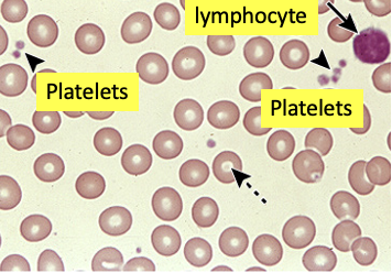
compare the two blood cells


in order for sickle cell anemia to happen, both parents need to be?
homozygous for the specific gene (HBB Gene, located on short (p) arm of Chromosome 11 at position 15.5)
(HBB Gene, located on short (p) arm of Chromosome 11 at position 15.5)
A: An abnormally high number of red blood cells (erythrocytes).
Q: How does polycythemia affect blood viscosity and blood flow?
A: Increases blood viscosity (thicker blood) → slows blood flow.
Q: What is a possible symptom of polycythemia due to increased blood viscosity?
A: Dizziness.
Q: What can cause primary polycythemia?
A: Abnormal RBC production associated with a bone marrow disorder/malignancy.
Q: What causes secondary polycythemia?
A: Low O₂ availability or increased erythropoietin (EPO) stimulates increased RBC production.
Q: Why can high altitude cause secondary polycythemia?
A: ↓ O₂ → ↑ EPO → ↑ RBC production.
Q: What is blood doping?
A: Removing and storing an athlete’s blood, then reinjecting it before an event to increase RBCs.