BPD - Structure and anatomy: Teeth

0.0(0)
Studied by 0 people
call kaiCall Kai
learnLearn
examPractice Test
spaced repetitionSpaced Repetition
heart puzzleMatch
flashcardsFlashcards
GameKnowt Play
Card Sorting

1/14

encourage image

There's no tags or description

Looks like no tags are added yet.

15 Terms

1
New cards

Tooth tissue

Hard tissue:

  1. Enamel - Hard outer covering of the anatomical crown

  1. Dentin - Forms most of the tooth underneath the enamel and cementum

  1. Cementum - Covers the anatomical root

  • Cellular vs. acellular cementum

  • Cells: cementoblasts and cementocytes

  1. Bone

Soft tissue:

  • Pulp - The living tissue inside the pulp cavity: pulp chamber (crown area) and root canal (root area)

    • Contains blood vessels, nerves, fibres, cells and stem cells

      • Blood vessels supply nutrients and oxygen, while nerves provide sensation

      • The pulp is connected to larger blood vessels and nerves outside the tooth

  • Periodontium

    • The tissue that support and hold the teeth in place

    • Consists of 4 main parts:

      • Cementum

      • Gingiva

      • Periodontal ligaments (Connective tissue, the Sparpay fibres - has different orientations to anchor the tooth to the alveolar bone)

      • Alveolar bone


2
New cards

Structure teeth

Corona dentis (crown)

  • Anatomical crown: Part of the tooth covered by enamel

  • Clinical crown: Part visible above the gingiva (gum)


Radix dentis (root)

  • Anatomical root: Part covered by cementum

  • Clinical root: Part below the gingival margin, located inside the alveolar socket


Margo cervicalis/CEJ/cemento/neck/enamel junction

  • Transition between crown and root, where the enamel meets cementum


The apex/apical

  • The tip of the tooth root. It usually has a small opening (apical foramen) where nerves and blood vessels enter the tooth.


<p><strong><u>Corona dentis</u></strong><u> (crown)</u></p><ul><li><p>Anatomical crown: Part of the tooth covered by <strong>enamel</strong></p></li><li><p>Clinical crown: Part visible above the<strong> gingiva (gum)</strong></p></li></ul><p>&nbsp;</p><p><strong><u>Radix dentis</u></strong><u> (root)</u></p><ul><li><p>Anatomical root: Part covered by <strong>cementum</strong></p></li><li><p>Clinical root: Part below the <strong>gingival margin</strong>, located inside the <strong>alveolar socket</strong></p></li></ul><p>&nbsp;</p><p><strong><u>Margo cervicalis</u></strong><u>/</u><strong><u>CEJ</u></strong><u>/cemento/neck/enamel junction</u></p><ul><li><p>Transition between crown and root, where the enamel meets cementum</p></li></ul><p></p><p><strong><u>The apex/apical</u></strong></p><ul><li><p>The tip of the tooth root. It usually has a small opening (<strong>apical foramen</strong>) where nerves and blood vessels enter the tooth.</p></li></ul><p></p>
3
New cards

Hard tissue: Enamel

  • The hardest tissue in the body

  • Covers the anatomical crown

  • 96% mineral - hydroxyapatite (Ca2+, PO43- and OH-)

  • 1% inorganic material (enamelling) and water

  • Contains enamel rods

  • Transperant

  • Produced by ameloblasts during development

  • Can’t be regenerated/rebuilt


4
New cards

Hard tissue: Dentin

  • 70% inorganic material (HAP)

  • 20% organic material (proteins and collagen)

  • 10% water

  • Dentin get produced during your whole life by the odontalblasts (placed in the outer layer of the pulp)

    • Dentinal tubules: The tubules extend from the odontalblasts through the dentin and exposes to the exposed dentinal tubeles

    • While the dentin gets bigger, the pulp gets smaller


5
New cards

Soft tissue - A part of periodontium: Gingiva

  • The part of the oral mucosa that surrounds the teeth and covers the alveolar bone. It consists of connective tissue covered by epithelium.


1. Free (marginal) gingiva

  • The gingiva surrounding the tooth without being firmly attached to its surface.

  • Forms the outer wall of the gingival sulcus.


2. Gingival sulcus

  • A shallow space between the tooth surface and free gingiva.

  • Normally approximately 1–3 mm deep when measured clinically.

  • An abnormally deepened sulcus due to periodontal tissue destruction may form a periodontal pocket.


3. Attached gingiva

  • Firmly attached to the underlying alveolar bone through connective tissue.

  • Provides stability and protection during chewing.


4. Junctional epithelium (epithelial attachment)

  • A specialised epithelium that attaches the gingiva to the tooth surface (enamel or cementum).

  • Forms a protective seal between the gingiva and tooth.


<ul><li><p>The part of the <strong>oral mucosa</strong> that surrounds the teeth and covers the alveolar bone. It consists of connective tissue covered by epithelium.</p></li></ul><p></p><p class="p2"><strong><u>1. Free (marginal) gingiva</u></strong></p><ul><li><p>The gingiva surrounding the tooth without being firmly attached to its surface.</p></li><li><p>Forms the outer wall of the <strong>gingival sulcus</strong>.</p></li></ul><p></p><p class="p2"><strong><u>2. Gingival sulcus</u></strong></p><ul><li><p>A shallow space between the <strong>tooth surface and free gingiva</strong>.</p></li><li><p>Normally approximately <strong>1–3 mm deep</strong> when measured clinically.</p></li><li><p>An abnormally deepened sulcus due to periodontal tissue destruction may form a <strong>periodontal pocket</strong>.</p></li></ul><p></p><p class="p2"><strong>3.<u> Attached gingiva</u></strong></p><ul><li><p>Firmly attached to the underlying <strong>alveolar bone</strong> through connective tissue.</p></li><li><p>Provides stability and protection during chewing.</p></li></ul><p></p><p class="p2"><strong><u>4. Junctional epithelium (epithelial attachment)</u></strong></p><ul><li><p>A specialised epithelium that <strong>attaches the gingiva to the tooth surface</strong> (enamel or cementum).</p></li><li><p>Forms a protective seal between the gingiva and tooth.</p></li></ul><p></p>
6
New cards

Surface of the teeth

Crown has 4 or 5 surfaces, and can be described in different ways.

  • Front teeth (incisors and canines): 4 surfaces

  • Distal teeth (premolars and molars): 5 surfaces


  • Oral surface - the tooth surface facing the inside of the oral cavity

    • Platinal: faces toward the palate - used for upper teeth

    • Lingual: faces toward the tongue - mainly used for lower teeth

  • Vesbibular/facial surface - the tooth surface facing outward

    • Labial - faces the lips, mainly for anterior teeth

    • Buccal - faces the cheeks, mainly for posterior teeth

  • Approximal surfaces - the surfaces facing neighoring teeth

    • Mesial - toward the midline/front of the dental arch

    • Distal - away from the midline, toward of the back of the dental arch

  • Occlusal surface - chewing surface of posterior teeth

  • Incisal edge - cutting edge of anterior teeth


7
New cards

Directions relative to a tooth: Vertical (axial)

  1. Apical (downwards)- toward the root apex

  2. Coronal/occlusal (upwards) - toward the crown/chewing surface

  3. Cervical/gingival (toward the middle) - toward the neck of the tooth/gingiva


<ol><li><p><strong><u>Apical</u></strong> (downwards)- toward the root apex</p></li><li><p><strong><u>Coronal/occlusal</u></strong> (upwards) - toward the crown/chewing surface</p></li><li><p><strong><u>Cervical/gingival</u></strong> (toward the middle) - toward the neck of the tooth/gingiva</p></li></ol><p></p>
8
New cards

Directions relative to a tooth: Horizontal

  1. Mesial ↔ distal - toward ↔ away from the dental midline

  2. Approximal - toward the neighoring teeth


  1. Vestibulo-oral - from the vestibular surface toward the oral cavity

  • Labial vestibular - facing the lips

  • Buccal vestibular - facing the cheeks


  1. Oro-vestibular - from the oral surface toward the lips/cheeks

  • Palatal - toward the upper jaw

  • Lingual - toward the tongue


<ol><li><p><strong>Mesial <span data-name="left_right_arrow" data-type="emoji">↔</span> distal </strong>- toward <span data-name="left_right_arrow" data-type="emoji">↔</span> away from the dental midline</p></li><li><p><strong>Approximal</strong> - toward the neighoring teeth</p></li></ol><p></p><ol start="3"><li><p><strong>Vestibulo-oral </strong>- from the vestibular surface toward the oral cavity</p></li></ol><ul><li><p><strong>Labial vestibular</strong> - facing the lips</p></li><li><p><strong>Buccal vestibular</strong> - facing the cheeks</p></li></ul><p></p><ol start="4"><li><p><strong>Oro-vestibular </strong>- from the oral surface toward the lips/cheeks</p></li></ol><ul><li><p><strong>Palatal</strong> - toward the upper jaw</p></li><li><p><strong>Lingual</strong> - toward the tongue</p></li></ul><p></p>
9
New cards

Permanent and deciduous teeth

  • Deciduous teeth = the first, temporary set of teeth (“baby teeth”)

  • Permanent teeth = the seond set that replaces the deciduous teeth and is intended to remain through adulthood


<ul><li><p><strong>Deciduous teeth</strong> = the first, temporary set of teeth (“baby teeth”)</p></li><li><p><strong>Permanent teeth</strong> = the seond set that replaces the deciduous teeth and is intended to remain through adulthood</p></li></ul><p></p>
10
New cards

Anatomy oral cavity

  • The oral cavity is divided into two main spaces:

    • Oral vestibule (vestibulum oris) - the space between the outside teeth/gingiva and the inside the lips and cheeks

      • Labial part - between the teeth/gingiva and lips

      • Buccal part - between the teeth/gingiva and cheeks (bucca)

  • Proper oral cavity - the space inside the dental arches (the curved arrangement of teeth in one jaw)

    • Upper part - mainly bound by the hard and soft palate

    • Floor of the mouth/lower part - mainly occupied by the tongue and structures underneath it


<ul><li><p>The oral cavity is divided into two main <strong>spaces</strong>:</p><ul><li><p><strong><u>Oral vestibule (vestibulum oris)</u></strong> - the space between the outside teeth/gingiva and the inside the lips and cheeks</p><ul><li><p><strong>Labial part </strong>- between the teeth/gingiva and <strong>lips</strong></p></li><li><p><strong>Buccal part </strong>- between the teeth/gingiva and <strong>cheeks (bucca)</strong></p></li></ul></li></ul></li><li><p><strong><u>Proper oral cavity</u></strong> - the space inside the dental arches (the curved arrangement of teeth in one jaw)</p><ul><li><p><strong>Upper part </strong>- mainly bound by the <strong>hard and soft palate </strong></p></li><li><p><strong>Floor of the mouth/lower part</strong> - mainly occupied by the <strong>tongue</strong> and structures underneath it</p></li></ul></li></ul><p></p>
11
New cards

Three main oral frenula

  • Upper labial vestibule:

    • Maxillary labial frenulum - connects the upper lip to the maxillary gingiva

    • Maxillary buccal frenulum - connects the cheeks to the maxillary gingiva (left and right)

  • Lower labial vestibule:

    • Mandibular labial frenulum - connects the lower lip to the mandibular gingiva

    • Mandibular buccal frenulum - connects the cheeks to the mandibular gingiva (left and right)

  • Lingual frenulum: connects the underside of the tongue to the floor of the mouth


The labial frenulum help attach and stabilize the lips, while the lingual frenulum helps anchor the tongue.


<ul><li><p><strong><em>Upper labial vestibule</em></strong>:</p><ul><li><p><strong><u>Maxillary labial frenulum</u></strong> - connects the <strong>upper lip</strong> to the maxillary gingiva</p></li><li><p><strong><u>Maxillary buccal frenulum</u></strong> - connects the <strong>cheeks</strong> to the maxillary gingiva (left and right)</p></li></ul></li><li><p><strong><em>Lower labial vestibule</em></strong>:</p><ul><li><p><strong><u>Mandibular labial frenulum</u></strong> - connects the<strong> lower lip</strong> to the mandibular gingiva</p></li><li><p><strong><u>Mandibular buccal frenulum</u></strong> - connects the <strong>cheeks</strong> to the mandibular gingiva (left and right)</p></li></ul></li><li><p><strong><u>Lingual frenulum</u></strong>: connects the <strong>underside of the tongue </strong>to the floor of the mouth</p></li></ul><p></p><p>The labial frenulum help attach and stabilize the lips, while the lingual frenulum helps anchor the tongue.</p><p></p>
12
New cards

Other important structures in the proper oral cavity

  • The palatle arches:

    • Arcus palatoglossus (palatoglossal arch) - anterior fold running from the soft palate toward the tongue (glossus)

    • Arcus palatopharyngeus (palatopharyngeal arch) - posterior fold running from the soft palate toward the pharynx

  • Tonsilla (palatine tonsil) - located between the palatoglossal and palatopharyngeal arches

  • Uvula - small projection hanging from the soft palate

  • Oral pharynx - throat region located posterior to the oral cavity
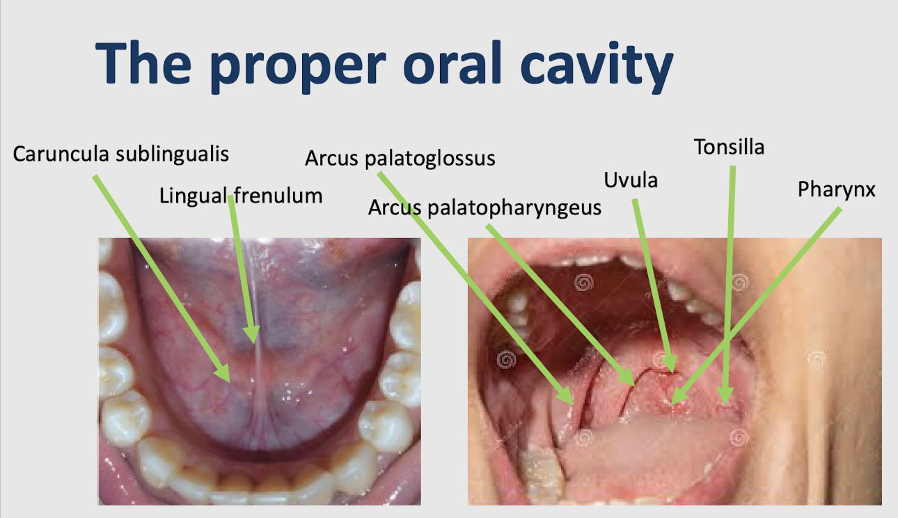

<ul><li><p><strong>The palatle arches:</strong></p><ul><li><p><strong><u>Arcus palatoglossus (palatoglossal arch)</u></strong> - anterior fold running from the <strong>soft palate</strong> toward the <strong>tongue</strong> (<strong>glossus</strong>)</p></li><li><p><strong><u>Arcus palatopharyngeus (palatopharyngeal arch)</u></strong> - posterior fold running from the <strong>soft palate</strong> toward the <strong>pharynx</strong></p></li></ul></li><li><p><strong><u>Tonsilla (palatine tonsil)</u></strong> - located <strong>between</strong> the palatoglossal and palatopharyngeal arches</p></li><li><p><strong><u>Uvula</u></strong> - small projection hanging from the <strong>soft palate</strong></p></li><li><p><strong><u>Oral pharynx</u></strong> - throat region located <strong>posterior to the oral cavity</strong></p></li></ul><p></p>
13
New cards

Four types of tonsils

  • Pharyngeal tonsil: Roof of the nasopharynx (behind the nasal cavity).

  • Tubal tonsils: Around the openings of the auditory tubes in the nasopharynx.

  • Palatine tonsils: On both sides of the oropharynx, behind the oral cavity.

  • Lingual tonsil: At the base (posterior part) of the tongue.


<ul><li><p><strong><u>Pharyngeal tonsil</u>:</strong> Roof of the nasopharynx (behind the nasal cavity).</p></li><li><p><strong><u>Tubal tonsils</u>:</strong> Around the openings of the auditory tubes in the nasopharynx.</p></li><li><p><strong><u>Palatine tonsils</u>:</strong> On both sides of the oropharynx, behind the oral cavity.</p></li><li><p><strong><u>Lingual tonsil</u>:</strong> At the base (posterior part) of the tongue.</p></li></ul><p></p>
14
New cards

Salivary glands and ducts

  • Caruncula sublingualis (sublingual caruncle) - small elevation beside the lingual frenulum where a major salivary duct (submandibular salivary duct) opens


<ul><li><p><strong><u>Caruncula sublingualis (sublingual caruncle) </u></strong>- small elevation beside the lingual frenulum where a <strong>major salivary duct (submandibular salivary duct) opens</strong></p></li></ul><p></p>
15
New cards

External anatomical structures of the mouth

  • Philtrum: Vertical groove between the nose and upper lip.

  • Nasolabial sulcus: Fold extending from the nose towards the corner of the mouth.

  • Oral commissure: Corner where the upper and lower lips meet.

  • Vermilion: Visible pink/red part of the upper and lower lips.

  • Oral orifice (rima oris): Opening between the lips.


<ul><li><p><strong><u>Philtrum</u>:</strong> Vertical groove between the nose and upper lip.</p></li></ul><ul><li><p><strong><u>Nasolabial sulcus</u>:</strong> Fold extending from the nose towards the corner of the mouth.</p></li></ul><ul><li><p><strong><u>Oral commissure</u>:</strong> Corner where the upper and lower lips meet.</p></li></ul><ul><li><p><strong><u>Vermilion</u>:</strong> Visible pink/red part of the upper and lower lips.</p></li><li><p><strong><u>Oral orifice (rima oris)</u>:</strong> Opening between the lips.</p></li></ul><p></p>



