Methods of Data Acquisition Part 2 (Helical)
 Call Kai
 Learn
 Practice Test
 Spaced Repetition
 Match
 Flashcards
 Knowt Play
1/47

There's no tags or description
Looks like no tags are added yet.
48 Terms
What are the AKA’s for helical scanning?
Spiral
Volumetric
Continuous Acquisition Scanning
What was eliminated with helical scanning compared to axial scanning?
Eliminated the interscan delay
How does helical scanning affect the scan time compared to axial scanning?
Increased scanning speed
What are the 3 defining characteristics of a helical scan
Continuously rotating x-ray tube
Constant x-ray output
Continuous table movement (uninterrupted)
What happens to image resolution when you increase the scan speed
Improved Image Resolution
What 3 things are improved when increasing the scan speed (that improves resolution)
Images with improved iodinated contrast concentration
Decreased respiratory and cardiac motion artifact
Superior multiplanar and 3D reformation capabilities
Why are using helical scanning multi row detector systems good when scanning the heart
Because a 320 row detector scanner can cover 160mm in one gantry rotation and this is enough to image the entire heart
Slip Ring Improvement
Gantries with a slip ring design eliminated the need for electrical cables
Tube Cooling Improvement
More efficient tube cooling
Helps dissipate heat
Helical scanning needs a constant x-ray output which creates a lot of heat
CT rooms should be kept cool to help dissipate heat
Higher x-ray output Improvement
Higher x-ray output (increased mA capabilities)
If you lower your time, you increase your mA to keep the optimal mAs
High frequency generators are now used instead of 3-phase to accommodate increased mA
Smoother Table Movement Improvement
Smoother table movement (not jerky)
Otherwise we would see motion artifact
Software that adjusts for table motion Improvement
Software that reduces artifact of the predictable table movement (we know the table speed)
Unfortunately, this software does not work for unpredictable motion (breathing, peristalsis, etc)
Improved raw data management Improvement
The scanner is acquiring a large data set which must be stored until the technologist has time to extract and create all image sets needed by the radiologist. The amount of time will depend on workload of the technologist and the radiologists
If the radiologist doesn’t read the exam for 3 days and then decided that he wants the tech to create a thinner slice, the raw data would still need to be stored in order to make that reconstruction
More efficient detectors Improvement
Manufacturers have also started using more efficient detectors because you can use less mAs when you increase the detector efficiency
What is interpolation
Complex statistical methods to take the slant and blur out of the helical image and create images that closely resemble those acquired in a traditional axial mode
What are the 2 disadvantages of interpolation
Loss of image resolution (minimal)
Negative impact on effective slice thickness
What needs to happen to interpolation as the slice becomes steeper
As the angle of the slice becomes steeper, the interpolation required to adjust the data increases
What factor controls the slice angle getting steeper
Increasing pitch increases the slice angle (steeper)
Effective Slice Thickness Definition
Thickness of the slice that is actually represented on the CT image, as opposed to the size selected by the collimator opening
Effective Slice Thickness AKA
Slice-sensitivity profile
How does helical scanning affect the effective slice thickness
In helical scanning, because of the interpolation process, the effective slice thickness may be wider than the selected slice thickness
What is blooming and degradation
When the effective slice thickness is wider than the selected slice thickness
What is pitch
Relation of table speed to slice thickness
The travel distance of the CT scan table per 360 degree rotation of the x-ray tube, divided by the x-ray beam collimation width
How is pitch calculated
Travel distance of table per 360 rotation/X-ray beam collimation width
What will pitch be when the table feed and beam collimation are equal/identical
1
What will the pitch be when the table feed is less than the beam collimation
Less than 1
When the table feed is less than the beam collimation, what will happen to the slices
Overlapping slices
When using helical scanning, what is table speed directly related to
Table speed and slice thickness are directly related
What happens when you increase pitch (when beam collimation/slice thickness remains the same)
Increase anatomic coverage
Decreased radiation dose
Improved tube heat limit (better)
What happens to the slant of the acquired data when you increase pitch
Increases
What happens to interpolation when you increase pitch
Increases
What happens to the negative effects of interpolation when you increase pitch
Increases
What are the 3 disadvantages of interpolation
Image Unsharpness
Effective slice thickness (blooming)
Image noise: fewer data being acquired (under-sampling)

What pitch would this represent
1
Requires less interpolation
Table moves the same distance as beam width

What pitch would this represent
1.5
Table moves 1.5 times the beam width

What pitch would this represent
2
Requires more interpolation
Table moves twice the beam width
What amount/setting should pitch not exceed so that the negative effects are minimized
1.5
What does it mean to change slice incrementation retrospectively
Changing where the slice lies over the anatomy (slice location)
When changing slice incrementation retrospectively, what data do you need
Raw data
What does changing slice incrementation retrospectively allow for the creation of
Allows for the creation of overlapping slices retrospectively
When changing slice incrementation retrospectively how is the patients radiation dose affected
When acquiring overlapping images in axial scanning, the patient is getting a higher dose because they are getting extra radiation where the overlap is
When changing slice incrementation retrospectively how is partial volume averaging is affected
May help reduce partial volume averaging
How does reconstructing slice thickness work in CT
MDCT Helical scanning allows for you to build a new set of images at a different slice thickness using retrospective reconstructions, without having to rescan the patient
What data do you need to reconstruct slice thickness in CT
Raw data
What will using thinner slices do
When reconstructing slice thickness into thinner slices it will reduce the amount of tissue in 1 voxel resulting in a Hounsfield unit with a more accurate representation on the image
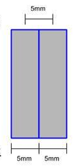
What type of slice interval is this
Contiguous 5/5 mm

What type of slice interval is this
Overlapped 5/2.5 mm

What type of slice interval is this
Non-Contiguous 5/7.5 mm