clinical 2 test 2
 Call Kai
 Learn
 Practice Test
 Spaced Repetition
 Match
 Flashcards
 Knowt Play
1/307

There's no tags or description
Looks like no tags are added yet.
308 Terms
what is an EGC?
An ECG or electrocardiogram is a device used to record the electrical activity of the heart from electrodes placed on the skin surface to monitor and diagnose.
the device records the changing electrical activity of the heart between positive an negative electrodes.
Echocardiogram
a moving ultrasound of the heart that tells us about the contractile nature of the heart
what is electrical activity in the heart caused by
it is caused by the influx/efflux or electrolytes such as sodium, potassium, and calcium ions through ion channels in heart cells.
Unoxygenated blood enters in the BLANK side of the heart and then goes out to the lungs
right side
oxygenated blood leaves the lungs and enters the BLANK side of the heart and goes to the body
left side
Veins carry blood BLANK the heart
Toward
Arteries carry blood BLANK from the heart
away
what do patients who are dealing with Heart issues look like upon PE
They have pale mucus membranes
weak
HR too fast to count easily
weak pulses
pulse deficits
sometimes they do not have heart murmurs
Diuretics
prevent sodium reabsorption ( water follows)
examples furosemide and spironolactone
Ace inhibitors
inhibit the conversion of angiotensin I to angiotensin II, helping to lower blood pressure and reduce heart workload.
positive inotropes
causes an increase in contractility of the heart making calcium more available to cardiac myocytes
examples: Digoxin and Pimobendane
Vasodilators/arterial dilators
medications that relax blood vessels, decreasing cardiac preload or afterload, thereby reducing the heart's workload. which then decreases the blood pressure
example nitroglycerine
what are some common drugs used for arrythmias
Beta-blockers ( lowers the HR and blood pressure, usually ends in lol)
sodium channel blockers ( lidocaine)
calcium channel blockers ( amlodipine, diliatlaizam)
what to do if you don’t have an ECG
use your stethoscope and palpate for pulse deficits
if you have a pulse deficit or if they are out of synchrony, then we have an abnormal electrical activity of the heart.
what is depolarization of the heart
Depolarization of the heart refers to the process by which cardiac muscle cells change their membrane potential, leading to contraction. This electrical activity initiates the heartbeat and coordinates heart rhythms.
it is the contract trigger
where should a patient be placed when performing a ECG?
The patient should be placed on a rubber mat and not directly onto the stainless steel table as this can reduce electrical activity and interfere with the ECG results.
how should an animal be positioned for an ECG?
The animal should be in right lateral position with the limbs perpendicular to the body
what should the paper speed be set at for ECG?
should be set at 50mm/sec can also be set at 25mm/sec
what lead should the ECG viewing device be set to?
it should be set to lead II
why should you not use alcohol on leads during emergency situations?
A possible fire can be caused by the defibrillator
what is a holter monitor
A Holter monitor is a portable device that continuously records the heart's electrical activity for 24 to 48 hours, allowing for long-term assessment of heart rhythm and detection of irregularities.
artifact
a wave that arises from sources other than the heart of brain
Cardiac arrhythmia
Any electrical activity of the heart that differs from that of a healthy awake patient. can be innocuous or life threatening
Deflection
movement from one side to the other
Depolarization
contractions as a result of cells stimulated by electricity
Ectopic
occurring in an abnormal position
VCP is an Ectopic
Lead
A pair of electrodes, connected by an axis that provides a particular view of the electrical activity of the heart.
Repolarization
relaxation of cells after depolarization
The p wave
the first waveform pulse of the ECG
this should be the first waveform you’re looking for on an ECG strip
Formed when the atria contract to pump blood into the ventricle
What does the P wave represent?
It represents atrial depolarization
any changes in the P wave could indicate atrial dysfunction
this wave is stimulated by the SA node
there should be a P wave for every QRS form
What are some abnormalities seen in the p wave?
Wide P waves and absent P waves
What is the P-R interval
Reflects the activation of the Av ( atrioventricular ) node
measured from the beginning of the p wave to the beginning of the QRS complex
time between atrial depolarization and ventricular depolarization, indicating conduction time through the AV node.
The QRS complex
Forms when the ventricles contract and reflects ventricular depolarization. It is measured from the beginning to the end of the QRS wave and is crucial for assessing heart rhythm and rate.
Q= 1st negative deflection
R= 1st positive deflection
S wave = negative deflection that follows the R wave
What does the QRS wave form indicate
it indicates ventricular disease
lack of a QRS wave could indicate a form of heart block
a wide QRS wave might indicate premature ventricular contractions
What can a tall and skinny QRS wave indicate
indicates problem above the ventricle i.e. problem in the atria ( supraventricular)
What does a fat/ wide QRS wave indicate
problem within the ventricle
The ST segment
Represents the time interval from the end of the QRS interval to the onset of the T-wave
contractions of the ventricles
this is early stage of ventricular repolarization
The T wave
Represents ventricular repolarization and occurs after the QRS complex.
Myocardial hypoxia, and electrolyte disturbances can be evident with changes in the T-wave
should not be more than ½ of the r wave
the t-wave can be positive or negative
Sometimes the T wave can deflect downwards depending on the patient ( this can be normal for them)
when can you see abnormal T waves
abnormal T-waves can be seen in relation to electrolyte disturbances
The QT interval
includes the ventricular depolarization and repolarization
QT interval is measure from the onset of the Q wave to the end of the T wave
Artifacts
something that is not natural ( man made) that may interfere with the interpretation of clinical data, often resulting from external influences or equipment malfunction.
this can be electrical interference by outside sources, electrical noise from elsewhere in the body, poor contact, and machine malfunction.
they are extremely common
What can be some artifacts seen on an ECG
Patient movement, the blood pressure machine, clippers, pressing on the stomach
Interference
Can be caused by other machinery such as a pulse-ox or BP monitor that is hook to the animal
What to do if the interference is coming from the patient ( panting, trembling/shaking)
Try moving the clips distally
try to calm the patient

what is this ECG reading?
Interference

What ECG abnormality?
electrical interference

what is this ECG abnormality?
Muscle tremors or purring

What is this ECG abnormality?
Patient movement

what is this ECG abnormality?
Loose electrodes
What are the steps to reading an ECG strip?
First determine the HR (is it too fast or too slow)
Look at the rhythm ( is it regular: every P-QRS- looks about the same, is it irregular)
Find the P wave ( there should be a P wave for every QRS, are they regular for each interval)
Next, measure the intervals (PR, QRS, QT) to ensure they are within normal limits. Finally, assess the ST segment and T wave for any abnormal changes that may indicate ischemia or other cardiac conditions.
Wandering Baseline
Resistance between the patient and the electrode can cause a wandering baseline
An example of this be would be respiratory movement, patient movement, clips not being clipped as good enough
placing the patient in sternal recumbency or standing position will help to reduce this resistance
holding the patient’s mouth closed for a few seconds could also help reduce resistance

What is this ECG reading?
wandering baseline
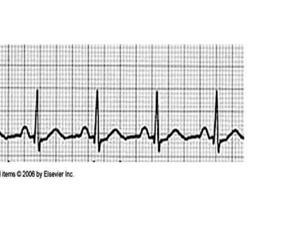
What is this ECG reading?
Normal sinus rhythm
normal ECG, depicting a normal rhythm of electrical conductivity through the heart
Respiratory sinus arrhythmia
non pathologic arrhythmia in dogs
HR increases during inspiration and decreases during expiration
THIS IS ABNORMAL IN CATS
normal in dogs can be seen more in respiratory illnesses

What is this ECG reading?
Respiratory sinus Arrhythmia
Sinus Bradycardia ( Bad for BRADY-cardia to be slow)
A normal sinus rhythm, but depolarization is occurring more slowly than usual
Originated from the SA node
Characterized by a rhythm less than 60 beats/min in dogs and 100 beats/ min in cats
This is based on an individual patient basis, and may be normal for some such as athletic dogs
Think it is bad for Tom Brady to be slow

What Is this ECG reading?
Sinus Bradycardia
Sinus tachycardia ( being fast is Tacky)
A normal sinus rhythm, but depolarization is occurring more quickly
Characterized by a rhythm of over 180 beats/min or 160 BPM for large dogs
Greater than 220-240 beats/min in cats
think being fast is tachy-cardia
What are some causes of Sinus tachycardia
painful, fever, hyperthyroidisms

What Is this ECG reading?
Sinus tachycardia
Ventricular Premature contraction ( VPC or PVCs)
is an abnormal heartbeat initiated in the ventricles, resulting in premature contractions before the left ventricle is adequately filled. This ectopic impulse often leads to insufficient tissue perfusion and is a common ECG abnormality, particularly in critically ill patients, necessitating treatment of the underlying disorder.
What is a ventricular premature contraction associated with?
can be associated with various conditions including weakness, exercise intolerance, electrolyte imbalances, syncope, and even sudden death when frequent. They may arise from hypoxic states like anemia or GDV, as well as trauma or damage to the heart, and are also observed in patients experiencing pain.
Hypoxic: a medical condition where your body tissues or organs do not receive enough oxygen to function properly.
Characteristics of VPCS
Seen on the ECG as wide, bizarre QRS complexes
Wil have a large amplitude of QRS complexes
Random P waves not associated with QRS complex ( or not seen)
When to treat VPCs
Intermittent VPCs will not be treated
they will be treated if a patient is clinical i.e. collapse, dyspnea and syncope
dyspnea: shortness of breath
Syncope: fainting

What is this ECG abnormality?
Ventricular premature contraction
Ventricular Tachycardia
occurs when three or more ventricular premature contractions (VPCs) occur consecutively, resulting in a rapid heart rate exceeding 180 beats per minute. This condition leads to inadequate ventricular filling, causing inefficient heart function and disconnection between the atria and ventricles.
essentially the ventricles are firing repeatedly without adequate filling of the ventricles
the heart does not have the time to relax and is not pumping correctly
there is no cohesion between the atria and the ventricles
When does ventricular tachycardia need to be treated by?
needs to be treated if they last longer than 30 seconds ( sooner would be better)
this is life threatening if it lasts longer than 30 seconds
Characterizes of Ventricular tachycardia
A series of three or more consecutive ventricular premature contractions that lead to a rapid heart rate and impaired cardiac function.
looks like Ghosts holding hands

What is this abnormality on the ECG strip?
Ventricular tachycardia
What is ventricular tachycardia treated with?
Usually treated with defibrillation and or lidocaine
Hyperkalemia
Causes really tall P waves on the ECG strip and is caused by excess potassium in the blood.
has peaked T waves
seen commonly in blocked cats
if not treated it is life threatening
what causes Hyperkalemia
Hyperkalemia is caused by an excess of potassium in the blood, which can result from conditions such as kidney dysfunction, tissue breakdown, or certain medications. It leads to characteristic changes on an ECG, including tall P waves and peaked T waves.
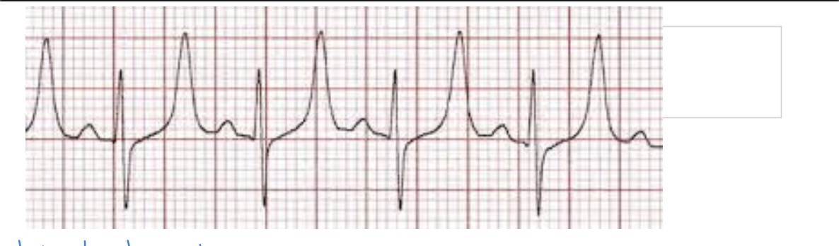
what is this ECG abnormality?
hyperkalemia
Ventricular fibrillation ( v-fib)
Chaotic depolarization of the ventricles that results in disorganized contractions and ineffective blood circulation. It is a life-threatening arrhythmia that requires immediate medical intervention.
both the atria and ventricles are firing with no coordinated rhythm.
has low Cardiac output and no adequate perfusion of blood
is a results of not treating ventricular tachycardia
the patient will die unless electrical defibrillation is preformed
What are the characteristics of a v-fib wave form
Ventricular fibrillation waveforms are irregular and chaotic, characterized by varying amplitudes and no discernible repeating pattern. There are no identifiable QRS complexes or P waves, indicating disorganized electrical activity.
looks like a bag of worms

what is the abnormality seen on this ECG strip?
ventricular Fibrillation
Atrial tachycardia ( supraventricular tachycardia)
The SA node is firing abnormally fast causing the atria to contract rapidly, often leading to an increased heart rate of over 220bpm, usually over 250.
The patient will come in collapsed with pale MM
super skinny and tall QRS waves will be seen
unable to tell the difference between a p wave and t waves
What can cause Atrial tachycardia
can be caused by congenital heart defects, structural abnormalities

What is this ECG abnormality?
Atrial tachycardia
Atrial Fibrillation ( A-fib)
A fast rhythm for the atria ( 350 -600 beats/min) and ventricles ( 220-240 beats/min)
It is a constant irregular rhythm
Commonly caused by atrial enlargement
Not as life threatening as v-fib
Fibrillation is the rapid, irregular and unsynchronized contraction of muscle fibers
Characteristics of Atrial Fibrillation
Lacks a p-wave ( looks like little bumps at the baseline)
Has an undulating baseline
Tall and skinny complex
Has a saw toothed look to the wave length

What is this ECG abnormality
Atrial Fibrillation
Atrial premature contraction
Is a premature impulse from the SA or has atrial origin
The signal is coming from the AV node too soon
Has a premature p wave
Seen in dogs with atrial enlargement
The reading is spacey short and has a tall skinny QRS complex

What is this ECG abnormality?
atrial premature contraction
Heart block
An electrical impulse that is not transmitted throughout the heart
There are 3 degrees of a heart block
The first degree of a heart block
A prolongation of the P-R interval, meaning there is a delay in conduction through out the heart
There is a P wave for every QRS complex
The least problematic
The rhythm is usually normal
The QRS interval is normal
Can be seen in young athletic breeds as well as brachycephalic breeds
Seen in older dogs
What drugs can cause a 1st degree AV block
Atropine, beta blockers and digoxin

What is this ECG abnormality
1st degree of AV block
Second degree of heart block
P waves occurring without QRS complexes ( dropped)
The signal is not getting thought to the ventricular to cause the ventricle to contract
Can be seen in anesthesia patients with bradycardia
A more serious heart block
Will usually have clinical signs
Syncope, exercise intolerance, coughing, dyspnea, heart failure signs
Has two types: Mobtiz type 1 and 2
Mobtiz type one
The PR interval progressively lengthens with each beat until the atrial impulses is not conducted and the QRS complex is dropped
The av node conduction resumes with the next beat and the sequence is repeated
Usually does not require treatment, however if it cases bradycardia it will be treated
If bradycardia occurs and patient is symptomatic, then a pacemaker placement may be considered
Mobtiz type 2
The PR interval remains constant. Beats are intermittently non-conducted and the QRS complex’s are dropped, usually in a repeating cycle of every 3rd or 4th P wave
P waves seen without QRs complexes
Always pathological
Patients may be asymptomatic. Or experience syncope, depending on the ratio of conducted to block beats
Patients are at risk of developing symptomatic high grade or complete and will be problematic to maintain systemic perfusion; there for a pacemaker is indicated
3rd degree AV Block
The heart is blocked completely making it the most severe heart block
The p wave is normal, but the QRS is wide and Bizarre
There is no electrical communication between the atria and ventricles and no relationship between p waves and QRS complexes
The atria and ventricles are not working together
Most patients require a pacemaker
If the block is caused by anti arrhythmic drugs, stopping the drug may be effective
Seen with aortic stenosis, HCM and digitalis toxicity

What is the abnormality seen on this ECG?
3rd degree AV block

What abnormality is seen on this ECG?
2nd degree AV block type 1

What abnormality is seen on this ECG?
2nd degree Av block type 2
Asystole ( flat line)
Cardiac arrest
No cardiac activity
CPR has already been initiated
Epinephrine and atropine have probably already been tried
Defibrillation probably won’t work
The prognosis is very poor

What is this ECG abnormality?
Asystole
Coagulation
The formation of a clot
Platelets
aka thrombocytes
Small, anucleate, discoid-shaped. Light blue-staining cells that may have multiple, fine, pink-to-purple granules in the cytoplasm. They play a crucial role in blood clotting and wound healing.
1/3 of the total blood platelets are located where
in the spleen