MSK Special Test NPTE
 Call Kai
 Learn
 Practice Test
 Spaced Repetition
 Match
 Flashcards
 Knowt Play
1/74

There's no tags or description
Looks like no tags are added yet.
75 Terms

Apprehension Test for anterior shoulder displacement
To test Anterior shoulder dislocation. Test is positive if the patient presents apprehensive

Apprehension Test for posterior shoulder displacement
To test posterior shoulder dislocation. Test is positive if the patient presents apprehensive

Sulcus Sign
Positive for inferior stability if a depression is seen between the acromion and humeral head

Ludington's test
Positive test indicated by absence of movement in the biceps tendon and may be indicative of a rupture
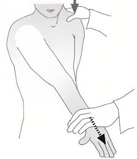
Speed’s Test
1 A positive test is indicated by pain over the bicep tendon in the bicipital groove and may be indicative for bicipital tendonitis

Yergason test shoulder
2 A positive test is indicated by pain over the bicep tendon in the bicipital groove and may be indicative for bicipital tendonitis

Drop Arm Test
To test rotator cuff tear. A positive test is indicated by the patient failing to slowly lower the arm to their side or in the presence of severe pain.
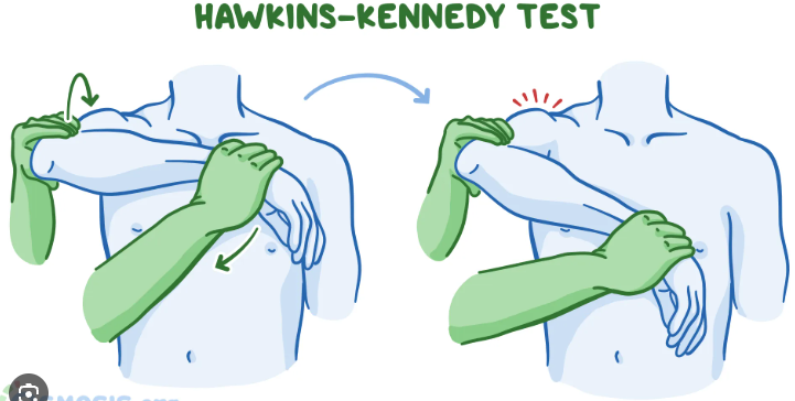
Hawkins Kennedy impingement test
A positive test is indicated by pain and may be indicative of shoulder impingement involving the supraspinatus tendon

Infraspinatus Test
Pain or weakness indicates the presence of infraspinatus strain or tear

Lateral Rotation Lag test
Test positive for infraspinatus and/or supraspinatus pathology if the patient can not hold the testing position
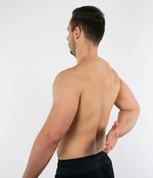
Lift off test (medial rotation lag sign)
if patient is not able to lift their hand off their back and/or hold the position is indictive of Subscapularis lesion may be present

Neer Impingement Test
A positive test is indicated by facial grimace or pain and may be due to shoulder impingement secondary to supraspinatus tendon
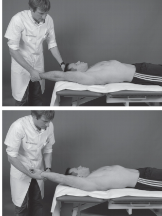
Supine Impingement Test
Positive if the patient experiences increase pain with medial rotation

Supraspinatus Test (empty can)
A positive test is indicated by weakness or pain meaning either tear of the supraspinatus tendon or suprascapular nerve involvement

acromioclavicular crossover test
Positive if feels pain over the AC joint

active compression test shoulder (O’Brien’s test)
Positive for a superior labrum tear if the patient has increase pain with medial rotation and relieved pain with lateral rotation
glenoid labrum tear test

Jerk Test
used to detect posteroinferior instability and posterior labral tears

Valgus Stress Test (Ulnar)
Evaluates the ulnar collateral ligament (UCL), also known as the medial collateral ligament (MCL)
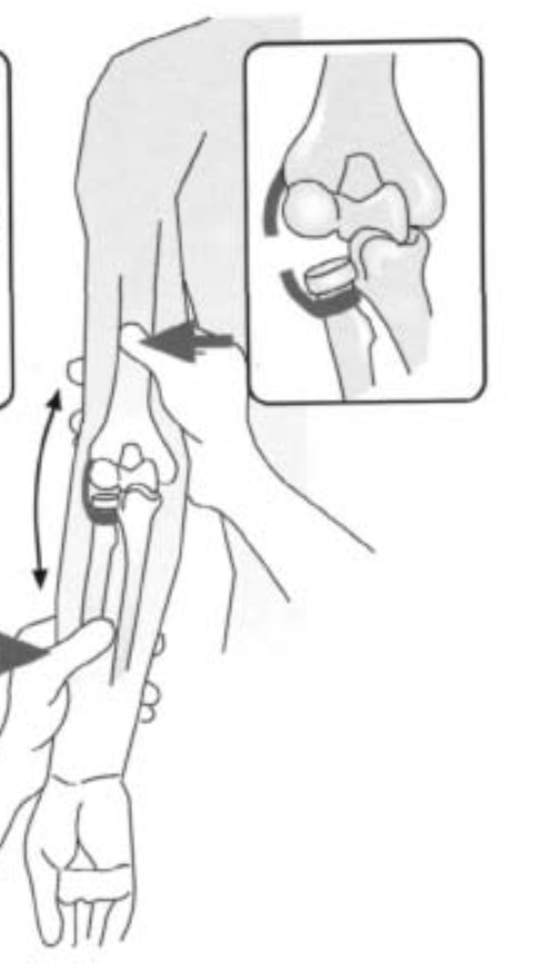
Varus Stress Test (radial)
Evaluates the radial collateral ligament (RCL) or lateral collateral ligament (LCL) complex.

Cozen’s test
a physical examination maneuver used to evaluate for lateral epicondylalgia, commonly known as tennis elbow, specifically the extensor carpi radialis brevis
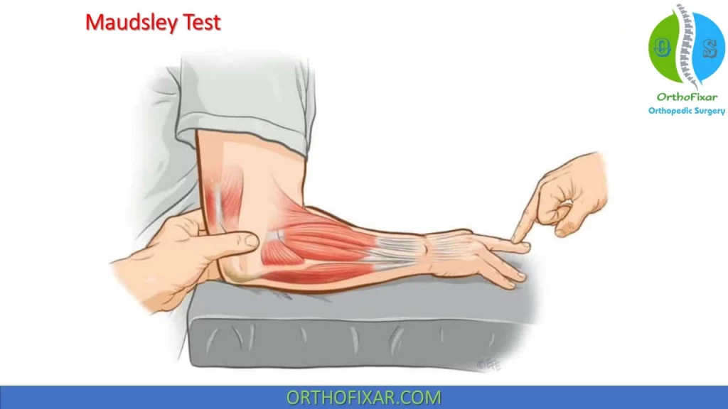
Lateral Epicondylitis (maudsley’s test)
The examiner applies downward pressure against the patient's middle finger (third digit) while the patient attempts to extend it.
Positive Test: Pain is reproduced at the outside of the elbow (lateral epicondyle) or along the common extensor tendon.

Medial Epicondylitis
is the Golfer's Elbow Test, which stretches or resists the flexor-pronator muscle group to reproduce pain at the inner elbow
Supinate the patient’s forearm, extend the elbow and the wrist
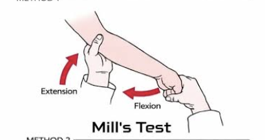
Mill’s Test
Primary Target: Identifies pain and tension associated with the common extensor tendon origin at the lateral humeral epicondyle
Passively pronate the forearm. the flex and extend the patients elbow fully

Bunnell-Littler Test
used to find the cause of stiffness or restricted flexion at the proximal interphalangeal (PIP) joint
Tight retinacular ligament test
Evaluates whether restricted movement in the finger's DIP joint is caused by tight soft tissue (the oblique retinacular ligament) or a stiff/contracted joint capsule

Ely’s Test
Test the spasticity or tightness of the rectus femoris

Ober’s test
used to check for tightness or contracture in the structures of the lateral hip and thigh—specifically the tensor fasciae latae (TFL) muscle, the iliotibial band (ITB), and related proximal hip structures like the gluteus medius/minimus and hip joint

Piriformis Test (FADIR)
a clinical assessment used to check for piriformis syndrome or deep gluteal syndrome. These conditions happen when the piriformis muscle irritates or traps the sciatic nerve
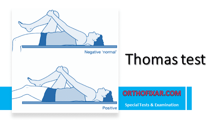
Thomas Test
measures hip flexor length and distinguishes tightness between one joint and two joint muscles.

Tripod sign
a quick orthopedic and physical therapy test used to evaluate hamstring muscle length (tightness or contracture) and screen for neural (sciatic nerve) irritation

90-90 straight leg test
Evaluates hamstring muscle length and flexibility
Positive Result (Tight Hamstrings): A deficit of greater than 20 to 30 degrees (meaning the knee cannot straighten past 20–30 degrees short of full extension).
Barlow’s Test
Ortolani’s Test
Anterior Labral tear test (FADER)

Craig’s Test
a clinical physical assessment used to estimate femoral anteversion (the forward twist or rotation of the femoral neck relative to the shaft)
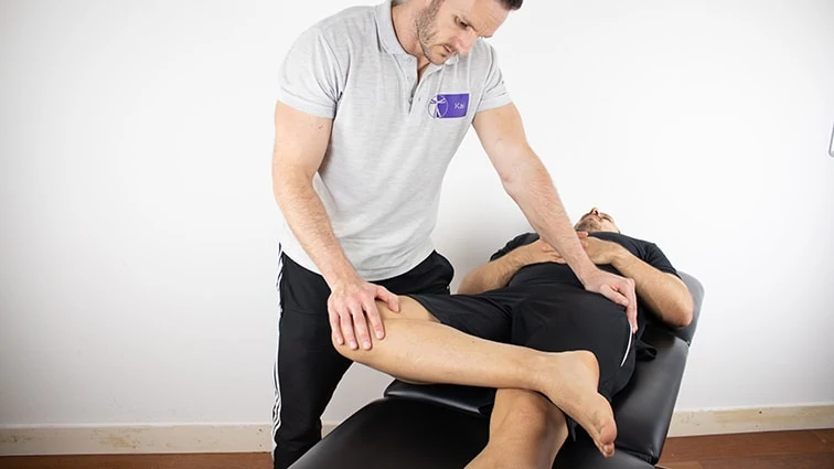
Patrick’s test (FABER test)
a physical examination tool used to screen for pathologies in the hip joint, sacroiliac (SI) joint, and lumbar spine

Quadrant scouring test
Used to screen for intraarticular hip pathologies
The test is positive if it reproduces the patient's familiar groin or anterior hip pain, catching, clicking, or grinding (crepitus)

Trendelenburg Test
an orthopedic assessment for hip abductor weakness

Anterior Drawer Test Knee
a physical examination maneuver used to evaluate the integrity of the anterior cruciate ligament (ACL) in the knee
High Hamstring involvement

Lachmen test
A clinical exam used to check for an injury to the anterior cruciate ligament (ACL) in the knee.
Low Knee Involvement
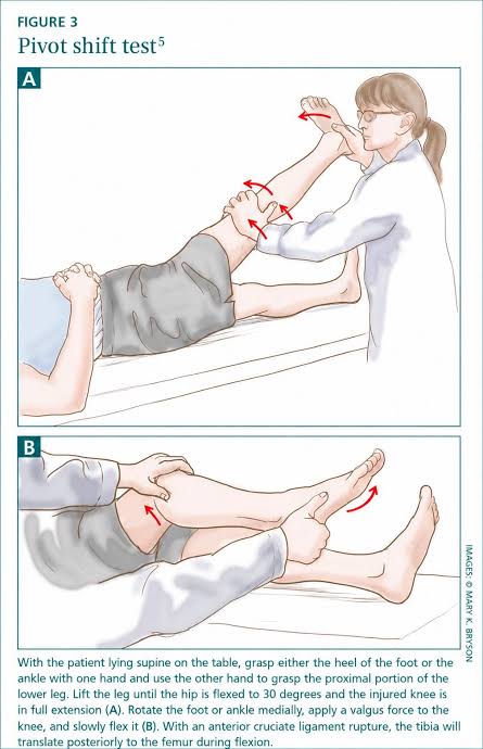
Lateral pivot shift test
a physical examination maneuver used to detect an Anterior Cruciate Ligament (ACL) tear and anterolateral rotatory instability of the knee
High specificity

Posterior Drawer Test
clinical physical examination used to evaluate the integrity of the posterior cruciate ligament (PCL) in the knee and detect posterior knee instability
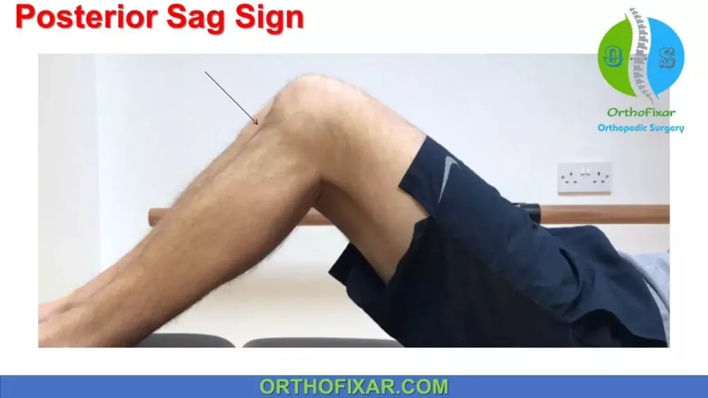
Posterior Sag sign (Godfrey’s Test)
PCL test
Positive sign: Gravity pulls the unsupported tibia backward (posteriorly) relative to the femur because the PCL is torn and no longer holds it in place, creating a visible "sag" or drop-off below the kneecap

Slocum test
a modified orthopedic examination used to evaluate rotatory instability of the knee, specifically anteromedial rotary instability (AMRI) and anterolateral rotary instability (ALRI)

Valgus Stress Test: Knee
a clinical physical examination used to evaluate the integrity of the medial collateral ligament (MCL) and medial knee structures

Varus Stress test: knee Test

Apley’s compression test
A physical examination maneuver used to help evaluate the knee for a suspected meniscus tear

Bounce home test
Abnormal (Positive) Test:
Incomplete Extension: The knee fails to straighten all the way.
Rubbery / Springy End Feel: The leg bounces slightly instead of locking firmly, which often points to a bucket-handle meniscus tear blocking the joint.
Hard End Feel: A rigid block can mean loose fragments (loose bodies) inside the joint.
Pain: Sharp pain around the joint line during extension is common
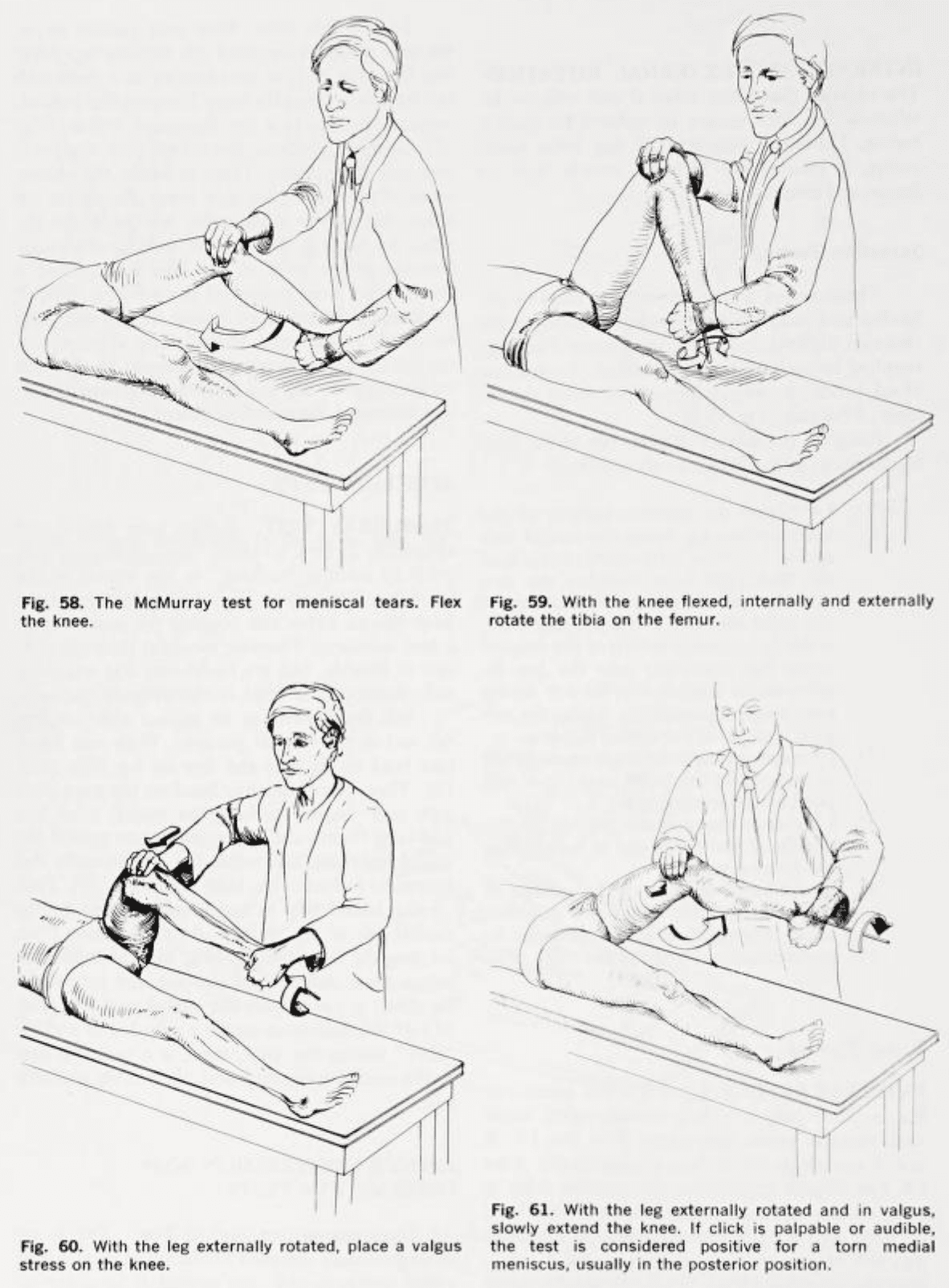
McMurray test
is a clinical orthopedic examination used to detect tears in the medial or lateral meniscus (the shock-absorbing cartilage of the knee)

Thessaly Test
Positive Test: Indicated by pain along the medial or lateral joint line, or a catching, locking, or popping sensation. This points toward a potential meniscus tear.

Brush Test
A visible fluid wave, bulge, or tissue change reappearing on the medial side indicates excess intra-articular fluid. This is graded from a trace amount up to a large 2+ or 3+ bulge.

Patella tap test (Ballottement)
The patella feels like it is "floating," pushes down through fluid, and rebounds or makes a palpable "tap" or "clunk" against the underlying bone. This indicates a moderate-to-large intra-articular effusion

Clarke’s sign (Patellar grind test)
an orthopedic physical examination maneuver used to evaluate anterior knee pain and conditions like patellofemoral pain syndrome (PFPS) or chondromalacia patellae

Hughston’s plica test
a physical exam maneuver used to detect medial knee plica syndrome, which occurs when a normal embryonic tissue fold (plica) in the knee becomes irritated, thickates, or gets pinched
Noble compression test
Patellar apprehension test

Anterior Drawer test (ankle)
If the foot slides forward excessively compared to the uninjured ankle, or if there is no firm "stop" (a soft end feel), it indicates damage or a tear to the ATFL.
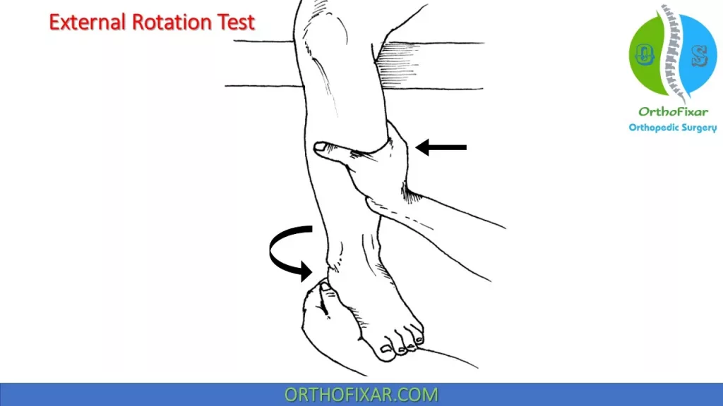
Lateral rotation stress test (kleiger test)
Syndesmosis injury (high ankle sprain), involving the distal tibiofibular ligaments and interosseous membrane.
Deltoid ligament disruption on the medial side of the ankle.

Talar tilt test
Lateral Ligament Complex (Inversion Stress): Primarily tests the calcaneofibular ligament (CFL) in a neutral position, the anterior talofibular ligament (ATFL) in plantarflexion, and the posterior talofibular ligament (PTFL) in dorsiflexion.
Medial Deltoid Complex (Eversion Stress): Stresses the medial deltoid ligaments when the foot and ankle are rolled outward

Thompson Test
Negative Test (Intact Tendon): Squeezing the calf causes the foot to naturally plantar flex because the force transmits through a healthy Achilles tendon.
Positive Test (Ruptured Tendon): Squeezing the calf results in no movement (or a lack of expected plantar flexion) at the ankle, indicating a complete tear.
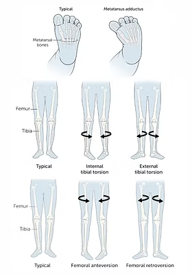
Tibial torsion test
evaluates the amount of twisting (rotation) in the shinbone (tibia) relative to the knee and ankle. It helps diagnose gait issues like intoeing ("pigeon-toed") or outtoeing in children and structural leg alignment problems in
True leg length discrepancy

Cervical Flexion rotation test
a clinical evaluation used to check for movement impairment and joint dysfunction in the upper cervical spine (specifically the C1-C2 or atlantoaxial joint), which is frequently linked to cervicogenic headaches

Distraction Test
The cervical distraction test is a physical exam maneuver used to help diagnose cervical radiculopathy (nerve root compression in the neck)
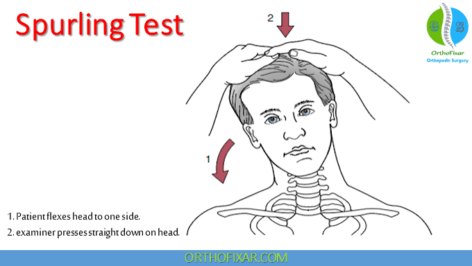
Foraminal compression test (Spurling’s)
The test is positive if the maneuver reproduces or intensifies radiating symptoms—such as pain, tingling, numbness, or weakness—traveling from the neck down into the shoulder, arm, or hand (radicular symptoms)

Vertebral Artery Test
a physical screening examination used to check the adequacy of blood flow through the vertebral and basilar arteries before performing cervical spine manipulation or mobilization
A positive test is indicated by the reproduction of neurological or vascular symptoms, commonly remembered by the 5 D’s and 3 N’s: [1, 2]
5 D’s: Dizziness/vertigo, Diplopia (double vision), Dysarthria (slurred speech), Dysphagia (difficulty swallowing), and Drop attacks (sudden loss of strength without fainting).
3 N’s: Nystagmus (involuntary eye twitching/flicker), Nausea/vomiting, and Numbness or sensory changes.
Other signs include unsteadiness (ataxia) or one arm drifting/dropping.

Gaenslen’s Test
A physical examination maneuver used to detect sacroiliac joint (SIJ) dysfunction, inflammation, or lower back and pelvic pathology by applying torsional stress to the pelvis.

Sacral Thrust test
a pain provocation assessment used to evaluate the sacroiliac (SI) joint for dysfunction or posterior pelvic pain

Sacroiliac compression test
Reproduces the patient's familiar posterior pelvic, buttock, or SI joint pain. Simply feeling localized pressure or general discomfort without familiar symptom replication does not count as a positive result

Sacroiliac distraction test
The Sacroiliac (SI) Distraction Test is a clinical pain-provocation maneuver used to evaluate the sacroiliac joint and stress the anterior sacroiliac ligaments
Positive Test: Reproduces the patient's familiar buttock, posterior pelvis, or low back pain

Slump Test
a sensitive neurodynamic physical examination used to assess abnormal neural tension, mechanosensitivity, or nerve root compression (such as lumbar disc herniation or sciatic nerve impingement)

Straight leg raise test
a physical examination maneuver used to check for lumbosacral nerve root irritation, sciatic nerve compression, or lumbar radiculopathy (sciatica)
Positive Sign: Pain, numbness, or tingling that shoots or radiates down the back of the leg, calf, or foot (below the knee).
Thigh thrust test
(also known as the posterior pelvic pain provocation or P4 test) is a physical examination maneuver used to evaluate the sacroiliac (SI) joint and determine if it is the source of a patient's buttock or lower back pain
Positive Test: Reproduces the patient's familiar unilateral posterior pelvic or buttock pain. General discomfort or soreness is not considered a true positive