Histology unit 1 objectives
 Call Kai
 Learn
Learn Practice Test
 Spaced Repetition
 Match
Match Flashcards
 Knowt Play
1/104

There's no tags or description
Looks like no tags are added yet.
Name | Mastery | Learn | Test | Matching | Spaced | Call with Kai | Chat |
|---|
No analytics yet
Send a link to your students to track their progress
105 Terms
what is the purpose of histological staining?
staining is done to visualize cellular components / micro anatomy of a cell
What are characteristics of hemotoxylin staining? (purple)
positive, basic
Binds (-) charged molecules
nuclei, histones
What are characteristics of Eosin staining? (Pink)
negative, acidic
binds (+) charged molecules
cytoplasm, cell membrane
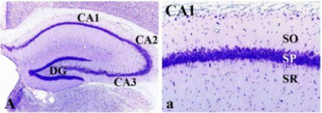
What does cresyl violet stain do?
basic dye, stains neuron cell bodies (rER in cell body of nucleus)
what does Fuelgen reaction stain do?
stains DNA hydrolysis, but NOT RNA sugar (ribose)
useful for DNA quantification/localization

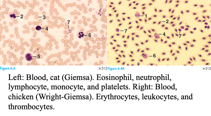
What does Giesma stain do?
Stains blood w/eosin, azure, + methylene blue
similar to Wright’s stain
classic lymphoma stainers
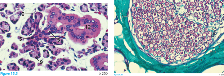
What does Gomori stain do?
Extremely broad, stains tons of stuff
group of different histochemical techniques
Example: the Gomori trichrome (mGT) uses multiple dyes to differentiate muscle fibers, connective tissue, and nuclei in tissue sections. An alternative method is called the Gomori methenamine silver (GMS), Gomori aldehyde fuchsin or Gomori reticulin stains.

What is H+E staining consist of?
Basophilic (blue/purple) + Acidophilic (pink) staining
used in combination with metal ions (mordent - aluminum or iron NOT SILVER)

What is the application for Mallory staining?
Viewing connective tissue
it contains aniline blue, Orange G, and azocarmine (or acid fuchsin)

What is characteristic of MASON trichrome staining?
2+ dyes of different colors
acid fuchsin, orange G, and light green
nuclei appear BLACK
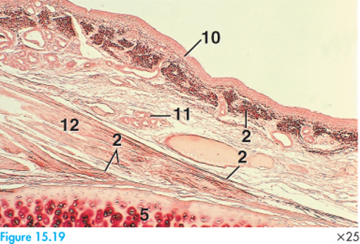
What is characteristic of orcein staining?
viewing elastic fibers
natural dye obtained from lichens
What is characteristic of period acid-schiff staining
viewing of proteins and glycogen/glycoproteins

What is characteristic of Toluidine blue?
basic stain that binds to nucleic acids, DNA & RNA

What is characteristic of wright blood stain?
Eosin + methylene blue to differentiate blood cell types
How does conventional histologic staining differ from immunohistochemistry as far as understanding and identifying molecules?
conventional staining looks at different types of molecules, while IMMUNOHISTOCHEMISTRY looks at specific molecules (usually a protein of RNA fragment)
What is the application for light microscopy?
cell-level visuals, is better for large visuals
What is the application for Transmission Electron Microscopy (TEM)?
Offers a higher resolution, finds smaller like proteins, organelle detail, etc.
electrons pass through ultra-thin specimens = image forms based on how electrons are scattered by atoms in a sample
What are characteristics of prokaryotic cells?
No true nucleus, organelles, or cytoskeleton
contain circular chromosomes (constructs nucleoid)
What are characteristics of Eukaryotic cells?
Think me and Eu-
Nucleus, organelles + cytoskeleton, nuclear membrane and nucleoli
linear chromosomes
How does compartmentation within eukaryotic cells contribute to cellular organization and function?
Compartmentation prevents mixing of biochemical reactions in the cell —> cells can have more specialized functions + grow to store molecules (hormones, neurotransmitters) for release
What are characteristics of passive diffusion?
moves easily through membrane directly, down concentration gradient
no energy required
What are characteristics of active transport?
requires energy
moves against concentration gradient
symporters - carries in same directions, sodium-glucose co-transporter)
antiporter - carries in opposite direction, sodium-potassium pump
What are characteristics of facilitated diffusion?
Moves easily via protein channels, as they are unable to dissolve in hydrophobic interior, down concentration gradient. Still passive
uniporters are passive
What are the functions of microfilaments?
provide cytoskeletal support
apical differentiations: projections (microvilli + stereo-cilia)
movement (filapoda + lamellapoda)
What is the structure of microfilaments? What are they composed of?
Helical F-actin formation, made of G-actin monomers
What is the function of intermediate filaments?
stable support
What is the function of microtubules?
mitotic spindle formation
form cilia + flagella
what is the structure of Intermediate filaments? What are they made of?
end-to-end ULFs made of tetramers (8)
filamentous monomers
What is the structure of microtubules? What are they made of
Tube of protofilaments (13 in complete tubule)
alpha- and beta- tubulin
what is the role of B4-Thymosin in actin-binding proteins?
gathers g-actin (monomer) but does NOT add them
what is the role of Profilins in actin-binding proteins?
Facilitates addition (polymerization) via ATP-->ADP
what is the role of Cofilin (or ADF) in actin-binding proteins?
Depolarizes - removing individual monomer
what is the role of Gelosin in actin-binding proteins?
Caps F-actin preventing loss or addition of actin monomers. when Ca2+ present fragements F-actin and binds to - end, preventing further filament growth.
What is the role of microvilli and how is it differentiated?
increases surface area for absorption
Formins (w/ FH1 + FH2 domain) on the cap extend the filament
Bundles of actin in the terminal web on cytoplasmic side to anchor filaments
Unbranched, unlike stereocilia
What is the role of microvilli and how is it differentiated?
long, branching projections
cross-linked core of actin in bundles
NO axoneme, unlike regular cilia
How does the structure and polarity of microtubules contribute to its organization and function?
it is made of tubulin heterodimers
polymerized on alpha-tubulin + beta-tubulin bound together via GTP
formed into rows called protofilaments
13 protofilaments make a hollow microtubule
Functions: intracellular transport, mechanical support, cell shape, flagella + cilia motility, and chromosome segregation
What contributes to stabilization in microtubules?
Microtubule associated proteins (MAPs) help stabilize by phosphorylating —> they preserve the GTP region + protect the tubule
classical MAPs
Non-classical MAPs
What are the functions of centrosomes?
nucleate (begin) the polymerization of tubulin into microtubules
organize microtubules into functional units (like mitotic spindle)
duplicate contriolar pair
become the bases for cilia (basal bodies)
Can microfilaments grow in different directions?
Yes
What are centrosomes?
organizers of microtubules (microtubule organizing center MTOC) - made of a pair of centrioles (mother+daughter)
MTOC- contains pericentriolar material (PCM) - where microtubules grow
how are centrioles organized?
organized in a 9 + 0 triplet arrangement

What are characteristics of “trichrome” staining?
2+ or more dyes (typically acidic)
to selectively color different basic tissue components within the same tissue section
many types of trichrome stains


What is direct IHC?
simpler, same host species can be used for multiple targets, there is no signal amplification. Not a sensitive, best for highly expressed antigens
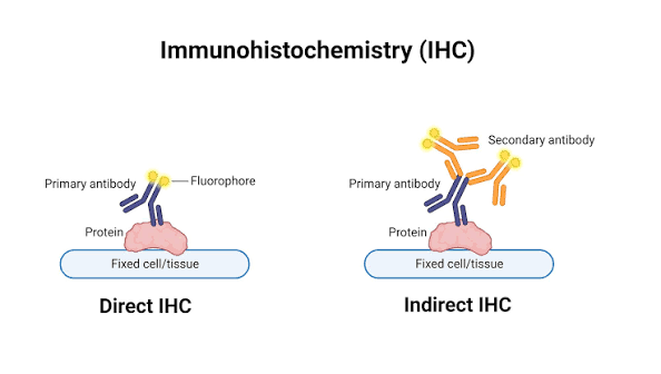
What is INdirect IHC?
more complex, considered more sensitive because MULTIPLE secondary antibodies can bind to single primary antibody, thus amplifying the detection signal
used to ensure you are fully able to visualize a color/fluorescent product when performing a staining procedure
What is immunohistochemistry (IHC) staining?
lab technique that enables the detection and visualization of specific antigens in tissue sections through the utilization of antibodies specific to those antigens/target proteins

What is this artifact (middle left)?
cracking artifact

What is this artifact (top left)?
stain precipitate
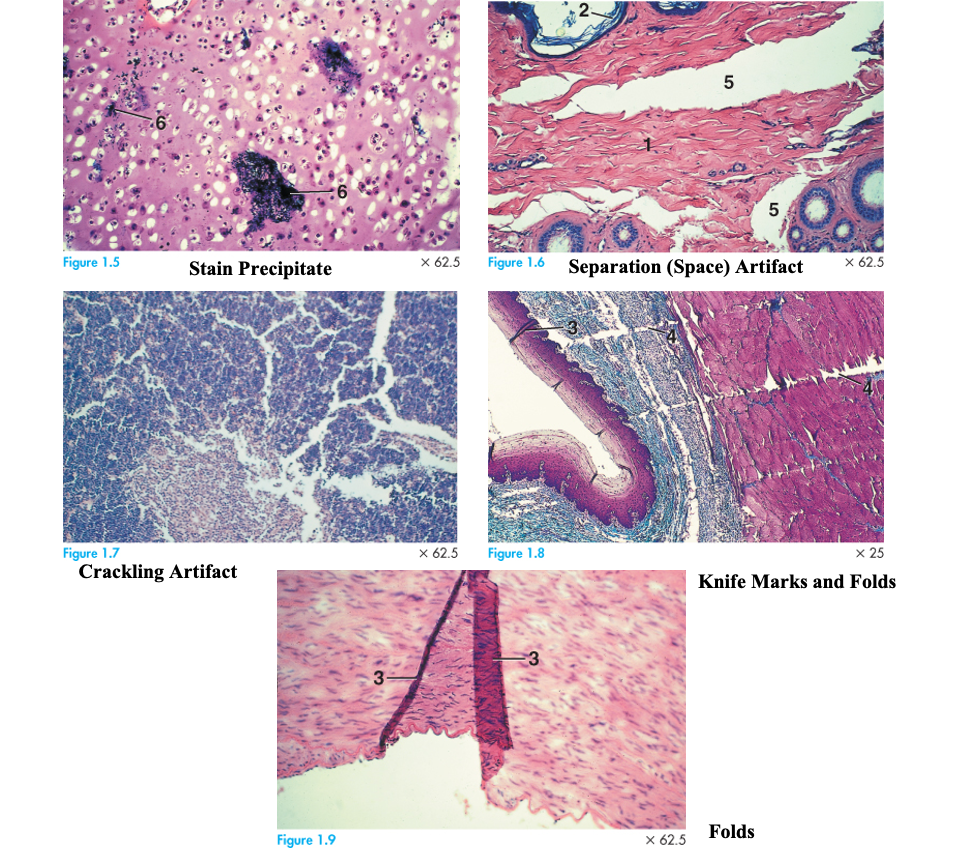
what is this artifact (top right)?
Separation (space) artifact

what is this artifact (middle right)?
Knife marks and folds
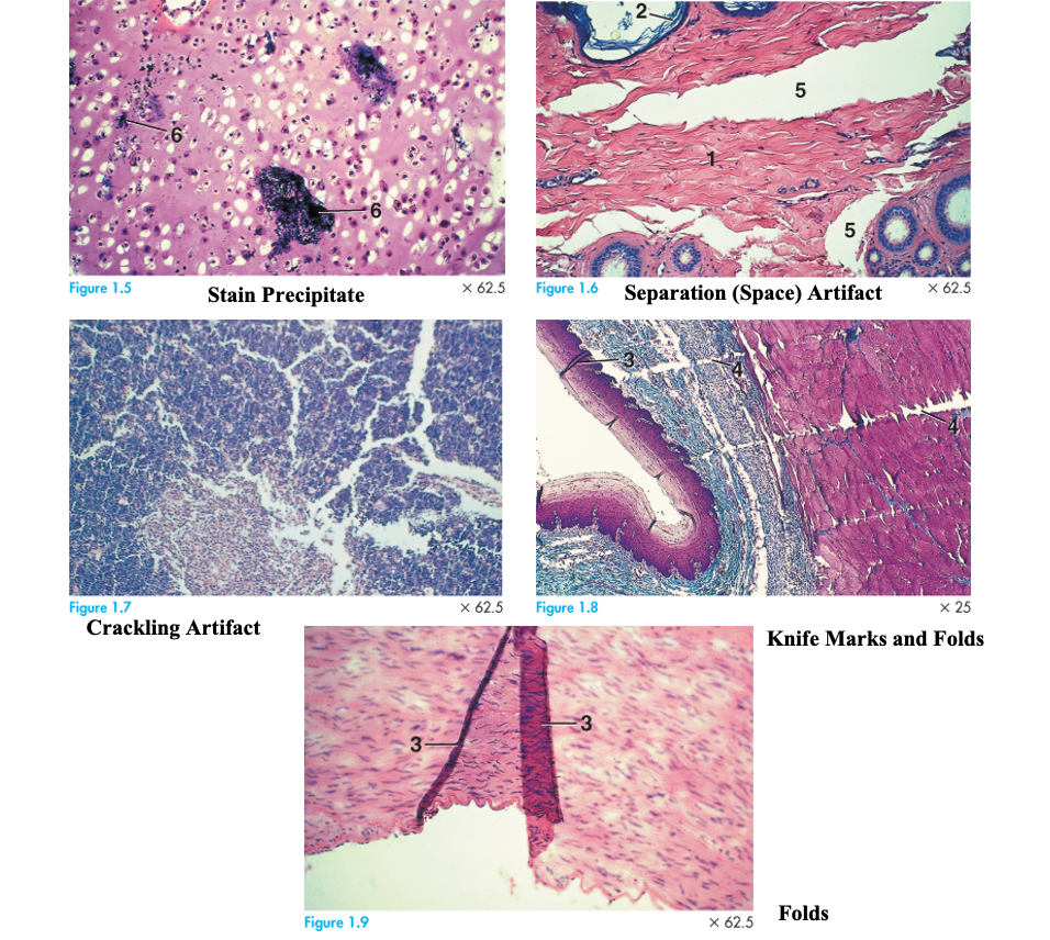
What is the artifact on the bottom?
folds
What is eukaryotic cell structure and function highly dependent on?
membranes and membrane-bound structures
What are peripheral proteins?
not inserted into the hydrophobic interior of the membrane —> indirectly associated with membranes through protein-protein ionic bond interactions
these interactions can be disrupted by high salt or extreme pH
What are integral proteins?
portions of integral proteins are inserted into the lipid bilayer and can only be released via solubilization using detergents
many are transmembrane proteins = span the entirety of the lipid bilayer, with segments exposed on both sides of the membrane
What prevents/hinders lateral movement of phospholipids and proteins?
can readily move in lateral direction, but is difficult to flip-flop from inner to outer membrane leaflets.
hindered by:
lipid rafts - proteins get trapped in them
attachment of membrane proteins to cytoskeletal and matrix proteins
cellular junctions - block lateral movement
Which cellular components exhibit polarity? (Microfilaments, intermediate filaments, and/or microtubules)
Microfilaments and Microtubules
What are the energy substrate(s) of Microfilaments, intermediate filaments, and microtubules?
ATPase (ATP/ADP) | NONE | GTPase |
respecitvely
How are microfilaments assembled and disassembled?
tread milling
How are intermediate filaments assembled and disassembled?
Regulated by phosphorylation and dephosphorylation
How are microtubules assembled and disassembled?
Dynamic Instability
What is treadmilling?
associated with microfilaments, G-actin monomers are assembled at one end (+) of the filament and concurrently disassembled at the other end (-)
what is dynamic instability?
associated with microtubules, switching of microtubules between states of persistent growth of GTP caps at the plus end and persistent unfolding at the GDP-rich minus end
3 steps:
polymerization phase
release of hydrolyzed phosphate (Pi)
Depolymerization phase
What is the transition from growth to shrinkage in dynamic instability called?
catastrophe
What is the transition from shrinkage to growth in dynamic instability called?
rescue (restore their length to about 20 um)
Which cellular component(s) exhibits stability? (Microfilaments, intermediate filaments, and/or microtubules)
Intermediate filaments are the only ones that exhibit stability
what is the cytochemical approach to localize DNA and RNA and evaluate nucleic acid synthesis within cells?
Fuelgen reaction stains DNA + RNA
what is the autoradiographic approach to localize DNA and RNA and evaluate nucleic acid synthesis within cells?
Radiographic precursors: radioactively labels DNA or RNA
[H3]thymadine vs [H3]uridine
how do Exportins regulate the directionality of macromolecule transport between the nucleus and cytoplasm?
Bring things out of the nucleus
how do Importins regulate the directionality of macromolecule transport between the nucleus and cytoplasm?
Bring things into the nucleus
how do NLS (nuclear localization signals) regulate the directionality of macromolecule transport between the nucleus and cytoplasm?
cargo proteins must be tagged w/this to go through pores
how do RAN-GTP regulate the directionality of macromolecule transport between the nucleus and cytoplasm?
Controls importins and exportins
How can small molecules diffuse through nuclear pore complexes?
small molecules can diffuse passively through the nuclear pore complex
How can proteins of ANY SIZE diffuse through nuclear pore complexes?
they must possess and NLS (nuclear localization amino acid sequence), they can be imported or exported into the cytoplasm through pore complex by an ENERGY-DEPENDENT mechanism requiring GTP
How is rRNA synthesized and processed within the nucleolus?
each gene has a Fibril: /cleaved/
each fibril= 45s rRNA // 18s rRNA // 28s rRNA // 5.8s rRNA
How does processed rRNAs contribute to the formation of small and large ribosomal subunits?
rRNA genes repeat, look like Xmas trees separated by spacers; each gene has a fibril
acts as the physical core and structural scaffolding that bind with ribosomal proteins to assemble the small and large ribosomal subunits
How does the Fibrillar Center of the nucleolus contribute to rRNA synthesis, processing, and ribosomal subunit assembly?
location of RNA pol I + SRP-RNA
How does the Fibrillar Component of the nucleolus contribute to rRNA synthesis, processing, and ribosomal subunit assembly?
Where nascent rRNA is present and undergoing processing (fibrillarin and nucleolin are found in the fibrillar dense component)
How does the Granular Component of the nucleolus contribute to rRNA synthesis, processing, and ribosomal subunit assembly?
where 18s rRNA + 28s RNA subunits are assembled/completed
DNA accessibility and transcriptional activity of less packed chromatin?
Transcription, S phase
DNA accessibility and transcriptional activity of more packed chromatin?
Inaccessible, cell division
what is the basic unit of chromatin condensation?
nucleosome: formed by 200 bp or 1.5 turns of double stranded DNA wrapped around a histone octamer core
What consists the histone octamer core?
composed of two molecules each of histone proteins H2A, H2B, H3, and H4
stabilized by linker histone (H1) protein cross-linking with DNA molecule which is wrapped around the octamer
What is the primary structure of chromatin characterized by?
(10 nm)consists of: nucleosomes + linker DNA
“beads on a string” appearance (nucleosome-nucleosome or fiber-fiber interactions) these allow for higher degrees of chromosomal condensation
What is the secondary structure of chromatin characterized by?
coils into 30 nm chromatin fiber: also stabilized by H1
fiber-fiber interactions result in coiling of chromatin
What is the Tertiary structure of chromatin characterized by?
further folding/coiling of secondary chromatin, also stabilized by H1, characteristic of condensed chromosomes seen during metaphase
gene transcription CAN OCCUR when chromatin is arranged in tertiary structure
Define euchromatin
diffuse chromatin; open for transcription - 10% of chromatin
electron lucent, stains light
Define heterochromatin
dense chromatin; transcriptionally inactive - 90% of chromatin
stains dark, electron-dense
what is the role of nuclear lamins?
Type V intermediate filaments: A1, B1, B2, + C
organize chromatin
space nuclear pores out
reassemble nucleus after cell division
what inner nuclear membrane proteins do nuclear lamins bind with?
LBR
Emerin
LAP1c
LAP 1 B
what is the role of the nuclear envelope?
enclosed + regulates nucleus
What is the role of chromatin?
DNA packaged w/ proteins
What is the role of nucleolus?
Produces rRNA + assembles ribosomes
How does the endomembrane system facilitate the compartmentalization, modification, packaging, and transport of cellular materials?
it separates the various processes + reactions into specific locations in the cell via membrane-bound organelles
What is the morphological characteristics, predominant functions, and tissue locations of simple squamous epithelium?
flat round nucleus; Barrier that allows for diffusion
alveoli, blood vessel lining, bowman’s capsule (kidney), serous
What is the morphological characteristics, predominant functions, and tissue locations of simple cuboidal?
round/square cell body: secretion
ducts and glands, PCT and DCT (kidney)
What is the morphological characteristics, predominant functions, and tissue locations of simple columnar?
long rectangular cell body; absorption
villi in the intestines
what are zonula adherens?
*belt desmosomes
forms circumferential belt around apical domain; associated w/ microfilaments via catenin complex to cytoplasmic desmosome cadherins
What are macula adherens?
*spot desmosomes
cytoplasmic plaques liek plakoglobin bind desmosome cadherins
What are hemidesmosomes?
link intermediate filaments to basal lamina
mode of:
inner cytoplasmic plate bound to tonofilaments
outer membrane plaque bound via laminins + integrins
What is the basal lamina?
Top layer of basal membrane; made of laminin, collagen, and proteoglycans
What is the reticular lamina?
the bottom layer of the basement membrane: made of Type III collagen