Cell Bio Exam 3 Flashcards

0.0(0)
Studied by 0 people
call kaiCall Kai
learnLearn
examPractice Test
spaced repetitionSpaced Repetition
heart puzzleMatch
flashcardsFlashcards
GameKnowt Play
Card Sorting

1/102

encourage image

There's no tags or description

Looks like no tags are added yet.

103 Terms

1
New cards

What is membrane transport?

the process by which substances are moved thru a cell or organelle membrane

2
New cards

The Rules of membrane transport are governed by diffusion and osmosis. What is the difference between them?

  • Diffusion – where a concentration gradient exists, substances dissolved in water will always move from an area of high concentration to an area of low concentration 

  • Osmosis – where a concentration gradient exists, water will diffuse from an area where solute concentration is low to an area where solute concentration is high 


<ul><li><p class="Paragraph SCXW113007820 BCX8" style="text-align: left;"><span style="line-height: 20.925px;">Diffusion – where a concentration gradient exists, substances dissolved in water will always move from an area of high concentration to an area of low concentration&nbsp;</span></p></li></ul><ul><li><p class="Paragraph SCXW113007820 BCX8" style="text-align: left;"><span style="line-height: 20.925px;">Osmosis – where a concentration gradient exists, water will diffuse from an area where solute concentration is low to an area where solute concentration is high&nbsp;</span></p></li></ul><p></p>
3
New cards

Diffusion and Osmosis occur bc of ______ which is the amount of randomness in the location of objects and energy w/i a system

Entropy

4
New cards

________ levels of entropy are found in highly organized systems, like a concentration gradient across a cell membrane. ______ levels of entropy are found where molecules or energy are equally and evenly distributed throughout a system

  • Lowest

  • Highest


5
New cards

What does the second law of thermodynamic state and what does it mean according to a cell’s concentration gradient?

  • The 2nd law of thermodynamics states that in a given system, entropy must always increase 

  • Bc of this, molecules in a cell’s concentration gradient will diffuse across the membrane until the conc of that molecule is = on both sides of the membrane 


<ul><li><p class="Paragraph SCXW242336479 BCX8" style="text-align: left;"><span style="line-height: 20.925px;">The 2<sup>nd</sup> law of thermodynamics states that in a given system, entropy must always increase&nbsp;</span></p></li></ul><ul><li><p class="Paragraph SCXW242336479 BCX8" style="text-align: left;"><span style="line-height: 20.925px;">Bc of this, molecules in a cell’s concentration gradient will diffuse across the membrane until the conc of that molecule is = on both sides of the membrane&nbsp;</span></p></li></ul><p></p>
6
New cards

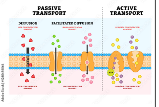
Membrane transport processes are split into 2 different types. What are they?

Passive and Active Transport
<p>Passive and Active Transport</p>
7
New cards

What is passive transport and what uses it?

  • Transport processes that follow the rules of entropy are passive transport processes 

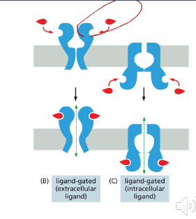
  • Molecules that are non-polar/hydrophobic can directly move thru the membrane by simple diffusion 

  • Molecules that are polar/hydrophilic must be moved thru the membrane by a transport protein (facilitated diffusion) 


<ul><li><p class="Paragraph SCXW177512649 BCX8" style="text-align: left;"><span style="line-height: 20.925px;">Transport processes that follow the rules of entropy are passive transport processes&nbsp;</span></p></li></ul><ul><li><p class="Paragraph SCXW177512649 BCX8" style="text-align: left;"><span style="line-height: 20.925px;">Molecules that are non-polar/hydrophobic can directly move thru the membrane by simple diffusion&nbsp;</span></p></li></ul><ul><li><p class="Paragraph SCXW177512649 BCX8" style="text-align: left;"><span style="line-height: 20.925px;">Molecules that are polar/hydrophilic must be moved thru the membrane by a transport protein (facilitated diffusion)&nbsp;</span></p></li></ul><p></p>
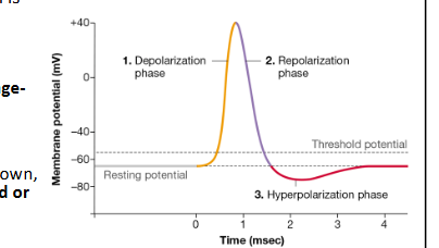
8
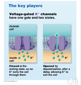
New cards

What is active transport and what is special about it?

  • Transport processes that “oppose” the rules of entropy are active transport processes 

  • The transport of substances from an area of low to high concentration 

  • Bc of the 2nd law of thermodynamics, active transport requires the cell to use transport proteins that expend cellular energy 


<ul><li><p class="Paragraph SCXW170829255 BCX8" style="text-align: left;"><span style="line-height: 20.925px;">Transport processes that “oppose” the rules of entropy are active transport processes&nbsp;</span></p></li></ul><ul><li><p class="Paragraph SCXW170829255 BCX8" style="text-align: left;"><span style="line-height: 20.925px;">The transport of substances from an area of low to high concentration&nbsp;</span></p></li></ul><ul><li><p class="Paragraph SCXW170829255 BCX8" style="text-align: left;"><span style="line-height: 20.925px;">Bc of the 2<sup>nd</sup> law of thermodynamics, active transport requires the cell to use transport proteins that expend cellular energy&nbsp;</span></p></li></ul><p></p>
9
New cards

How are entropy and energy tied together by the 2nd law of thermodynamics?

  • A system w/ low entropy has some amt of internal potential energy 

  • A system w/ high entropy has little or no internal potential energy 

  • The amt of potential energy is determined by how large the difference in concentration is 


10
New cards

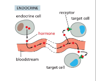
What drives the process of diffusion and how?

  • The internal potential energy of a system is what drives the process of diffusion 

  • This internal potential energy is converted into the movement of molecules down their concentration gradient 

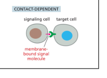
  • Organization is converted to randomness plus released energy


11
New cards

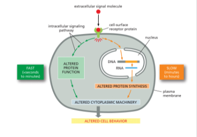
How does passive and active transport relate to the amount of entropy in a concentration gradient?

  • Passive transport processes like diffusion convert an organized concentration gradient into a more random (closer to =) distribution of a substance so passive transport releases energy that can be captured by the cell 

  • Active transport processes convert a more random (closer to equal) distribution of a substance into an organized conc gradient so the cells must spend energy to perform any active transport process 


12
New cards

What are the two types of active transport proteins based upon where cellular energy is spent?

  • Primary active transporters – transport proteins that spend cellular energy to transport substances across a membrane 

  • Secondary active transporters – transport proteins that use the intenral energy stored when primary transport proteins create a concentration gradient 


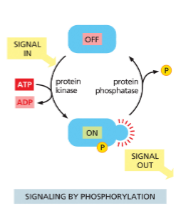
<ul><li><p class="Paragraph SCXW180248820 BCX8" style="text-align: left;"><span style="line-height: 20.925px;">Primary active transporters – transport proteins that spend cellular energy to transport substances across a membrane&nbsp;</span></p></li></ul><ul><li><p class="Paragraph SCXW180248820 BCX8" style="text-align: left;"><span style="line-height: 20.925px;">Secondary active transporters – transport proteins that use the intenral energy stored when primary transport proteins create a concentration gradient&nbsp;</span></p></li></ul><p></p>
13
New cards

What is transport coupling and how does it allow secondary active transporters to move molecules w/o breaking down ATP?

  • The sodium/potassium pump spends ATP to create concentration gradients of sodium and potassium 

  • The transport protein SGLT1 allows sodium to diffuse down its conc gradient 

  • This releases the internal energy in the sodium gradient 

  • That energy is then used or coupled to the transport of glucose from low to high conc 


<ul><li><p class="Paragraph SCXW61164752 BCX8" style="text-align: left;"><span style="line-height: 20.925px;">The sodium/potassium pump spends ATP to create concentration gradients of sodium and potassium&nbsp;</span></p></li></ul><ul><li><p class="Paragraph SCXW61164752 BCX8" style="text-align: left;"><span style="line-height: 20.925px;">The transport protein SGLT1 allows sodium to diffuse down its conc gradient&nbsp;</span></p></li></ul><ul><li><p class="Paragraph SCXW61164752 BCX8" style="text-align: left;"><span style="line-height: 20.925px;">This releases the internal energy in the sodium gradient&nbsp;</span></p></li></ul><ul><li><p class="Paragraph SCXW61164752 BCX8" style="text-align: left;"><span style="line-height: 20.925px;">That energy is then used or coupled to the transport of glucose from low to high conc&nbsp;</span></p></li></ul><p></p>
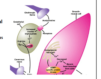
14
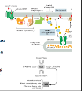
New cards

What are the 3 types of membrane proteins?

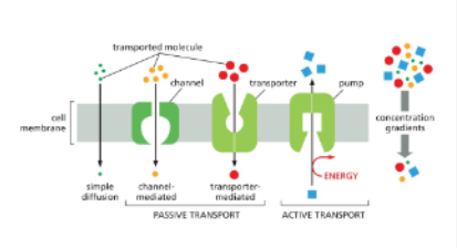
Channel proteins

Transporter (Carrier) Proteins

15
New cards

What are channel proteins and what do they have?

  • Proteins create a hydrophilic tunnel thru the membrane which allows polar molecules and ions a pathway to diffuse thru the membrane’s hydrophobic barrier 

  • Channels typically have a gate to regulate diffusion 

  • Conformational shifts in the protein can cause the gate to open to allow diffusion and close to block diffusion 


<ul><li><p class="Paragraph SCXW57945589 BCX8" style="text-align: left;"><span style="line-height: 20.925px;">Proteins create a hydrophilic tunnel thru the membrane which allows polar molecules and ions a pathway to diffuse thru the membrane’s hydrophobic barrier&nbsp;</span></p></li></ul><ul><li><p class="Paragraph SCXW57945589 BCX8" style="text-align: left;"><span style="line-height: 20.925px;">Channels typically have a gate to regulate diffusion&nbsp;</span></p></li></ul><ul><li><p class="Paragraph SCXW57945589 BCX8" style="text-align: left;"><span style="line-height: 20.925px;">Conformational shifts in the protein can cause the gate to open to allow diffusion and close to block diffusion&nbsp;</span></p></li></ul><p></p>
16
New cards

What are transporters and what do they do?

  • Binds to the substances they transport w/ high affinity 

  • Binding of the transported substances causes a conformational shift in the protein in two ways: 

  1. Closes the transporter on one side of the membrane and opens it on the other 

  2. Causes the affinity of the binding site to decrease 

  • Lowering the binding affinity allows the transported molecule to dissociate from the transporter and diffuse away 

<ul><li><p class="Paragraph SCXW153933858 BCX8" style="text-align: left;"><span style="line-height: 20.925px;">Binds to the substances they transport w/ high affinity&nbsp;</span></p></li></ul><ul><li><p class="Paragraph SCXW153933858 BCX8" style="text-align: left;"><span style="line-height: 20.925px;">Binding of the transported substances causes a conformational shift in the protein in two ways:&nbsp;</span></p></li></ul><ol><li><p><span style="line-height: 20.925px;">Closes the transporter on one side of the membrane and opens it on the other&nbsp;</span></p></li><li><p><span style="line-height: 20.925px;">Causes the affinity of the binding site to decrease&nbsp;</span></p></li></ol><ul><li><p class="Paragraph SCXW153933858 BCX8" style="text-align: left;"><span style="line-height: 20.925px;">Lowering the binding affinity allows the transported molecule to dissociate from the transporter and diffuse away&nbsp;</span></p></li></ul><p></p>
17
New cards

What are pumps and why and where do they get their energy?

  • Pumps bind to the substances they transport and use energy to transport them “up” a concentration gradient 

  • Pumps work similarity to carrier/transporters w/ one big exception: pumps require a source of energy for their conformational shifts 

  • 3 sources: 

  1. Gradient-driven pumps use the internal energy of diffusion down a gradient 

  2. ATP-driven pumps use the energy from the hydrolysis of ATP to ADP 

  3. Light-driven pumps have light-sensitive molecules incorporated into their structure  


<ul><li><p class="Paragraph SCXW261158521 BCX8" style="text-align: left;"><span style="line-height: 20.925px;">Pumps bind to the substances they transport and use energy to transport them “up” a concentration gradient&nbsp;</span></p></li></ul><ul><li><p class="Paragraph SCXW261158521 BCX8" style="text-align: left;"><span style="line-height: 20.925px;">Pumps work similarity to carrier/transporters w/ one big exception: pumps require a source of energy for their conformational shifts&nbsp;</span></p></li></ul><ul><li><p class="Paragraph SCXW261158521 BCX8" style="text-align: left;"><span style="line-height: 20.925px;">3 sources:&nbsp;</span></p></li></ul><ol><li><p><span style="line-height: 20.925px;">Gradient-driven pumps use the internal energy of diffusion down a gradient&nbsp;</span></p></li><li><p><span style="line-height: 20.925px;">ATP-driven pumps use the energy from the hydrolysis of ATP to ADP&nbsp;</span></p></li><li><p><span style="line-height: 20.925px;">Light-driven pumps have light-sensitive molecules incorporated into their structure &nbsp;</span></p></li></ol><p></p>
18
New cards

Why is the Na/K pump so important?

  • Uses active transport to pump 3 Na+ ions to the outside and 2K+ ions to the inside of the cell and the energy to power this comes from the creation of organization comes from ATP hydrolysis 

  • In animal cells, this protein sets up permanent gradients of sodium and potassium 

  • Sodium Concentration is always highest outside of the cell 

  • Potassium concentration is always highest in the cytoplasm 

  • These gradients are so critical for cellular function that a typical cell spends 1/3 of all its ATP on this one pump 


<ul><li><p><span style="line-height: 20.925px;">Uses active transport to pump 3 Na+ ions to the outside and 2K+ ions to the inside of the cell and the energy to power this comes from the creation of organization comes from ATP hydrolysis&nbsp;</span></p></li><li><p><span style="line-height: 20.925px;">In animal cells, this protein sets up permanent gradients of sodium and potassium&nbsp;</span></p></li></ul><ul><li><p class="Paragraph SCXW119565703 BCX8" style="text-align: left;"><span style="line-height: 20.925px;">Sodium Concentration is always highest outside of the cell&nbsp;</span></p></li></ul><ul><li><p class="Paragraph SCXW119565703 BCX8" style="text-align: left;"><span style="line-height: 20.925px;">Potassium concentration is always highest in the cytoplasm&nbsp;</span></p></li></ul><ul><li><p class="Paragraph SCXW119565703 BCX8" style="text-align: left;"><span style="line-height: 20.925px;">These gradients are so critical for cellular function that a typical cell spends 1/3 of all its ATP on this one pump&nbsp;</span></p></li></ul><p></p>
19
New cards

Why are epithelial cells unusual in their transport proteins?

  • Epithelial cells are unusual in that their transport proteins are different on each “side” of the cell 

  • The side that faces the environment is the apical membrane 

  • Side that faces the inside of the body is the basolateral membrane 

  • Two sides are separated from each other by anchoring proteins called tight junctions 


<ul><li><p class="Paragraph SCXW34299544 BCX8" style="text-align: left;"><span style="line-height: 20.925px;">Epithelial cells are unusual in that their transport proteins are different on each “side” of the cell&nbsp;</span></p></li></ul><ul><li><p class="Paragraph SCXW34299544 BCX8" style="text-align: left;"><span style="line-height: 20.925px;">The side that faces the environment is the apical membrane&nbsp;</span></p></li></ul><ul><li><p class="Paragraph SCXW34299544 BCX8" style="text-align: left;"><span style="line-height: 20.925px;">Side that faces the inside of the body is the basolateral membrane&nbsp;</span></p></li></ul><ul><li><p class="Paragraph SCXW34299544 BCX8" style="text-align: left;"><span style="line-height: 20.925px;">Two sides are separated from each other by anchoring proteins called tight junctions&nbsp;</span></p></li></ul><p></p>
20
New cards

What are the two ways that materials can enter/leave the body?

  • Transcellular pathway – transporting substances thru the membrane 

  • Paracellular Pathway – substances diffuse thru the tight junctions 


21
New cards

True or False: The Paracellular pathway can only be done by large molecules or ions due to the open effect of tight junctions

False; This pathway can typically only be done by very small molecules (water, gases) or ions due to the sealing effect of the tight junction 

22
New cards

Most of the epithelial cells in the body make and secrete what kind of solution? Where is one of the most important places it is done, and what are saline secretions used to do?

  • One of the most important places where this happens is the lungs 

  • Saline secretions are used to: 

  1. Create water-based solutions like digestive juices and bile 

  2. Keeps cells from becoming dehydrated by environmental exposure 

  3. Prevents mucus secretions from dying out and getting stuck in the lungs 


<ul><li><p class="Paragraph SCXW255393410 BCX8" style="text-align: left;"><span style="line-height: 20.925px;">One of the most important places where this happens is the lungs&nbsp;</span></p></li></ul><ul><li><p class="Paragraph SCXW255393410 BCX8" style="text-align: left;"><span style="line-height: 20.925px;">Saline secretions are used to:&nbsp;</span></p></li></ul><ol><li><p><span style="line-height: 20.925px;">Create water-based solutions like digestive juices and bile&nbsp;</span></p></li><li><p><span style="line-height: 20.925px;">Keeps cells from becoming dehydrated by environmental exposure&nbsp;</span></p></li><li><p><span style="line-height: 20.925px;">Prevents mucus secretions from dying out and getting stuck in the lungs&nbsp;</span></p></li></ol><p></p>
23
New cards

What are the 2 proteins that create the saline secretion and how?

  • NKCC – uses the sodium gradient created by the NA/K pump to transport chloride and potassium ions into the cytoplasm (Found in the basolateral membrane) 

  • CFTR – chloride ions then diffuse out of the cell thru the CFTR channel protein in the apical membrane 

  • STAGE 2

  1. Sodium ions leave by simply diffusing out of the body via the paracellular route due to the buildup of negative charged chloride ions will electrically attract positive sodium ions 

  2. The accumulation of salt on the surface of the cell creates an osmotic gradient that causes water to leave the body and build up on the surface of the cells


<ul><li><p class="Paragraph SCXW223369924 BCX8" style="text-align: left;"><span style="line-height: 20.925px;">NKCC – uses the sodium gradient created by the NA/K pump to transport chloride and potassium ions into the cytoplasm&nbsp;(Found in the basolateral membrane)&nbsp;</span></p></li></ul><ul><li><p class="Paragraph SCXW223369924 BCX8" style="text-align: left;"><span style="line-height: 20.925px;">CFTR – chloride ions then diffuse out of the cell thru the CFTR channel protein in the apical membrane&nbsp;</span></p></li><li><p class="Paragraph SCXW223369924 BCX8" style="text-align: left;">STAGE 2</p></li></ul><ol><li><p><span style="line-height: 20.925px;">Sodium ions leave by simply diffusing out of the body via the paracellular route due to the buildup of negative charged chloride ions will electrically attract positive sodium ions&nbsp;</span></p></li><li><p><span style="line-height: 20.925px;">The accumulation of salt on the surface of the cell creates an osmotic gradient that causes water to leave the body and build up on the surface of the cells</span></p></li></ol><p></p>
24
New cards

what is the ENaC and what does it maintain?

  • ENaC – protein that keep this saline secretion system in homeostasis 

  • Cells want to avoid creating tm saline and dehydrating the epithelial layer 

  • The ENaC is the apical membrane creates a sodium channel for sodium to diffuse back into the cell, which causes the absorption of chloride and water back to the body 

  • This channel can be closed when it binds to CFTR proteins bc CFTR causes a conformational shift in ENaC that stops sodium from being reabsorbed 


<ul><li><p class="Paragraph SCXW121956614 BCX8" style="text-align: left;"><span style="line-height: 20.925px;">ENaC – protein that keep this saline secretion system in homeostasis&nbsp;</span></p></li></ul><ul><li><p class="Paragraph SCXW121956614 BCX8" style="text-align: left;"><span style="line-height: 20.925px;">Cells want to avoid creating tm saline and dehydrating the epithelial layer&nbsp;</span></p></li></ul><ul><li><p class="Paragraph SCXW121956614 BCX8" style="text-align: left;"><span style="line-height: 20.925px;">The ENaC is the apical membrane creates a sodium channel for sodium to diffuse back into the cell, which causes the absorption of chloride and water back to the body&nbsp;</span></p></li></ul><ul><li><p class="Paragraph SCXW121956614 BCX8" style="text-align: left;"><span style="line-height: 20.925px;">This channel can be closed when it binds to CFTR proteins bc CFTR causes a conformational shift in ENaC that stops sodium from being reabsorbed&nbsp;</span></p></li></ul><p></p>
25
New cards

Why is Cystic Fibrosis (CF) increase the risk of serious lung infection and causes difficulty w/ breathing?

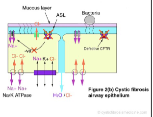
  • In CF, CFTR is misfolded and trapped in the ER 

  • Bc CFTR is absent from the apical membrane it destroys the production of saline in two ways: 

  1. Fewer chloride ions are transported onto the cell surface 

  2. Epithelial cells cannot stop sodium frm being reabsorbed 

  • Bc of this, the mucus lining of the lungs dries and becomes trapped in the lungs, which increases the risk of serious lung infections and causes difficulty w/ breathing 

<ul><li><p class="Paragraph SCXW164024144 BCX8" style="text-align: left;"><span style="line-height: 20.925px;">In CF, CFTR is misfolded and trapped in the ER&nbsp;</span></p></li></ul><ul><li><p class="Paragraph SCXW164024144 BCX8" style="text-align: left;"><span style="line-height: 20.925px;">Bc CFTR is absent from the apical membrane it destroys the production of saline in two ways:&nbsp;</span></p></li></ul><ol><li><p><span style="line-height: 20.925px;">Fewer chloride ions are transported onto the cell surface&nbsp;</span></p></li><li><p><span style="line-height: 20.925px;">Epithelial cells cannot stop sodium frm being reabsorbed&nbsp;</span></p></li></ol><ul><li><p class="Paragraph SCXW164024144 BCX8" style="text-align: left;"><span style="line-height: 20.925px;">Bc of this, the mucus lining of the lungs dries and becomes trapped in the lungs, which increases the risk of serious lung infections and causes difficulty w/ breathing&nbsp;</span></p></li></ul><p></p>
26
New cards

True or False: The primary job of intestinal epithelial cells is absorption of molecules 

True

27
New cards

What does the Na/K pump do and what does it allow?

  • The NA/K pump creates a low sodium environment in the cell by pumping sodium into the ECF inside the body 

  • Allows sodium to diffuse into the cell thru ENaC and nutrient transporters 

  • Accumulation of positively charged sodium in the ECF electrically attracts the chloride ions in thru the transcellular pathway 


<ul><li><p class="Paragraph SCXW165546438 BCX8" style="text-align: left;"><span style="line-height: 20.925px;">The NA/K pump creates a low sodium environment in the cell by pumping sodium into the ECF inside the body&nbsp;</span></p></li></ul><ul><li><p class="Paragraph SCXW165546438 BCX8" style="text-align: left;"><span style="line-height: 20.925px;">Allows sodium to diffuse into the cell thru ENaC and nutrient transporters&nbsp;</span></p></li></ul><ul><li><p class="Paragraph SCXW165546438 BCX8" style="text-align: left;"><span style="line-height: 20.925px;">Accumulation of positively charged sodium in the ECF electrically attracts the chloride ions in thru the transcellular pathway&nbsp;</span></p></li></ul><p></p>
28
New cards

What are aquaporins and what do they contribute to?

  • Accumulation of NaCl in the ECF causes water to enter the body by osmosis 

  • To do this efficiently, intestinal cells have water channels called aquaporins 

  • Water can diffuse through a membrane, but does it very slowly, so aquaporins associate tg to create a water-selective channel in the membrane 


<ul><li><p class="Paragraph SCXW29446965 BCX8" style="text-align: left;"><span style="line-height: 20.925px;">Accumulation of NaCl in the ECF causes water to enter the body by osmosis&nbsp;</span></p></li></ul><ul><li><p class="Paragraph SCXW29446965 BCX8" style="text-align: left;"><span style="line-height: 20.925px;">To do this efficiently, intestinal cells have water channels called aquaporins&nbsp;</span></p></li></ul><ul><li><p class="Paragraph SCXW29446965 BCX8" style="text-align: left;"><span style="line-height: 20.925px;">Water can diffuse through a membrane, but does it very slowly, so aquaporins associate tg to create a water-selective channel in the membrane&nbsp;</span></p></li></ul><p></p>
29
New cards

How do nutrient transporters use energy?

Use the energy in the sodium gradient to absorb nutrients

30
New cards

Why are sport drinks like Gatorade efficient at hydration?

  • The sugar and electrolyte solution enhances pumping of sodium into the ECF 

  • More sodium in the ECF means a stronger osmotic gradient and so absorption of water by osmosis is faster 


31
New cards

Remember: For _________ molecules like glucose, membrane transport is driven only by the _________ _______ for that molecule. Transport of ____ and other charged molecules is driven by ________ and _________ _______

  • Uncharged, Concentration Gradient

  • Ions, concentration and electrical gradients


<ul><li><p>Uncharged, Concentration Gradient</p></li><li><p>Ions, concentration and electrical gradients</p></li></ul><p></p>
32
New cards

Bc of the movement of ions is influenced by both concentration and charge we must combine the effects of both tg. What is this called?

An electrochemical gradient

33
New cards

How do we measure strength of an electrochemical gradient?

  • When the electrical and chemical gradient both drive movement of sodium in the same direction, the combined electrochemical gradient enhances diffusion of the ion (Ex: sodium across the cell membrane, from ECF to ICF)

  • When the electrical and chemical gradient drive movement of potassium in opposite directions, we must subtract the strength of the electrical gradient from the chemical gradient , so The combined electrochemical gradient is weaker and slows down ion diffusion (ex: Potassium across the cell membrane, from ICF to ECF)


<ul><li><p class="Paragraph SCXW184567737 BCX8" style="text-align: left;"><span style="line-height: 20.925px;">When the electrical and chemical gradient both drive movement of sodium in the same direction, the combined electrochemical gradient enhances diffusion&nbsp;of the ion (Ex: sodium across the cell membrane, from ECF to ICF)</span></p></li><li><p class="Paragraph SCXW184567737 BCX8" style="text-align: left;"><span style="line-height: 20.925px;">When the electrical and chemical gradient drive movement of potassium in opposite directions, we must subtract the strength of the electrical gradient from the chemical gradient&nbsp;, so The combined electrochemical gradient is weaker and slows down ion diffusion&nbsp;(ex: Potassium across the cell membrane, from ICF to ECF)</span></p></li></ul><p></p>
34
New cards

The difference in charge across a cell membrane is referred to as the cell’s…

Resting Membrane Potential

35
New cards

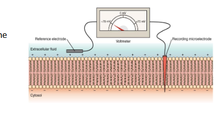
True or False: For most cells, the resting membrane potential is positive, so the inside leaflet of the plasma membrane is positive charged, and the outside leaflet is negatively charged

False; For most cells, the RMP is negative so the inside leaflet of the plasma membrane is negatively charged, and the outside leaflet is positively charged 
<p>False; <span style="line-height: 20.925px;">For most cells, the RMP is negative so the inside leaflet of the plasma membrane is negatively charged, and the outside leaflet is positively charged&nbsp;</span></p>
36
New cards

Charged membranes are referred to as…

polarized

37
New cards

The RMP of each cell comes from the combined effects of the ____ and ____ electrochemical gradients

Na+

K+

38
New cards

Where does the RMP begin to come from and how does it work?

  • While RMP requires several proteins, it starts w/ the Na/K pump 

  • Na/K pumps 3 Na+ to the ECF,  2 K+ to the ICF which creates a gradient of charge 

  • When it pumps 3 positive charges out and 2 positive charges in, the cell overall becomes more negative 

  • Most of the proteins of the cell have an overall negative charge and cannot leave the cell by diffusion so this pump removes positive charges and leaves negatively charged macromolecules behind


<ul><li><p class="Paragraph SCXW10342838 BCX8" style="text-align: left;"><span style="line-height: 20.925px;">While RMP requires several proteins, it starts w/ the Na/K pump&nbsp;</span></p></li></ul><ul><li><p class="Paragraph SCXW10342838 BCX8" style="text-align: left;"><span style="line-height: 20.925px;">Na/K pumps 3 Na+ to the ECF, &nbsp;2 K+ to the ICF&nbsp;which creates a gradient of charge&nbsp;</span></p></li></ul><ul><li><p class="Paragraph SCXW10342838 BCX8" style="text-align: left;"><span style="line-height: 20.925px;">When it pumps 3 positive charges out and 2 positive charges in, the cell overall becomes more negative&nbsp;</span></p></li></ul><ul><li><p class="Paragraph SCXW10342838 BCX8" style="text-align: left;"><span style="line-height: 20.925px;">Most of the proteins of the cell have an overall negative charge and cannot leave the&nbsp;cell by diffusion so this pump removes positive charges and leaves negatively charged macromolecules behind</span></p></li></ul><p></p>
39
New cards

What is the electrogenic pump?

the electrical driving force that the Na/K pump generates

40
New cards

True or False: Electrical potentials can exist where charges are not continuously flowing in a circuit

False;

  • It is a fundamental physical law that electrical potential can only exist where charges can continuously flow in a circuit 


41
New cards

How do cells complete their circuits of potassium and sodium flow?

  • Cells complete their circuits of potassium and sodium flow using leak channels 

  • Recall: Channels create hydrophilic tunnels thru the membrane 

  • The sodium and potassium leak channels allow Na+ and K+ to diffuse down their electrochemical gradients  


<ul><li><p class="Paragraph SCXW153784839 BCX8" style="text-align: left;"><span style="line-height: 20.925px;">Cells complete their circuits of potassium and sodium flow using leak channels&nbsp;</span></p></li></ul><ul><li><p class="Paragraph SCXW153784839 BCX8" style="text-align: left;"><span style="line-height: 20.925px;">Recall: Channels create hydrophilic tunnels thru the membrane&nbsp;</span></p></li></ul><ul><li><p class="Paragraph SCXW153784839 BCX8" style="text-align: left;"><span style="line-height: 20.925px;">The sodium and potassium leak channels allow Na+ and K+ to diffuse down their electrochemical gradients &nbsp;</span></p></li></ul><p></p>
42
New cards

What does the final part of RMP depend upon?

The ratio of potassium leak out to sodium leak in

43
New cards

True or False: Most cells leak more potassium than sodium, but each cell type in an organism can have a different RMP depending on their unique membrane transport physiology

True

44
New cards

What are the two purposes of the electrochemical gradients created by the Na/K pump?

  • The use of entropy to store energy for membrane transport processes 

  • Creating and processing electrical signals 


45
New cards

What are voltage-sensitive proteins?

Some of the proteins of the cell are voltage-sensitive proteins, meaning that they undergo conformational shifts when membrane potential changes 

46
New cards

What are excitable cells and what two types of channel proteins they have?

  • Some of the proteins of the cell are voltage-sensitive proteins, meaning that they undergo conformational shifts when membrane potential changes 

  • Electrical signaling is not commonly used in all cells, these cells must be excitable cells 


47
New cards

All Channel proteins have what 3 important functional domains?

  • Hydrophilic channel – a tunnel that is lined w/ hydrophilic amino acids 

  • Selectively Filter – a ring of amino acids at one end of the channel that only allows one type of ion to enter the channel  

  • Gate – the gate of a channel can open and close to regulate when ions are allowed to diffuse 


<ul><li><p class="Paragraph SCXW159667274 BCX8" style="text-align: left;"><span style="line-height: 20.925px;">Hydrophilic channel – a tunnel that is lined w/ hydrophilic amino acids&nbsp;</span></p></li></ul><ul><li><p class="Paragraph SCXW159667274 BCX8" style="text-align: left;"><span style="line-height: 20.925px;">Selectively Filter – a ring of amino acids at one end of the channel that only allows one type of ion to enter the channel &nbsp;</span></p></li></ul><ul><li><p class="Paragraph SCXW159667274 BCX8" style="text-align: left;"><span style="line-height: 20.925px;">Gate – the gate of a channel can open and close to regulate when ions are allowed to diffuse&nbsp;</span></p></li></ul><p></p>
48
New cards

What are the two kinds of membrane channels in excitable cells?

  1. Leak channels 

  • Gate of leak channels will cycle randomly btwn open and closed 

  • Bc no signal is required for this to happen, leak channels are always open 

  1. Gated channels 

  • These channels are normally closed and only open in response to a signal which trigger gated channels to open 


49
New cards

What are the 3 types of triggers to open gated membrane channels in excitable cells?

  1. Ligand Gated Ion channels

  2. Mechanically gated ion channels

  3. Voltage gated ion channels


50
New cards

How do ligand-gated ion channels trigger an electrical signal to open an gate in excitable cells?

  • These channels open when a chemical signal binds to them 

  • The chemical signal will attach to a binding site on the ion channel 

  • Binding of the signal causes a conformational shift that opens the gate 

  • This allows cells to convert chemical signals into electrical signals 

<ul><li><p class="Paragraph SCXW163697995 BCX8" style="text-align: left;"><span style="line-height: 20.925px;">These channels open when a chemical signal binds to them&nbsp;</span></p></li></ul><ul><li><p class="Paragraph SCXW163697995 BCX8" style="text-align: left;"><span style="line-height: 20.925px;">The chemical signal will attach to a binding site on the ion channel&nbsp;</span></p></li></ul><ul><li><p class="Paragraph SCXW163697995 BCX8" style="text-align: left;"><span style="line-height: 20.925px;">Binding of the signal causes a conformational shift that opens the gate&nbsp;</span></p></li></ul><ul><li><p class="Paragraph SCXW163697995 BCX8" style="text-align: left;"><span style="line-height: 20.925px;">This allows cells to convert chemical signals into electrical signals&nbsp;</span></p></li></ul><p></p>
51
New cards

How do mechanically-gated ion channels trigger an electrical signal to open an gate in excitable cells?

  • These channels open when a physical pressure is applied to their cell membrane 

  • Physical pressure bends the cell membrane, which causes the gate to open and allows cells to convert a physical signal to an electrical signal 


<ul><li><p class="Paragraph SCXW153624563 BCX8" style="text-align: left;"><span style="line-height: 20.925px;">These channels open when a physical pressure is applied to their cell membrane&nbsp;</span></p></li></ul><ul><li><p class="Paragraph SCXW153624563 BCX8" style="text-align: left;"><span style="line-height: 20.925px;">Physical pressure bends the cell membrane, which causes the gate to open and allows cells to convert a physical signal to an electrical signal&nbsp;</span></p></li></ul><p></p>
52
New cards

How do voltage-gated ion channels trigger an electrical signal to open an gate in excitable cells?

  • These channels are voltage-sensitive proteins bc their gate opens in response to a change in membrane potential 

  • Diff types of channels have gates that are tuned to open at a specific voltage 

  • These channels are special bc they can be activated in chains to produce complex electrical signals 


<ul><li><p class="Paragraph SCXW2106839 BCX8" style="text-align: left;"><span style="line-height: 20.925px;">These channels are voltage-sensitive proteins bc their gate opens in response to a change in membrane potential&nbsp;</span></p></li></ul><ul><li><p class="Paragraph SCXW2106839 BCX8" style="text-align: left;"><span style="line-height: 20.925px;">Diff types of channels have gates that are tuned to open at a specific voltage&nbsp;</span></p></li></ul><ul><li><p class="Paragraph SCXW2106839 BCX8" style="text-align: left;"><span style="line-height: 20.925px;">These channels are special bc they can be activated in chains to produce complex electrical signals&nbsp;</span></p></li></ul><p></p>
53
New cards

What is most common type of electrical signal and what is created by?

  • The most common type of electrical signal is the action potential 

  • Action potentials are created by the sequential opening and closing of voltage-gated channels 

  • Action potentials cannot begin on their own, they must be triggered by a ligand-gated or mechanically-gated channel 

<ul><li><p class="Paragraph SCXW72171207 BCX8" style="text-align: left;"><span style="line-height: 20.925px;">The most common type of electrical signal is the action potential&nbsp;</span></p></li></ul><ul><li><p class="Paragraph SCXW72171207 BCX8" style="text-align: left;"><span style="line-height: 20.925px;">Action potentials are created by the sequential opening and closing of voltage-gated channels&nbsp;</span></p></li></ul><ul><li><p class="Paragraph SCXW72171207 BCX8" style="text-align: left;"><span style="line-height: 20.925px;">Action potentials cannot begin on their own, they must be triggered by a ligand-gated or mechanically-gated channel&nbsp;</span></p></li></ul><p></p>
54
New cards

What are the two voltage-gated channels in neuronal and skeletal muscle action potentials?

  1. Voltage gated potassium channels 

  • Their gate can shift btwn open and closed states 

  1. Voltage-gated sodium channels 

  • These channels have a more complex gate that can shift btwn open, closed, and inactivated states 

<ol><li><p class="Paragraph SCXW129547975 BCX8" style="text-align: left;"><span style="line-height: 20.925px;">Voltage gated potassium channels&nbsp;</span></p></li></ol><ul><li><p class="Paragraph SCXW129547975 BCX8" style="text-align: left;"><span style="line-height: 20.925px;">Their gate can shift btwn open and closed states&nbsp;</span></p></li></ul><ol start="2"><li><p class="Paragraph SCXW129547975 BCX8" style="text-align: left;"><span style="line-height: 20.925px;">Voltage-gated sodium channels&nbsp;</span></p></li></ol><ul><li><p class="Paragraph SCXW129547975 BCX8" style="text-align: left;"><span style="line-height: 20.925px;">These channels have a more complex gate that can shift btwn open, closed, and inactivated states&nbsp;</span></p></li></ul><p></p>
55
New cards

Why are sodium channels voltage sensitive?

bc of a sensor alpha helix that contains positively charged amino acids

56
New cards

How does de[polarization effect the attraction of a helix?

  • At normal RMP (-70 to –90 mV) the sensor helix is attracted to the negatively charged inner leaflet 

  • This keeps the channel closed until an electrical signal arrives 

  • The channel undergoes a conformational shift when the cell membrane moves to a slightly more positive voltage (depolarization) 

  • Depolarization of the membrane weakens the electrical attraction of the sensor helix anf the helix twists the gate into the open state, which allows the action potential to begin  


<ul><li><p class="Paragraph SCXW104475120 BCX8" style="text-align: left;"><span style="line-height: 20.925px;">At normal RMP (-70 to –90 mV) the sensor helix is attracted to the negatively charged inner leaflet&nbsp;</span></p></li></ul><ul><li><p class="Paragraph SCXW104475120 BCX8" style="text-align: left;"><span style="line-height: 20.925px;">This keeps the channel closed until an electrical signal arrives&nbsp;</span></p></li></ul><ul><li><p class="Paragraph SCXW104475120 BCX8" style="text-align: left;"><span style="line-height: 20.925px;">The channel undergoes a conformational shift when the cell membrane moves to a slightly more positive voltage (depolarization)&nbsp;</span></p></li></ul><ul><li><p class="Paragraph SCXW104475120 BCX8" style="text-align: left;"><span style="line-height: 20.925px;">Depolarization of the membrane weakens the electrical attraction of the sensor helix anf the helix twists the gate into the open state, which allows the action potential to begin &nbsp;</span></p></li></ul><p></p>
57
New cards

Where does this initial depolarization come from? 

Depends on the cell

58
New cards

How do I predict how an opne channel will change membrane potential and some common examples?

  • Know the charge of the ion and direction of its diffusion 

  • Sodium is + and diffuses into the cell so the membrane potential becomes more positive 

  • Potassium is positive and diffuses out of the cell so the membrane potential becomes more negative 

  • Chloride is – and diffuses into the cell so the membrane potential becomes more negative 


59
New cards

What are the 4 (really 3 stages of an action potential)?

  1. Resting

  2. Depolarization

  3. Repolarization

  4. Hyperpolarization


<ol start="0"><li><p>Resting</p></li><li><p>Depolarization</p></li><li><p>Repolarization</p></li><li><p>Hyperpolarization</p></li></ol><p></p>
60
New cards

What happens during the resting stage of an action potential?

  1. At rest, all voltage gated channels are closed 

  1. These channels remain closed until a ligand or mechanically-gated channel depolarizes the cell above the threshold potential 

  • What determines threshold potential is the strength of the attraction btwn the inner membrane leaflet and the sensor helix 

  • The stronger the attraction, the greater the depolarization 

  1. Once threshold is reached, the gate shifts from closed to open 

  • This starts the action potential 


<ol><li><p class="Paragraph SCXW256661773 BCX8" style="text-align: left;"><span style="line-height: 20.925px;">At rest, all voltage gated channels are closed&nbsp;</span></p></li></ol><ol start="2"><li><p class="Paragraph SCXW256661773 BCX8" style="text-align: left;"><span style="line-height: 20.925px;">These channels remain closed until a ligand or mechanically-gated channel depolarizes the cell above the threshold potential&nbsp;</span></p></li></ol><ul><li><p><span style="line-height: 20.925px;">What determines threshold potential is the strength of the attraction btwn the inner membrane leaflet and the sensor helix&nbsp;</span></p></li><li><p><span style="line-height: 20.925px;">The stronger the attraction, the greater the depolarization&nbsp;</span></p></li></ul><ol start="3"><li><p class="Paragraph SCXW256661773 BCX8" style="text-align: left;"><span style="line-height: 20.925px;">Once threshold is reached, the gate shifts from closed to open&nbsp;</span></p></li></ol><ul><li><p class="Paragraph SCXW256661773 BCX8" style="text-align: left;"><span style="line-height: 20.925px;">This starts the action potential&nbsp;</span></p></li></ul><p></p>
61
New cards

What happens during the depolarization stage of an action potential?

  1. Open sodium channels cause the cell’s membrane potential to become more positive 

  1. The next phase begins when enough sodium has diffuse in that the membrane potential reaches +35 mV 

  1. Then: 

  • The reversal of the membrane potential to a positive voltage causes the sodium channel gate to shift from open to inactivated 

  • At the same time, the potassium channel gate shifts from closed to open 


<ol><li><p class="Paragraph SCXW137324940 BCX8" style="text-align: left;"><span style="line-height: 20.925px;">Open sodium channels cause the cell’s membrane potential to become more positive&nbsp;</span></p></li></ol><ol start="2"><li><p class="Paragraph SCXW137324940 BCX8" style="text-align: left;"><span style="line-height: 20.925px;">The next phase begins when enough sodium has diffuse in that the membrane potential reaches +35 mV&nbsp;</span></p></li></ol><ol start="3"><li><p class="Paragraph SCXW137324940 BCX8" style="text-align: left;"><span style="line-height: 20.925px;">Then:&nbsp;</span></p></li></ol><ul><li><p><span style="line-height: 20.925px;">The reversal of the membrane potential to a positive voltage causes the sodium channel gate to shift from open to inactivated&nbsp;</span></p></li><li><p><span style="line-height: 20.925px;">At the same time, the potassium channel gate shifts from closed to open&nbsp;</span></p></li></ul><p></p>
62
New cards

What is the inactivation gate and why is it important?

  • Voltage gated sodium channels have an additional domain called an inactivation gate that looks like a ball and chain 

  • The ball has several negatively charged amino acids 

  • At +35 mV, the now positively charged inner leaflet will attract the negatively charged ball into the opening of the gate and binds the inactivation gate to the channel w/ high affinity 

  • Binding of the inactivation gate prevents the gate from re-opening so the current action potential can finish before starting a new one 


<ul><li><p class="Paragraph SCXW106810844 BCX8" style="text-align: left;"><span style="line-height: 20.925px;">Voltage gated sodium channels have an additional domain called an inactivation gate that looks like a ball and chain&nbsp;</span></p></li></ul><ul><li><p class="Paragraph SCXW106810844 BCX8" style="text-align: left;"><span style="line-height: 20.925px;">The ball has several negatively charged amino acids&nbsp;</span></p></li></ul><ul><li><p class="Paragraph SCXW106810844 BCX8" style="text-align: left;"><span style="line-height: 20.925px;">At +35 mV, the now positively charged inner leaflet will attract the negatively charged ball into the opening of the gate and binds the inactivation gate to the channel w/ high affinity&nbsp;</span></p></li></ul><ul><li><p class="Paragraph SCXW106810844 BCX8" style="text-align: left;"><span style="line-height: 20.925px;">Binding of the inactivation gate prevents the gate from re-opening so the current action potential can finish before starting a new one&nbsp;</span></p></li></ul><p></p>
63
New cards

What happens during the repolarization stage?

  1. Voltage gated sodium channels inactivate 

  1. Voltage gated potassium channels open 

  1. This allows potassium to diffuse out of the cell, which makes the cell’s membrane potential more negative 

  1. The next phase begins when the membrane potential reaches the resting potential 


<ol><li><p class="Paragraph SCXW226355396 BCX8" style="text-align: left;"><span style="line-height: 20.925px;">Voltage gated sodium channels inactivate&nbsp;</span></p></li></ol><ol start="2"><li><p class="Paragraph SCXW226355396 BCX8" style="text-align: left;"><span style="line-height: 20.925px;">Voltage gated potassium channels open&nbsp;</span></p></li></ol><ol start="3"><li><p class="Paragraph SCXW226355396 BCX8" style="text-align: left;"><span style="line-height: 20.925px;">This allows potassium to diffuse out of the cell, which makes the cell’s membrane potential more negative&nbsp;</span></p></li></ol><ol start="4"><li><p class="Paragraph SCXW226355396 BCX8" style="text-align: left;"><span style="line-height: 20.925px;">The next phase begins when the membrane potential reaches the resting potential&nbsp;</span></p></li></ol><p></p>
64
New cards

What happens during the hyperpolarization stage?

  1. Potassium channels remain open until the cell reaches a potential below resting 

  • This is lower than the resting potential (hyperpolarization) 

  1. Voltage gated sodium channels reset from inactivated to closed 

  • The strong negative charge on the inner leaflet repels the inactivation gate away from the channel opening 

  1. Voltage gated potassium channels close 

  1. Then the membrane potential will slowly rise back up to resting 


<ol><li><p class="Paragraph SCXW225002094 BCX8" style="text-align: left;"><span style="line-height: 20.925px;">Potassium channels remain open until the cell reaches a potential below resting&nbsp;</span></p></li></ol><ul><li><p class="Paragraph SCXW225002094 BCX8" style="text-align: left;"><span style="line-height: 20.925px;">This is lower than the resting potential (hyperpolarization)&nbsp;</span></p></li></ul><ol start="2"><li><p class="Paragraph SCXW225002094 BCX8" style="text-align: left;"><span style="line-height: 20.925px;">Voltage gated sodium channels reset from inactivated to closed&nbsp;</span></p></li></ol><ul><li><p class="Paragraph SCXW225002094 BCX8" style="text-align: left;"><span style="line-height: 20.925px;">The strong negative charge on the inner leaflet repels the inactivation gate away from the channel opening&nbsp;</span></p></li></ul><ol start="3"><li><p class="Paragraph SCXW225002094 BCX8" style="text-align: left;"><span style="line-height: 20.925px;">Voltage gated potassium channels close&nbsp;</span></p></li></ol><ol start="4"><li><p class="Paragraph SCXW225002094 BCX8" style="text-align: left;"><span style="line-height: 20.925px;">Then the membrane potential will slowly rise back up to resting&nbsp;</span></p></li></ol><p></p>
65
New cards

What is the plasma membrane, in order to be a boundary and a transporter?

A receptor - a structure that can percieve a change in its environment

66
New cards

How does the plasma membrane become a receptor?

  • To perform this job, the cell membrane has a variety of receptor proteins 

  • Receptor proteins – proteins w/ a domain that can bind a signal ligand and can create a different signal in the cytoplasm while that ligand is bound 


<ul><li><p class="Paragraph SCXW265512187 BCX8" style="text-align: left;"><span style="line-height: 20.925px;">To perform this job, the cell membrane has a variety of receptor proteins&nbsp;</span></p></li></ul><ul><li><p class="Paragraph SCXW265512187 BCX8" style="text-align: left;"><span style="line-height: 20.925px;">Receptor proteins – proteins w/ a domain that can bind a signal ligand and can create a different signal in the cytoplasm while that ligand is bound&nbsp;</span></p></li></ul><p></p>
67
New cards

What are the types of signaling systems used?

  • Endocrine

  • Paracrine

  • Neuronal

  • Contact Dependent


68
New cards

What are the signaling molecules/ligand and target cells in endocrine signaling?

  • Signaling Molecule – a hormone that diffuses to its target cells thru the blood 

  • Target cells – typically far away from the hormone-producing cell 

<ul><li><p class="Paragraph SCXW49826838 BCX8" style="text-align: left;"><span style="line-height: 22.0875px;">Signaling Molecule – a hormone that diffuses to its target cells thru the blood&nbsp;</span></p></li></ul><ul><li><p class="Paragraph SCXW49826838 BCX8" style="text-align: left;"><span style="line-height: 22.0875px;">Target cells – typically far away from the hormone-producing cell&nbsp;</span></p></li></ul><p></p>
69
New cards

What are the signaling molecules/ligand and target cells in paracrine signaling?

  • Signaling Molecule – released into the ECF but does not circulate in the blood 

  • Target cells are usually nearby the signaling producing cell (w/i diffusion distance) 


<ul><li><p class="Paragraph SCXW41280917 BCX8" style="text-align: left;"><span style="line-height: 22.0875px;">Signaling Molecule – released into the ECF but does not circulate in the blood&nbsp;</span></p></li></ul><ul><li><p class="Paragraph SCXW41280917 BCX8" style="text-align: left;"><span style="line-height: 22.0875px;">Target cells are usually nearby the signaling producing cell (w/i diffusion distance)&nbsp;</span></p></li></ul><p></p>
70
New cards

What are the signaling molecules/ligand and target cells in neuronal signaling?

  • Signaling Molecule: a neurotransmitter released from the axon of a nerve cell 

  • The target cells are physically in contact w/ the axon terminal 


71
New cards

What are the signaling molecules/ligand and target cells in contact-dependent signaling?

  • Signaling molecule: Chemically bound to the membrane of the signaling cell 

  • Target Cells: typically in physical contact w/ the signaling cell 

<ul><li><p class="Paragraph SCXW186033570 BCX8" style="text-align: left;"><span style="line-height: 22.0875px;">Signaling molecule: Chemically bound to the membrane of the signaling cell&nbsp;</span></p></li></ul><ul><li><p class="Paragraph SCXW186033570 BCX8" style="text-align: left;"><span style="line-height: 22.0875px;">Target Cells: typically in physical contact w/ the signaling cell&nbsp;</span></p></li></ul><p></p>
72
New cards

True or False: The effects of a signal are determined by the action of the receptors

True

73
New cards

What are the two ways that receptors change physiology behavior?

  • Protein Regulation

  • Gene expression/protein production


74
New cards

How do receptors change cell physiology behavior by protein regulation?

  • Some receptors create an intracellular signal by changing the activity lvl of other existing proteins 

  • Receptors often interact w/ a variety of other cellular proteins as part of their intracellular signaling pathway 

  • Cellular changes caused by protein regulation are typically fast acting and temporary 

<ul><li><p class="Paragraph SCXW101087791 BCX8" style="text-align: left;"><span style="line-height: 22.0875px;">Some receptors create an intracellular signal by changing the activity lvl of other existing proteins&nbsp;</span></p></li></ul><ul><li><p class="Paragraph SCXW101087791 BCX8" style="text-align: left;"><span style="line-height: 22.0875px;">Receptors often interact w/ a variety of other cellular proteins as part of their intracellular signaling pathway&nbsp;</span></p></li></ul><ul><li><p class="Paragraph SCXW101087791 BCX8" style="text-align: left;"><span style="line-height: 22.0875px;">Cellular changes caused by protein regulation are typically fast acting and temporary&nbsp;</span></p></li></ul><p></p>
75
New cards

How do receptors change cell physiology behavior by gene expression/protein production?

  • Some receptors directly alter cell physiology by changing the expression of the cell’s genes 

  • One or more proteins in the Intracellular signaling pathway can enter the nucleus to either increase or decrease production of those genes’ proteins 

  • Cellular changes caused by changing gene expression are typically slow-acting but long-lasting 


<ul><li><p class="Paragraph SCXW112983768 BCX8" style="text-align: left;"><span style="line-height: 22.0875px;">Some receptors directly alter cell physiology by changing the expression of the cell’s genes&nbsp;</span></p></li></ul><ul><li><p class="Paragraph SCXW112983768 BCX8" style="text-align: left;"><span style="line-height: 22.0875px;">One or more proteins in the Intracellular signaling pathway can enter the nucleus to either increase or decrease production of those genes’ proteins&nbsp;</span></p></li></ul><ul><li><p class="Paragraph SCXW112983768 BCX8" style="text-align: left;"><span style="line-height: 22.0875px;">Cellular changes caused by changing gene expression are typically slow-acting but long-lasting&nbsp;</span></p></li></ul><p></p>
76
New cards

True or False: Receptors change cell physiology behavior in one of two ways, but the most important way depends upon the type of receptor used in the system

True

77
New cards

Cell Surface Receptors are located where, and which intracellular signaling pathway do they use?

  • Cell surface receptors are located in the plasma membrane of the cell 

  • They are used to bind to signaling molecules that are hydrophilic like protein-based or amino acid-based signals 

  • Cell surface receptors are nearly all associated w/ an intracellular signaling pathway 

  • Intracellular signaling pathways will either regulate proteins or use both means of control 


<ul><li><p class="Paragraph SCXW131711446 BCX8" style="text-align: left;"><span style="line-height: 22.0875px;">Cell surface receptors are located in the plasma membrane of the cell&nbsp;</span></p></li></ul><ul><li><p class="Paragraph SCXW131711446 BCX8" style="text-align: left;"><span style="line-height: 22.0875px;">They are used to bind to signaling molecules that are hydrophilic like protein-based or amino acid-based signals&nbsp;</span></p></li></ul><ul><li><p class="Paragraph SCXW131711446 BCX8" style="text-align: left;"><span style="line-height: 22.0875px;">Cell surface receptors are nearly all associated w/ an intracellular signaling pathway&nbsp;</span></p></li></ul><ul><li><p class="Paragraph SCXW131711446 BCX8" style="text-align: left;"><span style="line-height: 22.0875px;">Intracellular signaling pathways will either regulate proteins or use both means of control&nbsp;</span></p></li></ul><p></p>
78
New cards

Intracellular Receptors are located where, and which intracellular signaling pathway do they use?

  • Intracellular Receptors are located in the cytoplasm of the cell 

  • These are used to bind to signaling molecules that are hydrophobic like steroid based or lipid based signals 

  • Intracellular receptors are nearly all some sort of regulator of gene transcription 

  • This means that intracellular receptors work mostly by controlling gene expression 


<ul><li><p class="Paragraph SCXW28073595 BCX8" style="text-align: left;"><span style="line-height: 22.0875px;">Intracellular Receptors are located in the cytoplasm of the cell&nbsp;</span></p></li></ul><ul><li><p class="Paragraph SCXW28073595 BCX8" style="text-align: left;"><span style="line-height: 22.0875px;">These are used to bind to signaling molecules that are hydrophobic like steroid based or lipid based signals&nbsp;</span></p></li></ul><ul><li><p class="Paragraph SCXW28073595 BCX8" style="text-align: left;"><span style="line-height: 22.0875px;">Intracellular receptors are nearly all some sort of regulator of gene transcription&nbsp;</span></p></li></ul><ul><li><p class="Paragraph SCXW28073595 BCX8" style="text-align: left;"><span style="line-height: 22.0875px;">This means that intracellular receptors work mostly by controlling gene expression&nbsp;</span></p></li></ul><p></p>
79
New cards

What is the first type of cellular surface receptor proteins?

  • Receptor ion channels cause changes in membrane potential when signals bind 

  • Receptor ion channels are ligand-gated ion channels that open in response to the binding of a extracellular signal 

  • Changes in membrane potential can be used to activate voltage-gated channels or other voltage sensitive proteins 


<ul><li><p class="Paragraph SCXW205125899 BCX8" style="text-align: left;"><span style="line-height: 22.0875px;">Receptor ion channels cause changes in membrane potential when signals bind&nbsp;</span></p></li></ul><ul><li><p class="Paragraph SCXW205125899 BCX8" style="text-align: left;"><span style="line-height: 22.0875px;">Receptor ion channels are ligand-gated ion channels that open in response to the binding of a extracellular signal&nbsp;</span></p></li></ul><ul><li><p class="Paragraph SCXW205125899 BCX8" style="text-align: left;"><span style="line-height: 22.0875px;">Changes in membrane potential can be used to activate voltage-gated channels or other voltage sensitive proteins&nbsp;</span></p></li></ul><p></p>
80
New cards

What is the second type of cellular surface receptor proteins?

  • Enzyme-coupled receptors 

  • These receptors have an enzyme domain that becomes active when a signal binds 

  • These enzymes add chemical groups w/ covalent bonds to other proteins 

  • These chemical modifications cause conformational shifts that will either increase or decrease the activity of the modified proteins 


<ul><li><p class="Paragraph SCXW53003010 BCX8" style="text-align: left;"><span style="line-height: 22.0875px;">Enzyme-coupled receptors&nbsp;</span></p></li></ul><ul><li><p class="Paragraph SCXW53003010 BCX8" style="text-align: left;"><span style="line-height: 22.0875px;">These receptors have an enzyme domain that becomes active when a signal binds&nbsp;</span></p></li></ul><ul><li><p class="Paragraph SCXW53003010 BCX8" style="text-align: left;"><span style="line-height: 22.0875px;">These enzymes add chemical groups w/ covalent bonds to other proteins&nbsp;</span></p></li></ul><ul><li><p class="Paragraph SCXW53003010 BCX8" style="text-align: left;"><span style="line-height: 22.0875px;">These chemical modifications cause conformational shifts that will either increase or decrease the activity of the modified proteins&nbsp;</span></p></li></ul><p></p>
81
New cards

What is the most common type of enzymatic modification?

The most common type of enzymatic modification is adding a phosphate group (AKA phosphorylation) 

82
New cards

In a protein phosphorylation system, the enzyme that adds a phosphate group is a ________ and an enzyme that removes a phosphate group is a ________

Kinase

Phosphatase
<p>Kinase</p><p>Phosphatase</p>
83
New cards

True or False: It is essential that all parts of a signal pathway are non-regulated, because whichever enzyme has the lower activity controls the pathway

False; It is essential that all parts of a signal pathway are strictly regulated and so whichever enzyme has higher activity controls the pathway 

84
New cards

If the kinase is more active then the signal pathway will be more ______. If the phosphatase is more active then the signal pathway will be _____ active.

Active

Less

<p>Active</p><p>Less</p>
85
New cards

What is the third type of cellular surface receptor proteins?

  • G-protein coupled receptor

  • The receptor will bind to the signal and cause conformational shifts that activate the G-protein 

  • The activated G-protein (a type of regulator protein AKA molecular switch) will move on to activate other proteins in the signaling systems  


<ul><li><p class="Paragraph SCXW130548565 BCX8" style="text-align: left;">G-protein coupled receptor</p></li><li><p class="Paragraph SCXW130548565 BCX8" style="text-align: left;"><span style="line-height: 22.0875px;">The receptor will bind to the signal and cause conformational shifts that activate the G-protein&nbsp;</span></p></li></ul><ul><li><p class="Paragraph SCXW130548565 BCX8" style="text-align: left;"><span style="line-height: 22.0875px;">The activated G-protein (a type of regulator protein AKA molecular switch) will move on to activate other proteins in the signaling systems &nbsp;</span></p></li></ul><p></p>
86
New cards

How do molecular switch proteins bind and hydrolyze GTP?

  • G-proteins are active when bound to GTP  

  •  These proteins remain active as long as GTP is bound 

  • Hydrolysis of GTP deactivates the G-protein 

  • This essentially allows them to act like a molecular timer 

  • The timer is set to the amt of time is takes for the G-protein to hydrolyze GTP 


<ul><li><p class="Paragraph SCXW158621847 BCX8" style="text-align: left;"><span style="line-height: 22.0875px;">G-proteins are active when bound to GTP&nbsp;&nbsp;</span></p></li></ul><ul><li><p class="Paragraph SCXW158621847 BCX8" style="text-align: left;"><span style="line-height: 22.0875px;">&nbsp;These proteins remain active as long as GTP is bound&nbsp;</span></p></li></ul><ul><li><p class="Paragraph SCXW158621847 BCX8" style="text-align: left;"><span style="line-height: 22.0875px;">Hydrolysis of GTP deactivates the G-protein&nbsp;</span></p></li></ul><ul><li><p class="Paragraph SCXW158621847 BCX8" style="text-align: left;"><span style="line-height: 22.0875px;">This essentially allows them to act like a molecular timer&nbsp;</span></p></li></ul><ul><li><p class="Paragraph SCXW158621847 BCX8" style="text-align: left;"><span style="line-height: 22.0875px;">The timer is set to the amt of time is takes for the G-protein to hydrolyze GTP&nbsp;</span></p></li></ul><p></p>
87
New cards

The activation and deactivation of a G-protein can take longer than desired on its own so to speed these up, cells have 2 kinds of enzymes called…

  • GEF (guanine-nucleotide exchange factor) proteins - will remove GDP and help bind GTP which speeds up activation of the G-protein 

  • GAP or GTP-ase activating proteins – will speed up hydrolysis of GTP to GDP, which speeds up inactivation of the G-protein 


<ul><li><p class="Paragraph SCXW50217421 BCX8" style="text-align: left;"><span style="line-height: 22.0875px;">GEF (guanine-nucleotide exchange factor) proteins - will remove GDP and help bind GTP which speeds up activation of the G-protein&nbsp;</span></p></li></ul><ul><li><p class="Paragraph SCXW50217421 BCX8" style="text-align: left;"><span style="line-height: 22.0875px;">GAP or GTP-ase activating proteins – will speed up hydrolysis of GTP to GDP, which speeds up inactivation of the G-protein&nbsp;</span></p></li></ul><p></p>
88
New cards

What is the process of G-protein coupled receptors work?

  1. Both of the activated subunits can change the activity lvl of other proteins in the intracellular signaling system 

  1. As long as the signal remains bound to the receptor the G-protein remains active 

  1. After the signal goes away, the G-protein will hydrolyze GTP and become inactive 

  1. The alpha-subunit and beta-gamma subunit will bind tg again 

  1. The system remains inactive until a new signal binds to the receptor 


<ol><li><p class="Paragraph SCXW210853501 BCX8" style="text-align: left;"><span style="line-height: 22.0875px;">Both of the activated subunits can change the activity lvl of other proteins in the intracellular signaling system&nbsp;</span></p></li></ol><ol start="2"><li><p class="Paragraph SCXW210853501 BCX8" style="text-align: left;"><span style="line-height: 22.0875px;">As long as the signal remains bound to the receptor the G-protein remains active&nbsp;</span></p></li></ol><ol start="3"><li><p class="Paragraph SCXW210853501 BCX8" style="text-align: left;"><span style="line-height: 22.0875px;">After the signal goes away, the G-protein will hydrolyze GTP and become inactive&nbsp;</span></p></li></ol><ol start="4"><li><p class="Paragraph SCXW210853501 BCX8" style="text-align: left;"><span style="line-height: 22.0875px;">The alpha-subunit and beta-gamma subunit will bind tg again&nbsp;</span></p></li></ol><ol start="5"><li><p class="Paragraph SCXW210853501 BCX8" style="text-align: left;"><span style="line-height: 22.0875px;">The system remains inactive until a new signal binds to the receptor&nbsp;</span></p></li></ol><p></p>
89
New cards

What is the one of the main ways that the body regulates blood pressure and what is the anatomy of that structure?

  • One of the main ways that the body regulates blood pressure is regulating the diameter of blood vessels 

  • Endothelium – inner lining of the blood vessel 

  • Outside of the Endothelium, is a layer of smooth muscle that regulates blood vessel diameter 


90
New cards

When the smooth muscle on the outside of the blood vessel contracts the vessel gets _______ (Vasoconstriction) which leads to ______ blood pressure. When the muscle relaxes the vessel gets _____ (vasodilation) which leads to ________ blood pressure

  • smaller, higher

  • larger, lower


<ul><li><p>smaller, higher</p></li><li><p>larger, lower</p></li></ul><p></p>
91
New cards

What are two signaling pathways in vasodilation?

  1. Neuronal signaling pathway where the neurotransmitter ACh is the signal 

  • ACh binding to cells in the blood vessel endothelium causes production of nitric oxide gas 

  1. Paracrine signaling where nitric oxide diffuses to nearby smooth muscle 

  • Nitric oxide activates the pathways that lead to muscle relaxation and vasodilation 

<ol><li><p class="Paragraph SCXW103907331 BCX8" style="text-align: left;"><span style="background-color: transparent; line-height: 22.0875px;">Neuronal signaling pathway where the neurotransmitter ACh is the signal</span><span style="line-height: 22.0875px;">&nbsp;</span></p></li></ol><ul><li><p class="Paragraph SCXW103907331 BCX8" style="text-align: left;"><span style="background-color: transparent; line-height: 22.0875px;">ACh binding to cells in the blood vessel endothelium causes production of nitric oxide gas</span><span style="line-height: 22.0875px;">&nbsp;</span></p></li></ul><ol start="2"><li><p class="Paragraph SCXW103907331 BCX8" style="text-align: left;"><span style="background-color: transparent; line-height: 22.0875px;">Paracrine signaling where nitric oxide diffuses to nearby smooth muscle</span><span style="line-height: 22.0875px;">&nbsp;</span></p></li></ol><ul><li><p class="Paragraph SCXW103907331 BCX8" style="text-align: left;"><span style="background-color: transparent; line-height: 22.0875px;">Nitric oxide activates the pathways that lead to muscle relaxation and vasodilation</span><span style="line-height: 22.0875px;">&nbsp;</span></p></li></ul><p></p>
92
New cards

What is the process of the first signaling pathway in vasodilation?

  1. Binding of acetylcholine to the receptor activates the receptor’s GEF domain

  • Small molecular signals that are created during a signaling pathway are known as second messengers 

  1. The first messenger is the original signaling molecule 

  1. Receptor GEF activates the G-protein by swapping GDP for GTP on the G-protein

3. Activated G-protein then binds to and activates phospholipase C

4. Phospholipase C enzymes cut PIP2 phospholipids into IP3 and diacylglycerol

  1. IP3 Binds to and opens ligand-gated calcium channels in teh smooth ER 

  2. Calcium binds to an Nitric oxide Synthase – enzyme that reduces arginine amino acids to produce nitric oxide gas 

<ol><li><p class="Paragraph SCXW123783238 BCX8" style="text-align: left;"><span style="background-color: transparent; line-height: 22.0875px;">Binding of acetylcholine to the receptor activates the receptor’s GEF domain</span></p></li></ol><ul><li><p class="Paragraph SCXW123783238 BCX8" style="text-align: left;"><span style="background-color: transparent; line-height: 22.0875px;">Small molecular signals that are created during a signaling pathway are known as </span><span style="line-height: 22.0875px;">second messengers&nbsp;</span></p></li></ul><ol start="2"><li><p class="Paragraph SCXW175310188 BCX8" style="text-align: left;"><span style="background-color: transparent; line-height: 22.0875px;">The </span><span style="line-height: 22.0875px;">first messenger</span><span style="background-color: transparent; line-height: 22.0875px;"> is the original signaling molecule</span><span style="line-height: 22.0875px;">&nbsp;</span></p></li></ol><ol><li><p class="Paragraph SCXW123783238 BCX8" style="text-align: left;"><span style="background-color: transparent; line-height: 22.0875px;">Receptor GEF activates the G-protein by swapping GDP for GTP on the G-protein</span></p></li></ol><p><span style="background-color: transparent; line-height: 22.0875px;">3. Activated G-protein then binds to and activates phospholipase C</span></p><p><span style="background-color: transparent; line-height: 22.0875px;">4. Phospholipase C enzymes cut PIP2 phospholipids into IP3 and diacylglycerol</span></p><ol start="5"><li><p class="Paragraph SCXW177702068 BCX8" style="text-align: left;"><span style="background-color: transparent; line-height: 22.0875px;">IP3 Binds to and opens ligand-gated calcium channels in teh smooth ER</span><span style="line-height: 22.0875px;">&nbsp;</span></p></li><li><p class="Paragraph SCXW177702068 BCX8" style="text-align: left;"><span style="background-color: transparent; line-height: 22.0875px;">Calcium binds to an </span><span style="line-height: 22.0875px;">Nitric oxide Synthase</span><span style="background-color: transparent; line-height: 22.0875px;"> – enzyme that reduces arginine amino acids to produce nitric oxide gas</span><span style="line-height: 22.0875px;">&nbsp;</span></p></li></ol><p></p>
93
New cards

What is the process of second signaling pathway in the process of vasodilation?

  • Where nitric oxide diffuses to nearby smooth muscle 

  1. Nitric oxide activates the enzyme guanlyl cyclase (an intracellular receptor) 

  1. Activation of the receptor results in production of the second messenger signal cyclic GMP 


94
New cards

Enzyme coupled receptors come in what two main types and what is similar about them?

  • Receptor Tyrosine Kinases (RTK) – have a kinase enzyme in the receptor itself 

  • Receptor – associated tyrosine kinase (RATK) – have a separate kinase enzyme bound ot the receptor 

  • Both have a very similar pathway, for receptor activation 


<ul><li><p class="Paragraph SCXW49256195 BCX8" style="text-align: left;"><span style="line-height: 22.0875px;">Receptor Tyrosine Kinases (RTK) – have a kinase enzyme in the receptor itself&nbsp;</span></p></li></ul><ul><li><p class="Paragraph SCXW49256195 BCX8" style="text-align: left;"><span style="line-height: 22.0875px;">Receptor – associated tyrosine kinase (RATK) – have a separate kinase enzyme bound ot the receptor&nbsp;</span></p></li></ul><ul><li><p class="Paragraph SCXW49256195 BCX8" style="text-align: left;"><span style="line-height: 22.0875px;">Both have a very similar pathway, for receptor activation&nbsp;</span></p></li></ul><p></p>
95
New cards

What is the structure of RTKs and RTAKs?

  • One domain in the ECF that binds to signal ligands 

  • A single transmembrane alpha helix 

  • One intracellular domain that either is a : Tyrosine kinase enzyme  or Binds to an associated tyrosine kinase 

  • A tail domain w/ several tyrosine amino acids 


<ul><li><p class="Paragraph SCXW119880183 BCX8" style="text-align: left;"><span style="line-height: 22.0875px;">One domain in the ECF that binds to signal ligands&nbsp;</span></p></li></ul><ul><li><p class="Paragraph SCXW119880183 BCX8" style="text-align: left;"><span style="line-height: 22.0875px;">A single transmembrane alpha helix&nbsp;</span></p></li></ul><ul><li><p class="Paragraph SCXW119880183 BCX8" style="text-align: left;"><span style="line-height: 22.0875px;">One intracellular domain that either is a :&nbsp;Tyrosine kinase enzyme&nbsp; or Binds to an associated tyrosine kinase&nbsp;</span></p></li></ul><ul><li><p class="Paragraph SCXW119880183 BCX8" style="text-align: left;"><span style="line-height: 22.0875px;">A tail domain w/ several tyrosine amino acids&nbsp;</span></p></li></ul><p></p>
96
New cards

How do RTK and RTAKs activate?

  • Two RTK proteins must first associate tg 

  • Signaling molecules that bind to RTKs are symmetrical and small protein dimers (dimerization or cross-linking) 

  • Once the two proteins are brought in close proximity, their intracellular domains can bind tg and activate both halves of the receptor 


<ul><li><p class="Paragraph SCXW161383146 BCX8" style="text-align: left;"><span style="line-height: 22.0875px;">Two RTK proteins must first associate tg&nbsp;</span></p></li></ul><ul><li><p class="Paragraph SCXW161383146 BCX8" style="text-align: left;"><span style="line-height: 22.0875px;">Signaling molecules that bind to RTKs are symmetrical and small protein dimers (dimerization or cross-linking)&nbsp;</span></p></li></ul><ul><li><p class="Paragraph SCXW161383146 BCX8" style="text-align: left;"><span style="line-height: 22.0875px;">Once the two proteins are brought in close proximity, their intracellular domains can bind tg and activate both halves of the receptor&nbsp;</span></p></li></ul><p></p>
97
New cards

How do RTK and RTAKs activate other intracellular signaling pahtways, after they activate themselves?

  • RTKs phosphorylate the tyrosines in e/o tail domain 

  • These phosphorylated tyrosines act as binding sites for proteins called adaptor proteins 

  • Adaptor proteins all have a region (SH2 domain) that causes the adaptors to activate when bound to phosphorylated tyrosines 

  • These will then activate other intracellular signaling pathways 


<ul><li><p class="Paragraph SCXW46226707 BCX8" style="text-align: left;"><span style="line-height: 22.0875px;">RTKs phosphorylate the tyrosines in e/o tail domain&nbsp;</span></p></li></ul><ul><li><p class="Paragraph SCXW46226707 BCX8" style="text-align: left;"><span style="line-height: 22.0875px;">These phosphorylated tyrosines act as binding sites for proteins called adaptor proteins&nbsp;</span></p></li></ul><ul><li><p class="Paragraph SCXW46226707 BCX8" style="text-align: left;"><span style="line-height: 22.0875px;">Adaptor proteins all have a region (SH2 domain) that causes the adaptors to activate when bound to phosphorylated tyrosines&nbsp;</span></p></li></ul><ul><li><p class="Paragraph SCXW46226707 BCX8" style="text-align: left;"><span style="line-height: 22.0875px;">These will then activate other intracellular signaling pathways&nbsp;</span></p></li></ul><p></p>
98
New cards

What is the biomolecular condensate and what does it affect?

  • Adaptor proteins extensively interact w/ e/o and other proteins near the cell membrane 

  • This forms a gel-like network (biomolecular condensate) which increases signaling efficiency by concentrating the proteins of the signaling pathway in one physical location 


<ul><li><p class="Paragraph SCXW220484395 BCX8" style="text-align: left;"><span style="line-height: 22.0875px;">Adaptor proteins extensively interact w/ e/o and other proteins near the cell membrane&nbsp;</span></p></li></ul><ul><li><p class="Paragraph SCXW220484395 BCX8" style="text-align: left;"><span style="line-height: 22.0875px;">This forms a gel-like network (biomolecular condensate) which increases signaling efficiency by concentrating the proteins of the signaling pathway in one physical location&nbsp;</span></p></li></ul><p></p>
99
New cards

What is Angiogenesis and what uses it to release what?

  • Angiogenesis – the process of growing new blood vessels from existing vessels 

  • Damaged tissue that needs repair and regrowth releases pro-angiogenic signals like VEGF 


<ul><li><p class="Paragraph SCXW201631773 BCX8" style="text-align: left;"><span style="line-height: 22.0875px;">Angiogenesis – the process of growing new blood vessels from existing vessels&nbsp;</span></p></li></ul><ul><li><p class="Paragraph SCXW201631773 BCX8" style="text-align: left;"><span style="line-height: 22.0875px;">Damaged tissue that needs repair and regrowth releases pro-angiogenic signals like VEGF&nbsp;</span></p></li></ul><p></p>
100
New cards

What is VEGF?

  • Growth Factors – protein based cell-growth promoting signals that are either paracrine or endocrine signals 

  • Typically released during wound healing and muscle regeneration bc it promotes growth of blood vessels into newly healed or repaired tissues



