Muscles of the head, neck, and torso
 Call Kai
 Learn
 Practice Test
 Spaced Repetition
 Match
 Flashcards
 Knowt Play
1/40

Earn XP
Description and Tags
Chapter 24
41 Terms

What is highlighted in blue?
The frontalis of the Epicranius; the anterior belly

What is highlighted in red?
The Occipitalis of the Epicranius; the posterior belly
What tissue connects the frontal and occipital belly of the epicranius?
The aponeurosis
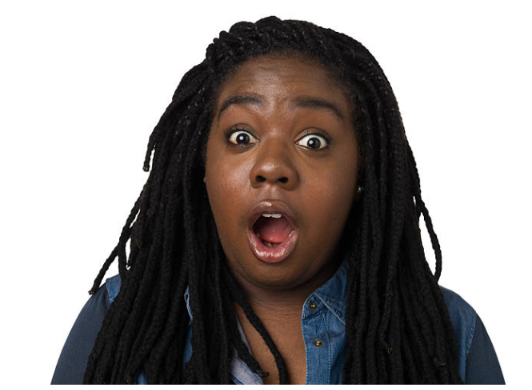
What muscle is being used in this image?
The epicranius

What muscle is highlighted?
The orbicularis oculi.

What muscle is highlighted
The orbicularis oculi

What muscle is being used in this image?
Left orbicularis oculi to wink

What muscle is highlighted
Orbicularis oris

What muscle is being used in this picture
Orbicularis oris

What is this muscle? name the origin and insertion
This the zygomaticus major. It originates from the zygomatic bone and inserts into the superolateral skin of the mouth

What muscle is being used here?
The zygomaticus major

What muscle is this? Name the origin and insertion point
The temporalis. It originates from the temporal lines of the parietal bone and inserts into the coronoid process of the mandible

What muscle is this? Name the origin and insertion point
The Masseter muscle. Originates from the zygomatic arch and inserts into the ramus and angle of the mandible

What muscle is being used here?
The Masseter and temporalis muscle

What muscle is this?
The Platysma

What muscle is being used here?
The Platysma

What muscle is this?
The superior rectus

What muscle is this?
The inferior rectus

What muscle is this?
The Lateral rectuse

What is this muscle?
The medial rectus

What muscle is being used here?
The medial rectus on the left eye
The lateral rectus on the right eye
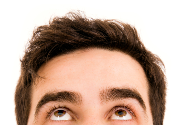
What muscle is being used here?
The superior rectus
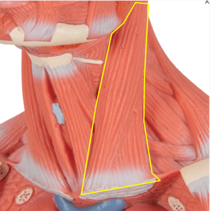
What muscle is this? Name the origin and insertion point
The sternocleidomastoid muscle. Originates from the manubrium of the sternum and the medial clavicle. Inserts in the mastoid process of the temporal bone

What muscles are being used in this image?
Both sternocleidomastoid muscles
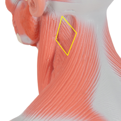
What muscle is this?
The splenius capitis

What muscles are being used in this image?
Both splenius capitis

What muscles are being used in this picture?
Left sternocleidomastoid and right splenius capitis
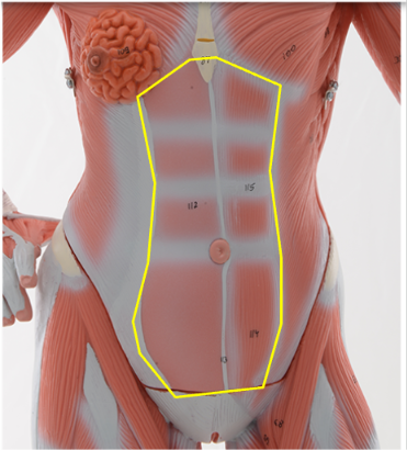
What muscle is this? Name the origin and insertion point
The Rectus abdominis.
It originates from the pubic crest of the coxal bone and inserts into the Xiphoid process of the sternum and the costal cartilage of ribs 5-7

What muscle is this?
The external oblique

What muscle is this?
The internal oblique

What are the muscles that are used in this image
Rectus abdominis
External oblique
Internal oblique
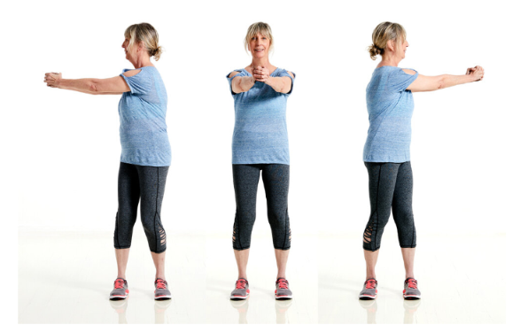
What muscles are being used in this image?
Internal oblique
External oblique
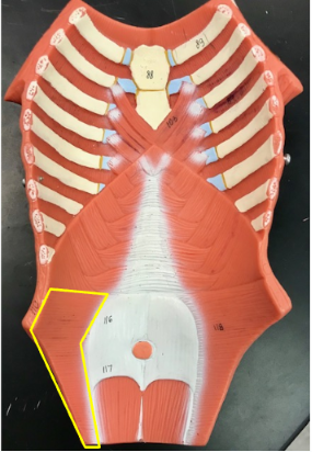
What muscle is this?
Transverse abdominis
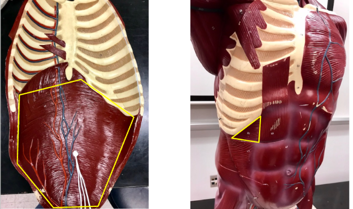
What muscles are highlighted in this picture?
The transverse abdominis

What muscle is highlighted in this picture?
The diaphragm

What muscle is being highlighted in this picture?
The diaphragm

What muscles are highlighted in this picture?
External intercostals
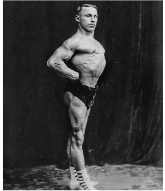
What muscles are being used in this image
External intercostals

What muscle is highlighted in this picture?
Internal intercostals
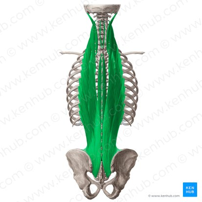
What muscle is this?
The erector spinae muscle

What muscle is being used in this image?
The erector spinae muscle