Lecture 14 - Muscle Contraction and Introduction to Enzymes
 Call Kai
 Learn
 Practice Test
 Spaced Repetition
 Match
 Flashcards
 Knowt Play
1/85

There's no tags or description
Looks like no tags are added yet.
Name | Mastery | Learn | Test | Matching | Spaced | Call with Kai | Chat |
|---|
No analytics yet
Send a link to your students to track their progress
86 Terms

Muscle hierarchy (largest to smallest)
Muscle → bundle of muscle fibers → individual muscle fiber (multinucleated cell) → myofibril → sarcomere → thick and thin filaments

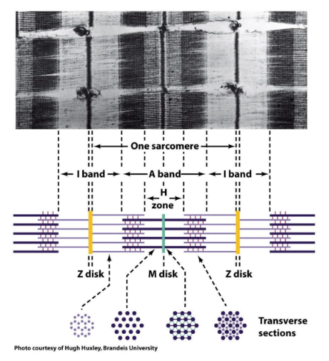
Sarcomere
The repeating contractile unit of a myofibril, bounded by two Z disks
contains I bands, A band, H zone, M disk, and thick and thin filaments
I band
Light band containing only thin (actin) filaments
spans across the Z disk
shrinks during contraction

A band
Dark band spanning the full length of the thick (myosin) filaments, including where they overlap thin filaments
length stays constant during contraction
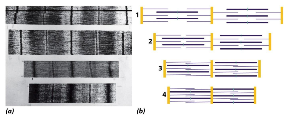
Sliding filament model of contraction
Sarcomere shortens as thin and thick filaments slide past each other and their overlap increases
I band and H zone shrink, A band length stays the same
filaments themselves do not shorten

Thick filament composition
Made of myosin
each myosin = 6 polypeptides: 2 heavy chains (~220 kD each) + 2 pairs of light chains (essential, ELC, and regulatory, RLC)

Myosin head
Globular N-terminal portion of the myosin heavy chain
binds actin and hydrolyzes ATP
each myosin molecule has two heads

Myosin tail
C-terminal coiled-coil rod formed by the two heavy chains' α-helices (~1600 Å long: 950 Å + 650 Å segments, ~20 Å wide)
tails bundle together to form the thick filament

Myosin Head Groups interaction
many head groups that interact with the thin chain that is composed of mostly actin. Here the micrograph depicts the complete thick filament with many myosin proteins interweaved

Thin filament composition
Mainly actin (F-actin), plus tropomyosin and troponin

G-actin vs F-actin
G-actin = globular monomer
F-actin = filamentous polymer of G-actin

Actin monomer binding and polymerization
Each actin monomer binds ATP and a divalent cation (Ca2+)
ATP hydrolysis —→ polymerization

Actin grooves
Grooves along the F-actin filament that contain the myosin-binding sites
tropomyosin lies in these grooves

Myosin head–actin binding
Each myosin head binds in the actin groove where several binding sites lie close together
many myosin heads attach along one thin filament at once
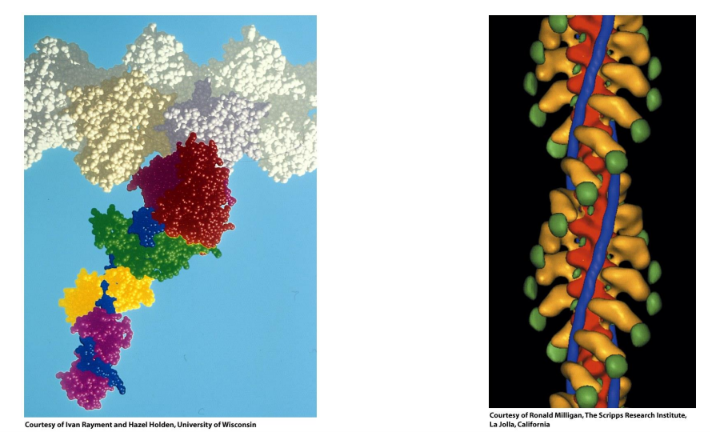
Tropomyosin
Long coiled-coil protein that lies in the grooves of the actin filament
at rest it blocks the myosin-binding sites on actin

Troponin
Three-subunit regulatory complex on the thin filament: TnC, TnI, TnT
TnC: Troponin subunit that binds Ca2+ (the calcium sensor)
TnI: Troponin subunit that binds actin and inhibits myosin binding (contains the "inhibitory" and "switch" regions)
TnT: Elongated troponin subunit that binds tropomyosin at its head-to-tail junction
Titin
Largest known polypeptide (34,350 residues) made of ~300-residue repeats
associated with thick filaments
acts as a spring that resists sarcomere overextension
Nebulin
Thin-filament-associated protein that sets the length of the thin filament
Dystrophin
Protein on the inner surface of the muscle plasma membrane that prevents tearing from the mechanical stress of contraction

Myosin cross-bridge cycle step 1
ATP binds the myosin head
actin-binding site opens and the head releases actin
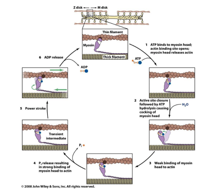
Myosin cross-bridge cycle step 2
Active site closes and ATP is hydrolyzed (ADP + Pi stay bound), "cocking" the head into a high-energy, extended state like a set mousetrap

Myosin cross-bridge cycle step 3
Cocked head binds weakly to a new actin site closer to the Z disk

Myosin cross-bridge cycle step 4
Release of Pi causes the head to bind actin tightly

Myosin cross-bridge cycle step 5 (power stroke)
Myosin head returns to its low-energy, compressed conformation and moves ~100 Å toward the Z disk along actin
this pulls the thin filament toward the M disk and shortens the sarcomere
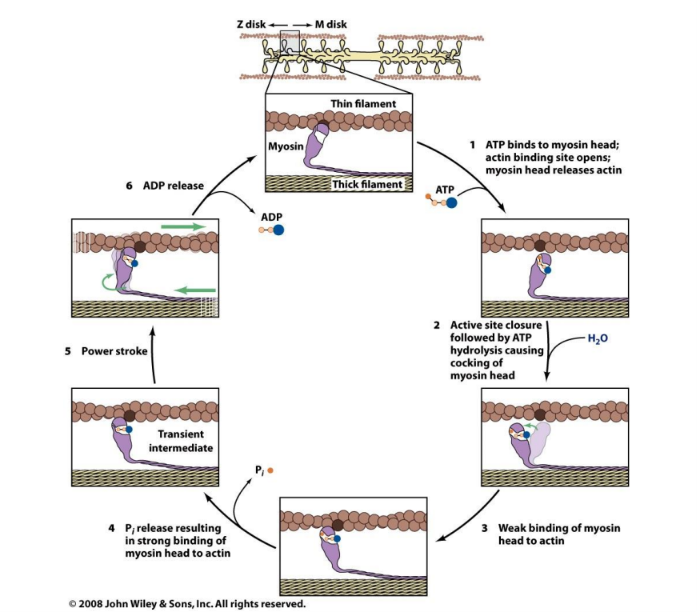
Myosin cross-bridge cycle step 6
ADP is released
myosin stays tightly bound to actin until a new ATP binds, completing the cycle

Role of Ca2+ in muscle contraction
Ca2+ binds troponin C, causing a conformational change that shifts tropomyosin within the actin groove, exposing the myosin-binding sites
without Ca2+, contraction cannot occur

Source of Ca2+ for contraction
The sarcoplasmic reticulum (specialized ER) releases stored Ca2+, raising cytosolic [Ca2+] from ~10^-7 M to ~10^-5 M

Relaxation and Ca2+
ATP-driven Ca2+ pumps return Ca2+ to the sarcoplasmic reticulum, lowering cytosolic Ca2+ so tropomyosin blocks myosin sites again
Four ways enzymes differ from chemical catalysts
1) Much higher reaction rates (10^6 to 10^12-fold)
2) Milder reaction conditions
3) Greater reaction specificity
4) Capacity for regulation

Do enzymes change ΔG?
No; enzymes change only the reaction rate (by lowering ΔG‡), not ΔG or the equilibrium position
Enzyme common names
Usually end in "-ase" and describe the reaction or substrate (e.g., alcohol dehydrogenase)
EC nomenclature
Enzyme Commission system giving each enzyme a 4-part number:
class.subclass.sub-subclass.serial number
so activity is unambiguous (e.g., aconitate hydratase, EC 4.2.1.3)

Catalytic power of enzymes
Understand the main idea here


Classes of Enzymes
Classes of Enzymes


Substrate specificity basis
Enzymes bind substrates through maximized noncovalent interactions:
van der Waals forces
electrostatic (ionic) bonds
hydrogen bonds
and hydrophobic interactions

Stereospecificity
Enzyme selectivity for one stereoisomer or one face/group of a molecule, because the chiral enzyme active site binds substrates asymmetrically

Aconitase stereospecificity example
Aconitase converts citrate to isocitrate
citrate is symmetric, yet the reaction occurs on only one of its two identical -CH2COO- arms
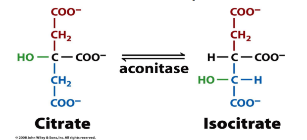
Prochiral center
An atom with two identical substituents (like citrate's C3) that becomes distinguishable—behaves like a chiral center—when bound to an asymmetric enzyme surface

Three-point attachment
A substrate binding an enzyme at three or more specific sites can only fit one way, so the enzyme can tell apart identical groups (e.g., pro-R vs pro-S H) and enantiomers

Chiral specificity figure
The substrate's hydrophobic, charged, and H-bonding groups fit complementary groups in the enzyme pocket
because binding uses multiple specific contact points, only one orientation or stereoisomer fits

Geometric specificity
Enzymes are selective about the identity of chemical groups but are generally not specific for one molecule
a small range of related compounds can bind or react

Chymotrypsin geometric specificity example
Chymotrypsin hydrolyzes both peptide (amide) bonds and ester bonds

Similar-shaped molecules can be toxic
Molecules resembling natural substrates/coenzymes can bind enzymes or receptors and disrupt them
example contrast: nicotinamide (NAD+ ring) vs tobacco nicotine

Niacin naming
Nicotinic acid was renamed niacin (by bread manufacturers) to avoid confusion with nicotine
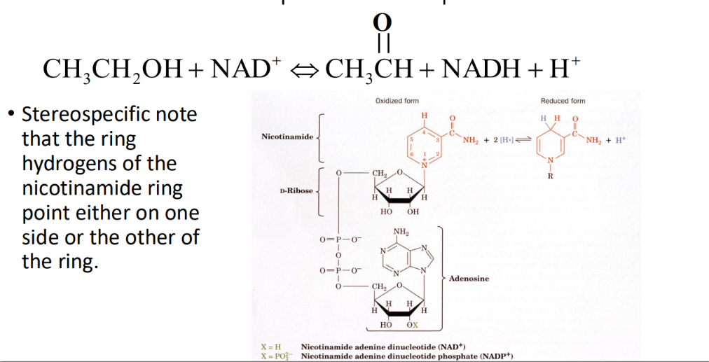
Yeast alcohol dehydrogenase (YADH) reaction
CH3CH2OH + NAD+ ⇌ CH3CHO (acetaldehyde) + NADH + H+

NAD+ structure
Nicotinamide + D-ribose + two phosphates + adenosine
NADP+ has an extra phosphate on the adenosine ribose 2'-OH

NAD+ vs NADH
NAD+ = oxidized (aromatic pyridinium ring)
NADH = reduced, with a hydride added at C4 of the nicotinamide ring, giving two C4 hydrogens on opposite faces

Re-side and si-side addition
The two faces of the nicotinamide ring
hydride added to the re face becomes H pro-R, to the si face becomes H pro-S

Deuterium experiment with YADH
CH3CD2OH + NAD+ → CH3CDO + NADD
the D is transferred to one specific face of NAD+, showing stereospecific hydride transfer

YADH deuterium experiment (reverse direction)
NADD + CH3CHO gives one enantiomer of CH3CHDOH, while NADH + CH3CDO gives the opposite enantiomer, proving YADH always transfers the same (pro-R) hydrogen
YADH stereospecificity
Yeast alcohol dehydrogenase is stereospecific for the pro-R hydrogen: it adds to (forward) or removes from (reverse) the pro-R position; with the other enantiomer, D is not transferred
Pro-R vs pro-S use by dehydrogenases
Both give identical products, but reactions with Keq for reduction >10^-12 tend to use pro-R H, while those with Keq <10^-10 tend to use pro-S H
reason unclear
Liver ADH Leu182 mutation
Wild-type error rate 1 in 7 billion
Leu182→Ala raises it to 1 in 850,000 (~8000-fold increase), showing amino acid side chains maintain stereospecificity
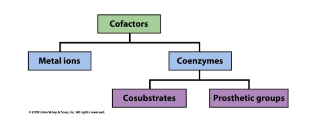
Cofactor
Non-protein component required for enzyme activity
two types: metal ions and coenzymes

Coenzyme
Small organic molecule that aids enzyme chemistry
subdivided into cosubstrates and prosthetic groups

Prosthetic group vs cosubstrate
Prosthetic group = permanently (tightly/covalently) associated with the enzyme
cosubstrate = transiently associated, binds and leaves like a substrate (e.g., NAD+)
Holoenzyme
Catalytically active enzyme with its cofactor bound
Apoenzyme
Enzyme without its cofactor (inactive)
What enzymes CAN do alone
Acid-base reactions
transient covalent bonds
charge-charge interactions
What enzymes CANNOT do alone (need coenzymes)
Oxidation-reduction reactions
carbon group transfers
Coenzyme regeneration
Coenzymes must be returned to their original form to keep functioning
e.g., glycolysis converts NAD+ to NADH, and without regenerating NAD+, glycolysis stops and death occurs

Common Coenzymes
Common Coenzymes


Vitamins and coenzymes
Water-soluble vitamins are coenzyme precursors
humans can't synthesize them and must get them from the diet (fat-soluble vitamins include A and D, as well as E and K)
Niacin (niacinamide) deficiency
leads to pellagra characterized by diarrhea, dermatitis and dementia
Pellagra was endemic is Southern United States in the early 20th century
Niacin can be synthesized from the essential amino acid, tryptophan
A corn diet prevalent at the time restricted the absorption of tryptophan causing a deficiency
Treatment of corn with base could release the tryptophan (Mexican Indians treated corn with Ca(OH)2 before making tortillas!)
Two general ways to regulate enzymes
1) Control the amount/availability of the enzyme (synthesis and degradation rates)
2) Control the enzyme's catalytic activity
Control of enzyme amount
Depends on synthesis rate (transcriptional or translational control) and degradation rate
Ways catalytic activity is altered
Positively or negatively, by conformational changes (e.g., small-molecule allosteric effectors altering the catalytic site) or covalent modifications (e.g., phosphorylation)
Allosteric effector examples (hemoglobin)
O2, CO2, and BPG binding alter hemoglobin's activity—an example of conformational regulation
Phosphorylation as regulation
Phosphorylation of Ser residues can activate or deactivate enzymes
usually hormonally controlled for a coordinated effect across tissues
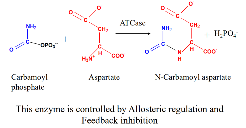
Aspartate transcarbamoylase (ATCase)
Catalyzes the first committed step of pyrimidine biosynthesis:
carbamoyl phosphate + aspartate → N-carbamoyl aspartate + H2PO4-
regulated allosterically and by feedback inhibition

ATCase kinetics curve
Sigmoidal (S-shaped) rate vs [aspartate], showing positive homotropic cooperative binding of aspartate

Homotropic vs heterotropic effects
Homotropic = substrate itself affects binding at other sites (aspartate on ATCase)
heterotropic = a different molecule acts as effector (CTP, ATP on ATCase)
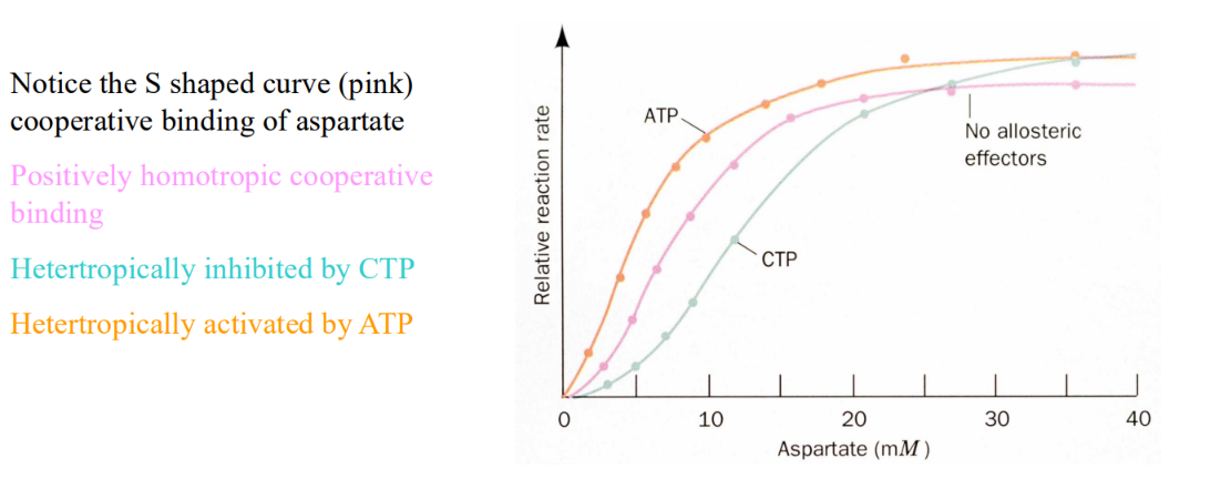
CTP and ATCase
CTP is a heterotropic allosteric inhibitor of ATCase (shifts curve right)
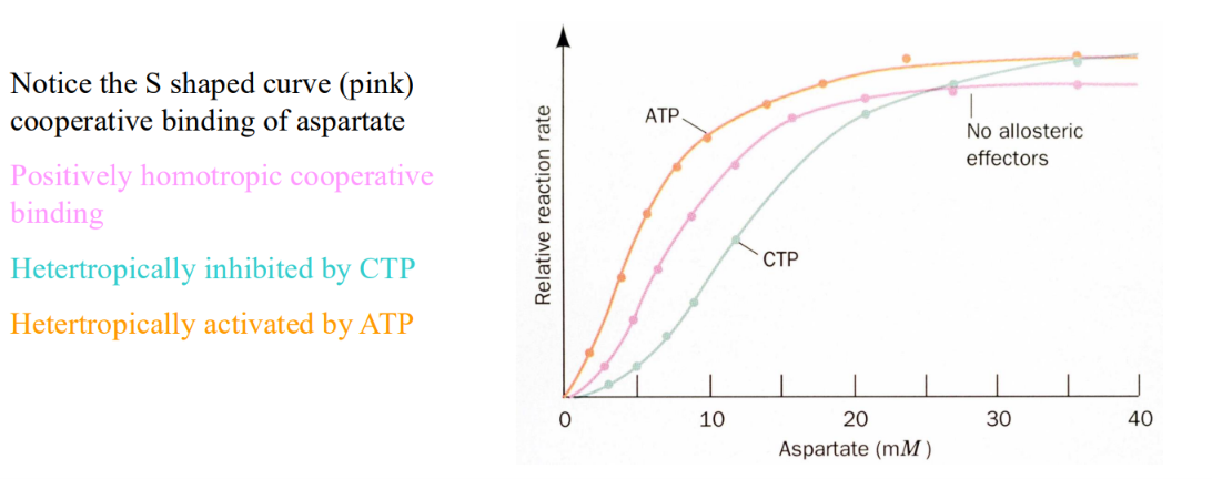
ATP and ATCase
ATP is a heterotropic allosteric activator of ATCase (shifts curve left)

Feedback inhibition
The end product of a metabolic pathway inhibits its own synthesis at the first committed step
e.g., CTP inhibits ATCase (6 enzymatic steps upstream)
Why CTP regulates ATCase
When DNA/RNA synthesis uses up CTP, CTP leaves ATCase and activity rises
when CTP is in excess, it rebinds and inhibits
Why ATP activates ATCase
Purines and pyrimidines are needed in equal amounts
when ATP exceeds CTP, ATP activates ATCase until ATP and CTP levels balance
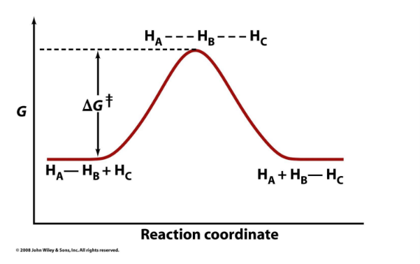
Activation energy (ΔG‡)
The free-energy barrier between reactants and the transition state
determines reaction rate
it is not the same as ΔG

Transition state (X‡)
Highest-energy, unstable arrangement along the reaction coordinate (e.g., HA---HB---HC) that all reactants must pass through

Negative ΔG and activation energy
Even if ΔG is negative (products lower in G than reactants), the reaction must still pass through the transition state, so ΔG‡ still controls the rate

Reaction intermediate
A relatively stable species (local energy minimum) between two transition states in a multistep reaction

Rate-limiting step
The step with the highest-energy transition state
the slowest step that controls the overall rate

Rate-limiting step example (two-step reaction A → I → P)
The step whose transition state is highest in free energy limits the rate:
in one profile A‡ is highest (first step limits)
in another, I‡ is highest (second step limits)

ΔΔG‡cat
The reduction in activation energy produced by a catalyst (uncatalyzed ΔG‡ minus catalyzed ΔG‡)

How enzymes speed reactions
Lower the activation energy (ΔG‡) without changing ΔG, so the reaction reaches equilibrium faster

Equilibrium and enzymes
Because an enzyme lowers ΔG‡ equally for the forward and reverse reactions, it speeds the approach to equilibrium without shifting the equilibrium constant