A2-A3 Digestive Organs
 Call Kai
 Learn
 Practice Test
 Spaced Repetition
 Match
 Flashcards
 Knowt Play
1/10

There's no tags or description
Looks like no tags are added yet.
11 Terms

Relationship of digestive organs to pertitoneum
Peritoneum:
The peritoneum is a serous membrane in the abdominopelvic cavity. It has two layers:
Parietal peritoneum: Lines the inside of the body wall.
Visceral peritoneum: Covers the external surface of most digestive organs.
Peritoneal cavity: The space between the two layers.
Mesentery:
A mesentery is the 2 layers of peritoneum fused back to back, it connects the body wall to the digestive system.
Its functions are to:
Provide routes for blood vessels, lymphatics, and nerves to reach digestive organs/viscera.
Hold organs in place.
Store fat.
(some mesenteries are called omenta or ligaments)
Types of Mesentary
1. Dorsal mesentery
Most mesenteries are dorsal.
They attach the digestive organs to the posterior (back) abdominal wall.
2. Ventral mesentery
Attaches certain organs to the anterior (front) abdominal wall.
(Example: The mesentery that extends from the liver to the anterior abdominal wall.)
3. Retroperitoneal organs
During development, some organs become attached directly to the posterior abdominal wall.
As a result:
They lose their mesentery.
They end up behind the peritoneum.
Examples: pancreas, duodenum, large intestine
4. Intraperitoneal organs
These organs Keep their mesentery. Remain suspended within the peritoneal cavity.
Example: Stomach, jejunum, ileum
General Parts of the Stomach
Esophagus:
Extends from the pharynx to the stomach
Transports food and liquids from the throat to the stomach
Stomach
Function: acidic and mechanical digestion
Converts food into chyme
Small intestine
Extending from the pyloric sphincter to the ileocecal sphincter
3 subdivisions: duodenum, jejunum, & ileum
Function: digestion is completed and virtually all absorption occurs
Large intestine
Extends from the ileocecal valve to the anus
Subdivisions: cecum, appendix, colon, rectum, anal canal
Absorbs most of the remaining water, eliminates Indigestible food resides from the body

Parts of greater and lesser onemtum
Greater omentum
• Extends superiorly, laterally to the left, and inferiorly from the greater curvature of the stomach and the proximal part of the duodenum
-Gastrophrenic ligament: between the greater curvature of the stomach and the diaphragm
-Gastrosplenic ligament: between the greater curvature of the stomach and the spleen
-Gastrocolic ligament: from the inferior portion of the greater curvature of the stomach, descends anteriorly of the transverse colon and then ascending posteriorly, fusing with the visceral peritoneum of the transverse colon and the superior layer of its mesentery
Lesser omentum
• Connects the lesser curvature of the stomach and the proximal part of the duodenum to the liver
-Hepatogastric ligament
-Hepatoduodenal ligament

Peristalsis
Definition: Adjacent segments of the alimentary canal contract and relax alternating
Function: Moves food distally (forward) along the digestive tract.
Main purpose: Propulsion (pushing food forward), although some mixing may occur.
Bolus: A ball of chewed food mixed with saliva that moves through the esophagus.
Upper esophageal sphincter (UES): food enters forms pharynx to esophagus
Lower esophageal sphincter (LES): Relaxes for food entry to stomach and helps prevent stomach contents from flowing backward.

Curvature of Stomach
Cardia: surrounds the cardial orifice, the opening of the esophagus into the stomach
Fundus: dome-shaped part, tucked beneath the diaphragm
Body: the midportion of the stomach
Pylorus (Antrum & Canal): the distal part of the stomach; continues to the pyloric sphincter
Rugae: These longitudinal folds unfold to expands the stomach volume from 50ml to 4L
The stomach has two curvatures:
Greater curvature: the convex lateral surface of the stomach
Lesser curvature: the concave medial surface of the stomach
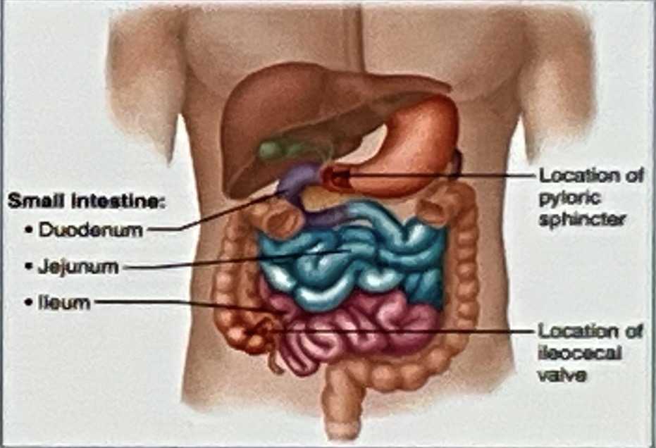
Small Intestine
extending from the pyloric sphincter to the ileocecal valve (sphincter)
the longest part of the alimentary canal
Although 6-7m long in a cadaver, it is only 2-4 m long du ng life because of muscle tone
about half the diameter of the large intestine (2.5-4cm)
Duodenum: the shortest intestinal subdivision; retroperitoneal
Jejunum: 2.5m long; intraperitoneal
Ileum: approximately 3.6m in length, intraperitoneal

Layers of Small Intestine
Superior part (1st part)
Receives chyme from the stomach through the pyloric sphincter.
2. Descending part (2nd part)
(Major Duodenal Papilla: small opening in the inner duodenum for entry of digestive juices (enzymes from pancreas & bile from gallbladder))
3. Horizontal part (3rd part)
Travels horizontally from right to left across the abdomen.
4. Ascending part (4th part)
Travels upward and joins the jejunum, the next part of the small intestine.

Parts of the Duodenum
Superior part (1st part)
Receives chyme from the stomach through the pyloric sphincter.
2. Descending part (2nd part)
(Major Duodenal Papilla: small opening in the inner duodenum for entry of digestive juices (enzymes from pancreas & bile from gallbladder))
3. Horizontal part (3rd part)
Travels horizontally from right to left across the abdomen.
4. Ascending part (4th part)
Travels upward and joins the jejunum, the next part of the small intestine.

Overview of large intestine
Teniae coli: three thickened bands of longitudinal smooth muscle fibers
Haustra: sacculations/pouches of the colon between the teniae coli
Omental appendices: small and fatty appendices
Semilunar folds: fold that help form the haustra
ileocecal valve: connects from ilieum to cecum
Cecum: the first part of the large intestine, continues along ascending colon
Appendix: The appendix is a small, closed-ended pouch attached to the back-inner side of the cecum, below, the small intestine connects to it.
Ascending colon — travels upward on the right side.
Right colic flexure (hepatic flexure) — the bend near the liver.
Transverse colon — travels across the abdomen from right to left.
Left colic flexure (splenic flexure) — the bend near the spleen.
Descending colon — travels downward on the left side.
Sigmoid colon — an S-shaped section that leads to the rectum.


Rectum and anal canal
1. Rectum
The final storage area for feces before it leaves the body.
2. Anal canal
The last segment of the large intestine.
Opens to the body exterior at the anus
3. Internal anal sphincter
Made of smooth muscle and works Involuntary
Relaxes during a bowel movement.
4. External anal sphincter
Made of skeletal muscle and works Voluntary
Helps you hold in feces until you are ready to have a bowel movement.