Heart anatomy
 Call Kai
 Learn
Learn Practice Test
 Spaced Repetition
 Match
Match Flashcards
 Knowt Play
1/58

There's no tags or description
Looks like no tags are added yet.
Name | Mastery | Learn | Test | Matching | Spaced | Call with Kai | Chat |
|---|
No analytics yet
Send a link to your students to track their progress
59 Terms
Costal cartilage
Segments of hyaline cartilage that connect the medial ends of ribs 1-7 to the sternum
Costal margin
Inferior edge of the rib cage

Intercostal space
The space between two adjacent ribs wherein nerves, arteries, and veins run
Nerves, arteries, veins
What structures are in the neurovascular bundle? (3)
Mid-axillary line
Imaginary line from the middle of the armpit that divides the body into the anterior and posterior halves
Manubrium, body, xiphoid process
Three parts of the sternum:
Manubrium
Most superior part of the sternum; contains the sternal notch on the superior end and attaches to the body of the sternum at the sternal angle (green part)

Sternal angle
Place where the manubrium and body of the sternum meet; around the costal cartilage of the 2nd ribs

Body
Middle section of the sternum (blue part)

Xiphoid process
Inferior end of the sternum, made of cartilage (purple part)

Thoracoabdominal diaphragm
What is deep to the costal margin?
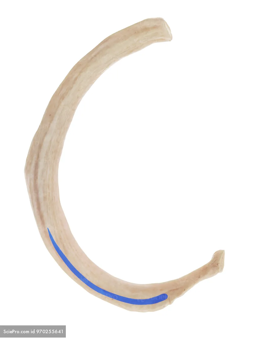
Costal groove
Space on the inferior surface of each rib wherein the intercostal nerve, artery, and vein run
Clavicle
Collarbone; attaches the upper limb to the rib cage
2nd
Which rib articulates at the sternal angle?
Internal thoracic, internal mammary arteries
Two names for the arteries that travel vertically on either side of the deep surface of the sternum, supplying blood to the sternum and intercostal spaces
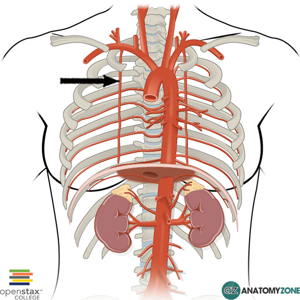
Subclavian arteries
Where do the internal mammary arteries originate from?
round, rigid
In differentiating between arteries and veins, arteries are more: (2)
flat, floppy
In differentiating between arteries and veins, veins are more: (2)
Left internal thoracic artery
Structure that might be missing in patients that have received coronary artery bypass grafting:
Costal groove
Where do the neurovascular bundle run?
vein, nerve
Which structure is the most superior (1) and inferior (2) in each neurovascular bundle?
Internal thoracic vein, internal mammary vein
Two names for the large vein that runs alongside the internal mammary artery and drains deoxygenated blood from the thoracic region
Anterior intercostal artery
Arteries that supply the muscles and skin within the intercostal spaces; part of the neurovascular bundle
Phrenic nerves
Pair of nerves that descend the thoracic region, runs between the pericardial and pleural sacs to control the movement of the diaphragm
Yellow in diagram

Somatomotor, somatosensory, sympathetic
Phrenic nerves have these types of axons: (3)
Right ventricle
Most anterior chamber of the heart; makes up most of the inferior edge of the heart and is just deep to the sternum
Left atrium
Chamber of the heart that is most posterior
Coronary sulcus
Green line; most prominent on posterior side of the heart

Pulmonary veins, left atrium, coronary sinus
Landmarks on the posterior side of the heart: (3)
Coronary sinus
From the posterior view of the heart, what is the structure with the arrow pointing at it?

Anterior
The pulmonary trunk is _________ and to the right of the aorta (from our view)
right, left brachiocephalic veins
Veins that empty into the superior vena cava (blue structures)

Brachiocephalic trunk, left common carotid artery, left subclavian artery
Name the branches of the aortic arch from right to left (patient perspective)

Pulmonary trunk
Great vessel that exits from the right ventricle; divides into the right and left pulmonary arteries
Ligamentum arteriosum
Fibrous cord that connects the pulmonary trunk/left pulmonary artery to the aortic arch
Remnant of ductus arteriosis, a crucial part of fetal circulation that closes at birth and becomes fibrous
Pulmonary trunk, aortic arch
The ligamentum arteriosum connects these structures: (2)
Brachiocephalic trunk
From the patient’s perspective, the right-most branch of the aortic arch (our left-most)
Left common carotid artery
Middle branch of the aortic arch

Left subclavian artery
From the patient’s perspective, the left-most branch off of the aortic arch (our right-most)

Right coronary artery

Marginal artery

Posterior interventricular, posterior descending artery
Two names for the artery that usually branches off of the right coronary artery in the posterior side of the heart
Determines right/left/co-dominant
Left dominant

Codominant

Right dominant

Circumflex artery
Number 4

Left anterior descending, anterior interventricular artery
Number 6; two names

Great cardiac vein
Vein that runs with the left coronary arteries

Posterior interventricular artery
The middle cardiac vein runs with this artery:
Left coronary arteries
The great cardiac vein runs with:
Marginal artery
The small cardiac vein runs with this artery:
Coronary sinus
The three cardiac veins empty into:
Superfiscial
The coronary veins are _________ to the coronary arteries
Pectinate muscles
Rough muscle that is characteristic of the right atrium; merges with smooth muscle at the crista terminalis
Right ventricle
The septomarginal band is characteristic of the:
Right atrium
The fossa ovalis is characteristic of the:
Coronary sulcus
Divides the atria and ventricles on the exterior of the heart
Ascending aorta
First part of the aorta that comes immediately out of the left ventrical PRIOR to the aortic arch
AV
The papillary muscles and chordae tendineae in the ventricles moderate the opening and closing of these valves: