Respiratory Tract Infections
 Call Kai
 Learn
 Practice Test
 Spaced Repetition
 Match
 Flashcards
 Knowt Play
1/103

There's no tags or description
Looks like no tags are added yet.
Name | Mastery | Learn | Test | Matching | Spaced | Call with Kai | Chat |
|---|
No analytics yet
Send a link to your students to track their progress
104 Terms
In host cells viruses cause
Tissue damage and Inflammation
Examples of physcial manifestations of viral infections
Pharyngitis
Hepititis
Pharyngitis is the
inflammation/ulceration of the tonsillar/pharyngeal tissue
Hepititis is
inflammation of the liver, progressing to cirrhosis/carinoma
Immunosuppressants can
reduce an exaggerated inflammatory response
Immunosuppressants reduce repsonse by
Reduction anibody productions
inhibit production of immune cells
Block chemical messengers
Lysogenic Viruses
are non-virulent and do not cause disease
Lytic Viruses
are virulent and cause disease
Step 1 of a bacteriophage life cycle - virulent/non-virulent
Attachment and injection - The phage attaches to the host bacterial cell and injects its double-stranded DNA into the cell.
Step 2 of a bacteriophage life cycle - virulent/non-virulent
Circularization and decision point The injected phage DNA circularizes. At this point, the virus "chooses" one of two paths: the lytic cycle or the lysogenic cycle.
Step 3A - bacteriophage life cycle - virulent
Synthesis and assembly New phage DNA and proteins are synthesized using the host cell's machinery, then assembled into complete virus particles (virions).
Step 4A - bacteriophage life cycle - virulent
Lysis and release The host cell bursts (lyses), releasing the newly formed phage virions to infect other cells.
Step 3B - bacteriophage life cycle - non-virulent
Integration The phage DNA integrates into the bacterial chromosome by recombination, becoming a prophage.
Step 4B - bacteriophage life cycle - non-virulent
Normal reproduction The lysogenic bacterium reproduces normally, copying the prophage along with its own DNA through many cell divisions.
Step 5B - bacteriophage life cycle - non-virulent
Excision (occasionally) Occasionally, the prophage excises itself from the bacterial chromosome by another recombination event, initiating the lytic cycle and starting the process over.
Step 1 - General Viral Reproductive Cycle
Attachment The virus (a capsid enclosing its DNA) binds to the surface of the host cell.
Step 2 - General Viral Reproductive Cycle
Entry and uncoating The virus enters the cell and its DNA is released (uncoated) from the capsid.
Step 3 - General Viral Reproductive Cycle
Replication The viral DNA is replicated, producing multiple copies of the viral genome.
Step 4 - General Viral Reproductive Cycle
Transcription The viral DNA is transcribed into mRNA.
Step 5 - General Viral Reproductive Cycle
Translation of viral proteins The mRNA is translated to produce capsid proteins (and other viral components).
Step 6 - General Viral Reproductive Cycle
Self-assembly and exit New virus particles self-assemble from the viral DNA and capsid proteins, then exit the cell (typically by budding or lysis) to infect new host cells.
Fomite is
an inanimate object that can transmit infection between people
Myeloperoxidase - found in neutrophils
makes mucus green due to increase neutrophil numbers during infection
Croup
Childhood infection causing swelling inside trachea
Otitis media
Inflammation of the middle ear
How many transmission modes are there?
Three: direct, aerosol, indirect
Direct transmission
Contact with infected person
Aerosol transmission
Inhalation of infected droplets
Indirect transmission
contact with infected fluids on surfaces/objects
The most common cause of the common cold are
Rhinoviruses ~40%
Rhinoviruses have how many virus types
100 distinct types
The common cold is caused by how many specific types
~200 specific viruses
Other major causes of the cold
Coronavirus
adenovirus
respiratory synytial virus (RSV)
Symptoms of the common cold
runny/stuffy nose
sneezing
sore throat
cough
cogestion
mild body aches/headache
fever is rare and mild
Common colds are typically
upper respiratory tract infections
Influenza is caused by
Type A, B and C viruses
Flu symptoms eppear
Quickly and severly
Flu symptoms
Runny/stuff nose, sneezing, sore throat, cough, muscle aches, fatigue, usually high fever
Flu can affect
upper and lower respiratory tract
Mucus functions
Lubricates, protects (antimicrobial), and blocks (prevents spread of infections)
Viruses cause what percent of RTI
70%
Upper Respiratory Tract
Nose
Sinuses
Throat
Lower Respiratory Tract
Airways and lungs
LRTI’s
Influenza, croup, bronchiolitis, pneumonia
URTI’s
Connon cold, infulenza
URT viruses
Rhinovirus, Coronavirus, Influenza Virus, RSV, parainfluenza virus, herpes virus, adenovirus, boca virus, coxsackievirus
LRT
Influenza Virus, Parainfluenza Virus, RSV, Adenovirus, Bocavirus, MEtapneumovirus
Rhinitis is
Inflamation of the mucus membrane of the nose
Allergic Rhinitis is
caused by an allergen (seasonal/year round)
Viral non-allergic rhinitis
caused by rhinoviruses, coronavirus, adenovirus, RSV
Viral rhinitis treatment
No specific tratment required
Coricosteroid nasal spray - anti-inflammatory
Deongestant nasal spray - reduce swelling of nasal blood vessels
Allergic rhinitis treatment
Anti-histamine compound
Pharyngitis
erythema (redness) of the oropharynx, with or without tonsillar inflammation.
Tonsils
pair of soft tissue masses at the rear of the pharynx
Laryngitis
hoarse or gravelly voice (inflamed vocal cords altering airflow).
Laryngitis Viruses
Rhinovirus, parainfluenza, RSV, adenovirus
Pharyngitis and Tonsilitis are
commonly viral (70-80%)
Bacterial Pharyngitis and Tonsilitis are caused by
20–30% caused by group A beta-haemolytic streptococcus.
How many LRTI’s are there?
Four
Tracheitis is
Inflamation of the trachea
Bronchitis is
Inflamation of the bronchus
Bronchiolitis is
Inflamation of the bronchioles
Pneumonia is
Inflamation of the alveoli
Pnemonia cause
The air sacs (alveoli) become inflamed and fill with fluid instead of air, making breathing difficult; the lungs may not get enough oxygen.
Typical Pneumonia viruses in adults
Influenza A and B
Typical pneumonia virus in children
RSV
Pneumonia complications
Respiratory failure – low oxygen / high carbon dioxide in the lungs.
Sepsis – overwhelming inflammatory response → multiple organ failure, life-threatening.
Pleural effusion – fluid builds up around the lung.
Influenza Viruses Family
Orthomyxoviridae
Types of Influenza Viruses
A & B – cause seasonal flu epidemics each year.
C – mild respiratory illness, not thought to cause epidemics.
D – primarily affects cattle; not known to infect humans.
All influenza viruses are
envloped viruses with segmented RNA
Influenza A and B have
8 RNA segemnts and 2 types of surface proteins
Influenza C has
7 RNA segments and 1 type of surface proteins
Type A target species
Birds, swine, humans, seals, horses
Type B
Humans
Type C
Humans and swine
Influenza A and B surface protiens
Spikes of glycoproteins: Haemagglutinin (HA, ~80%) and Neuraminidase (NA, ~20%) — targets for the host immune response.
HA (H1–H18) – role in viral attachment to host cells.
NA (N1–N11) – role in release of new virus particles by budding.
Surface proteins determine the subtype (e.g., H1N1, H3N2).
Influenza C surface proteins
Hemagglutinin-esterase-fusion, HEF
Pathogenesis of Influenza A Virus
Transmission and Entry
Attachment and Cell Entry
Replication
Cell damage and Tissue Injury
Immune Response and Cytokine Storm
Systemic Effects and Complications
Immune Evasion
Step 1 - Pathogenesis of IAV
Transmission and Entry: Spreads via aerosol droplets and fomites (contaminated surfaces).
Enters through the respiratory tract, targeting epithelial cells of the airways (trachea, bronchi, and bronchioles).
Step 2 - Pathogenesis of IAV
Attachment and Cell Entry
The viral hemagglutinin (HA) protein binds to sialic acid receptors on the host cell surface.
The virus is taken up by receptor-mediated endocytosis.
The low pH inside the endosome triggers a conformational change in HA, causing fusion of the viral envelope with the endosomal membrane.
The viral RNA is released into the cytoplasm.
Step 3 - Pathogenesis of IAV
Replication
The viral RNA is transported to the nucleus, where it is transcribed and replicated by the viral RNA-dependent RNA polymerase.
The neuraminidase (NA) protein later cleaves sialic acid, allowing newly formed virions to bud off and release from the cell.
Step 4 - Pathogenesis of IAV
Cell Damage and Tissue Injury
Infection causes necrosis and apoptosis of epithelial cells, stripping the ciliated airway lining.
This impairs mucus clearance and exposes the basement membrane, leading to inflammation and secondary bacterial infections.
Step 5 - Pathogenesis of IAV
Immune Response and Cytokine Storm
Infected cells release cytokines and chemokines, recruiting neutrophils, macrophages, and lymphocytes.
In severe cases, an excessive response causes a "cytokine storm" — high levels of TNF-α, IL-6, and interferons — leading to widespread tissue damage.
Cytotoxic T cells (CD8+) kill infected cells, contributing to both viral clearance and tissue pathology.
Step 6 - Pathogenesis of IAV
Systemic Effects and Complications
Local damage plus inflammatory mediators cause fever, myalgia, and malaise.
Complications include:
Primary viral pneumonia
Secondary bacterial pneumonia (e.g., Streptococcus pneumoniae, S. aureus)
Acute respiratory distress syndrome (ARDS)
Exacerbation of asthma/COPD, and in rare cases myocarditis or encephalitis
Step 7 - Pathogenesis of IAV
Immune Evasion
Antigenic drift (small mutations in HA/NA) allows the virus to evade pre-existing immunity.
Antigenic shift (reassortment of genome segments between strains) can create novel viruses with pandemic potential.
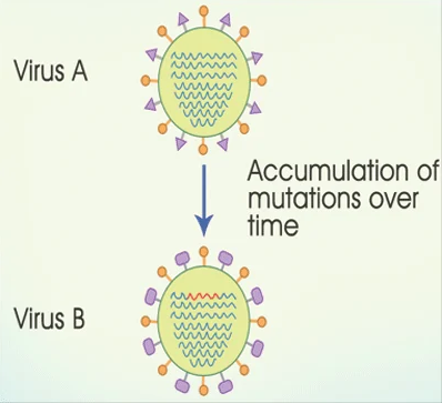
Antigenic drift
accumulation of mutations in the genes that code for regions on HA and NA
the virus becomes unrecognisable in the host due to changes in surface proteins
leads to an infection and successful transmission since it is a new strain
Antigenic drift is a
slow, gradual, frquent process
Antigentic Drift can
cause an epidemic
Influenza A, B and C undergo
Antigenic drift

Antigenic Shift
intermixing of two or more viral genomes inside the host cell
a new combination of genes
a novel subtype of the virus
not recognized by the host genome
Only inflenza A undgergoes
Antigenic shift
Antigenic shift is
a rare, sudden process
Antigenic shift can cause
a pandemic
Pandemic
Across international boarders
Epidemic
Contained within an area
Antigenic shift can jump
from one species to another
Examples of antigenic shift
Spanish flu (H1N1), Hong Kong flu (H3N2, 1968 pandemic)
Examples of Antigenic Drift
outbreaks of H3N2.
Influenza B has
two evolutionary lineages
Influenza B is more
antigenically stable than influenza A