Venous Physiologic
 Call Kai
 Learn
Learn Practice Test
 Spaced Repetition
 Match
Match Flashcards
 Knowt Play
1/75

There's no tags or description
Looks like no tags are added yet.
Name | Mastery | Learn | Test | Matching | Spaced | Call with Kai | Chat |
|---|
No analytics yet
Send a link to your students to track their progress
76 Terms
Anterior Accessory Vein
Runs parallel and anterior to femoral vein
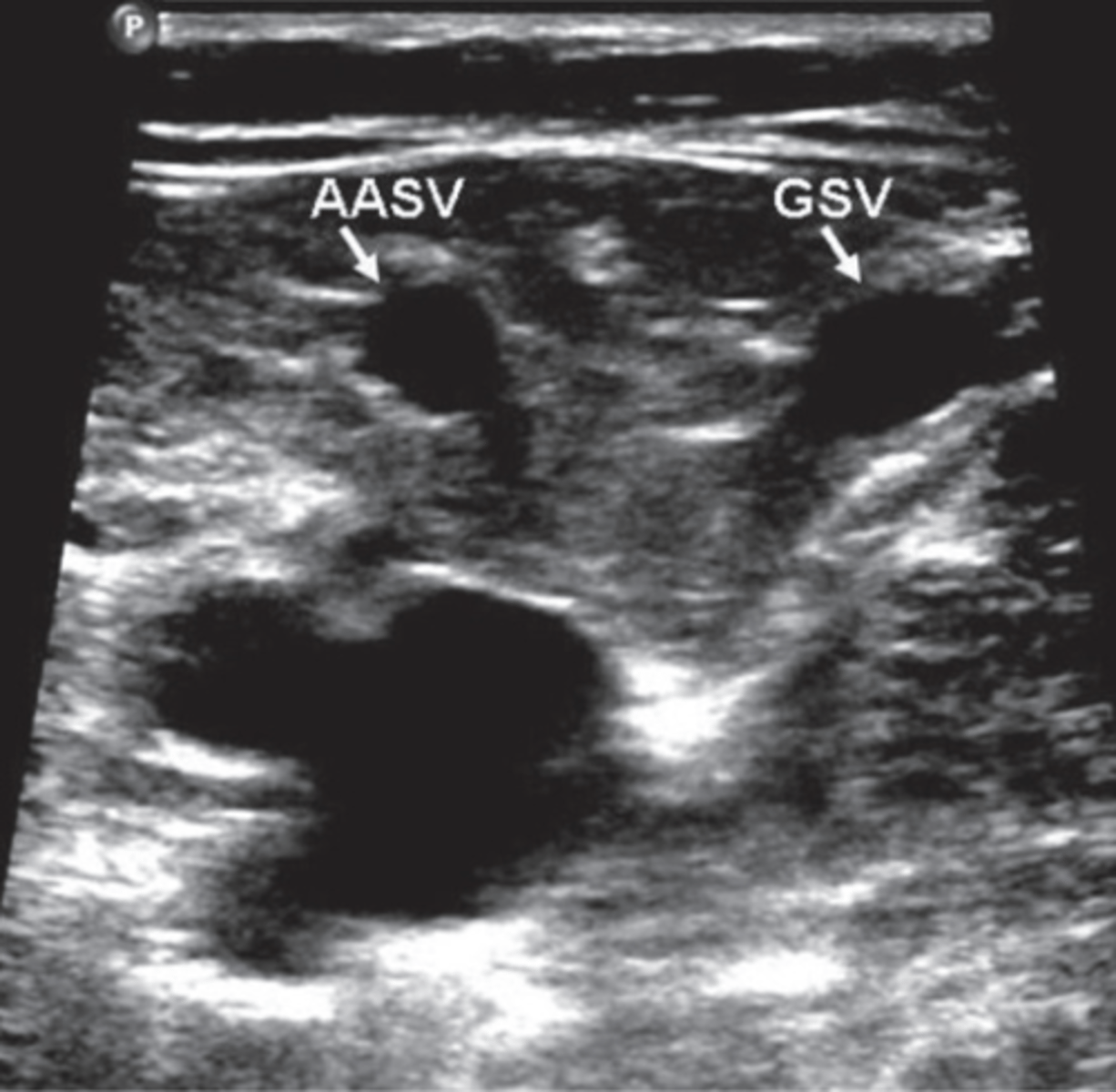
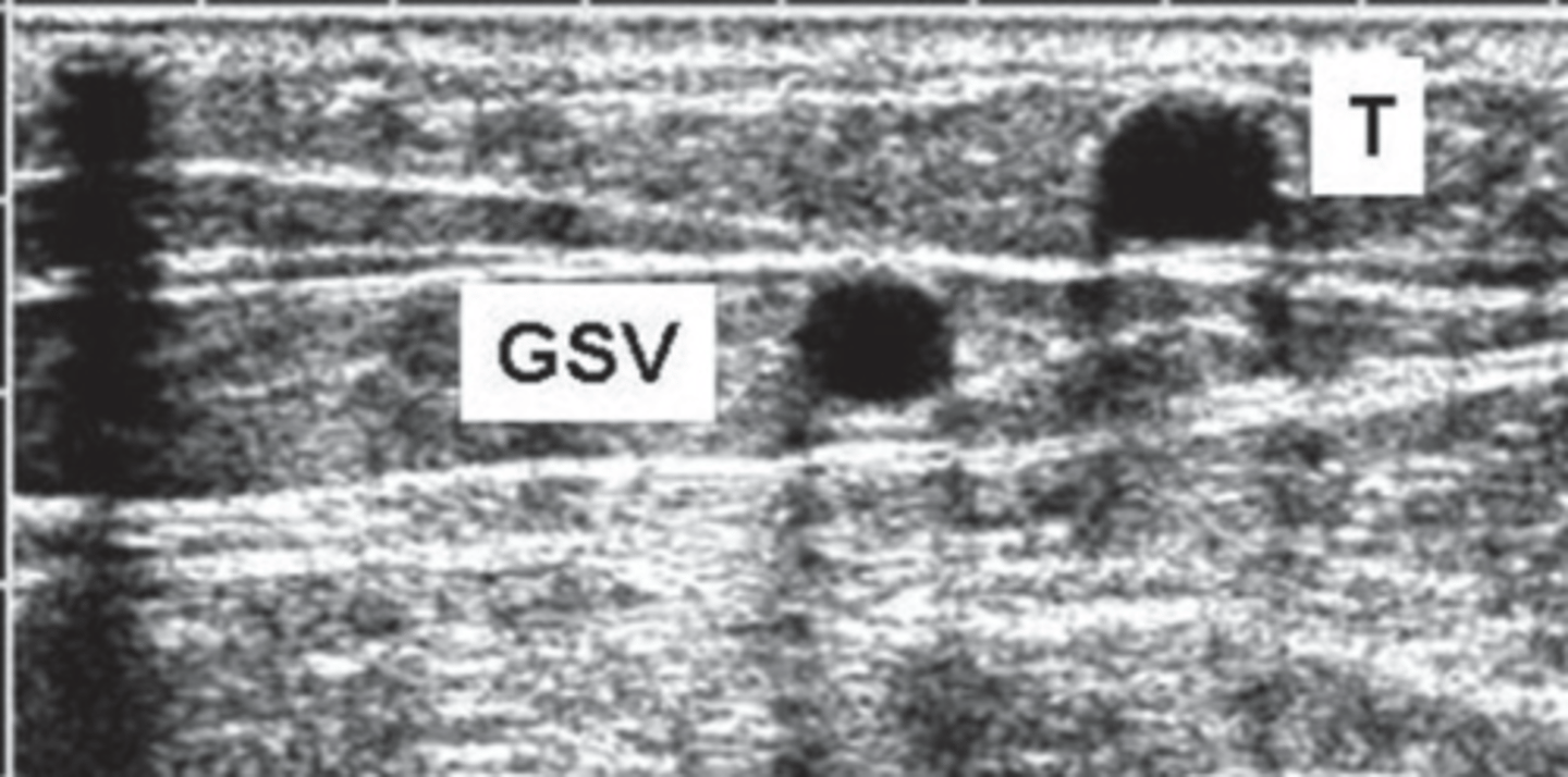
GSV
Saphenous Eye
Between facial layers
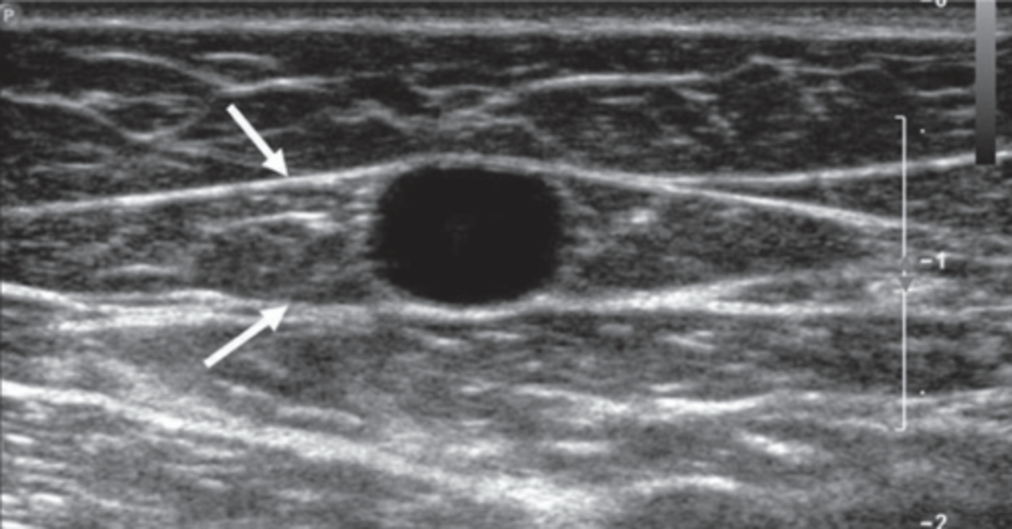
GSV in Calf
Angle Sign
Triangular form between gastrocnemius muscle and tibial bone
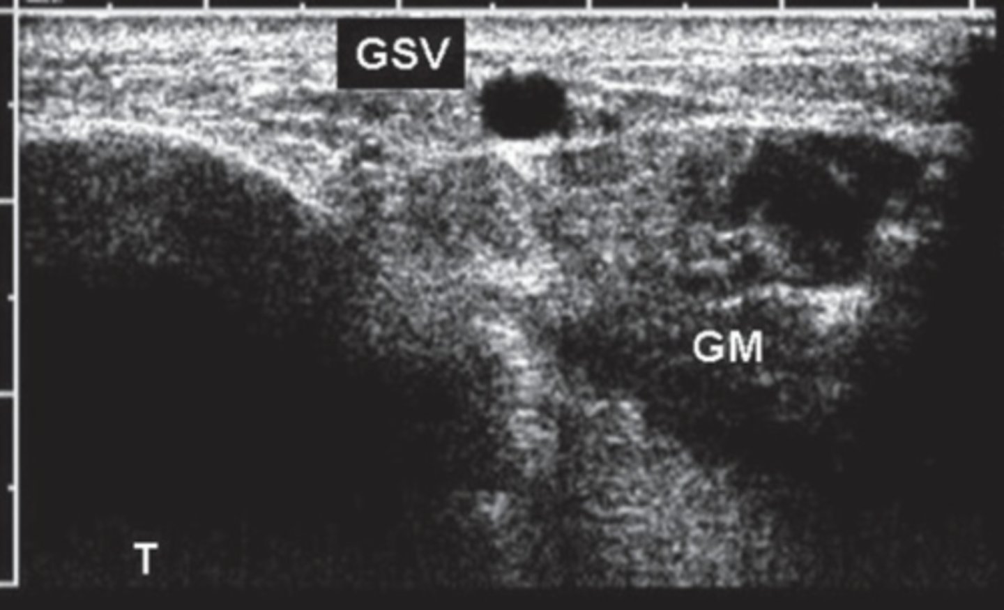
Tributary
Outside fascial plane
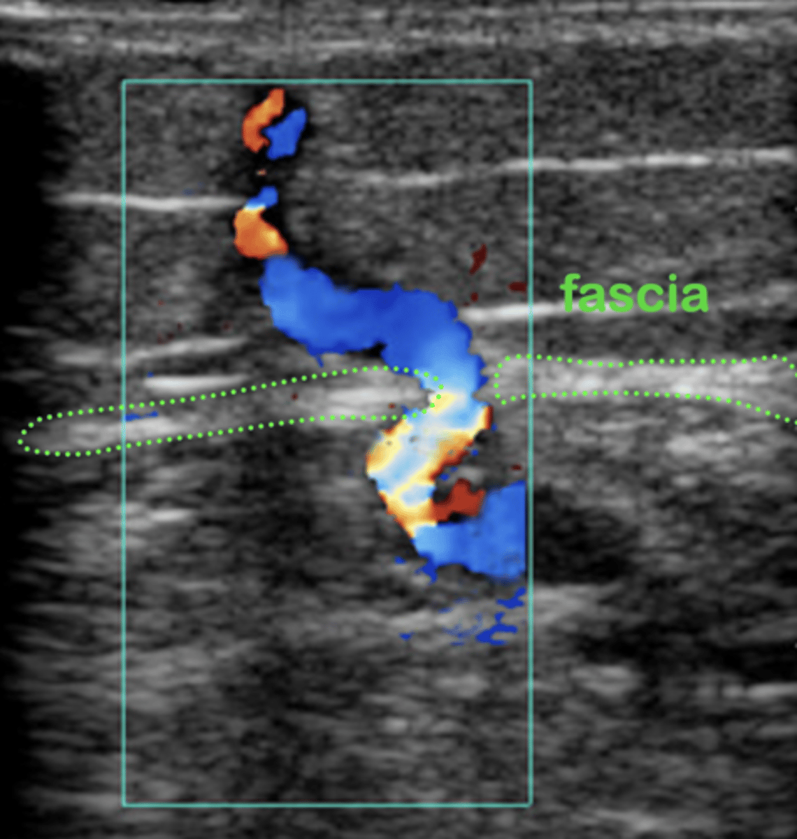
Perforator
Pass through muscular fascia layers - connect deep system to superficial system
Venous Valves in Lower Extremity
Femoral 1-6 valves
Pop 0-4 valves
GSV 10-12 valves
SSV 7-9 valves
Calf veins 8-19 (every 2 cm)
Perforations 1 each
Veins with No Valves
Soleal
Internal iliac
Common iliac
IVC
Subclavian VV
Innominate
SVC
Distribution of Venous Valves in Deep System
2.1 %
Distribution of Venous Valves in Superficial System
16.8 %
Distribution of Venous Valves in Perforator System
8.4 %
Risk Factors for Leg Swelling
Heart dysfunction (CHF)
Kidney disease
Liver disease
Lymphatic system/Lymphedema
Pulmonary HTN
High BMI
Medications (calcium channel blockers)
Multifactorial
Age
Family History
Obesity
Oral Contraceptive use
Smoking/Tobacco
Pregnancy
History of DVT
Prolonged standing or sitting
Sedentary lifestyle
Gender (women)
Primary Valvular Insufficiency
Congenital absence of veins
Secondary Valvular Insufficiency
Damaged valves caused by thrombotic events
Chronic Venous Insufficiency
Obstruction & valve insufficiency
Chronic Venous Valvular Insufficiency
Impaired superficial or deep flow
Causes venous hypertension
Duplex is primary modality for diagnosing
Symptoms of DVT
Swelling
Shortness of breath - emboli
Redness
Warmth
Painful
Throbbing
Phlebitis/Thrombophlebitis
Symptoms of CCVI
Swelling
Dull ache/heaviness or painful
Skin changes
Phlebitis/Thrombophlebitis
Ulcers
Varicose veins
Post-Thrombotic Syndrome
Venous stress disorder
Chronic pain and swelling at the site of a previous DVT
Symptoms of Post-Thrombotic Syndrome
Edema
Chronic pain
Skin changes
Heaviness of affected limb
Swelling
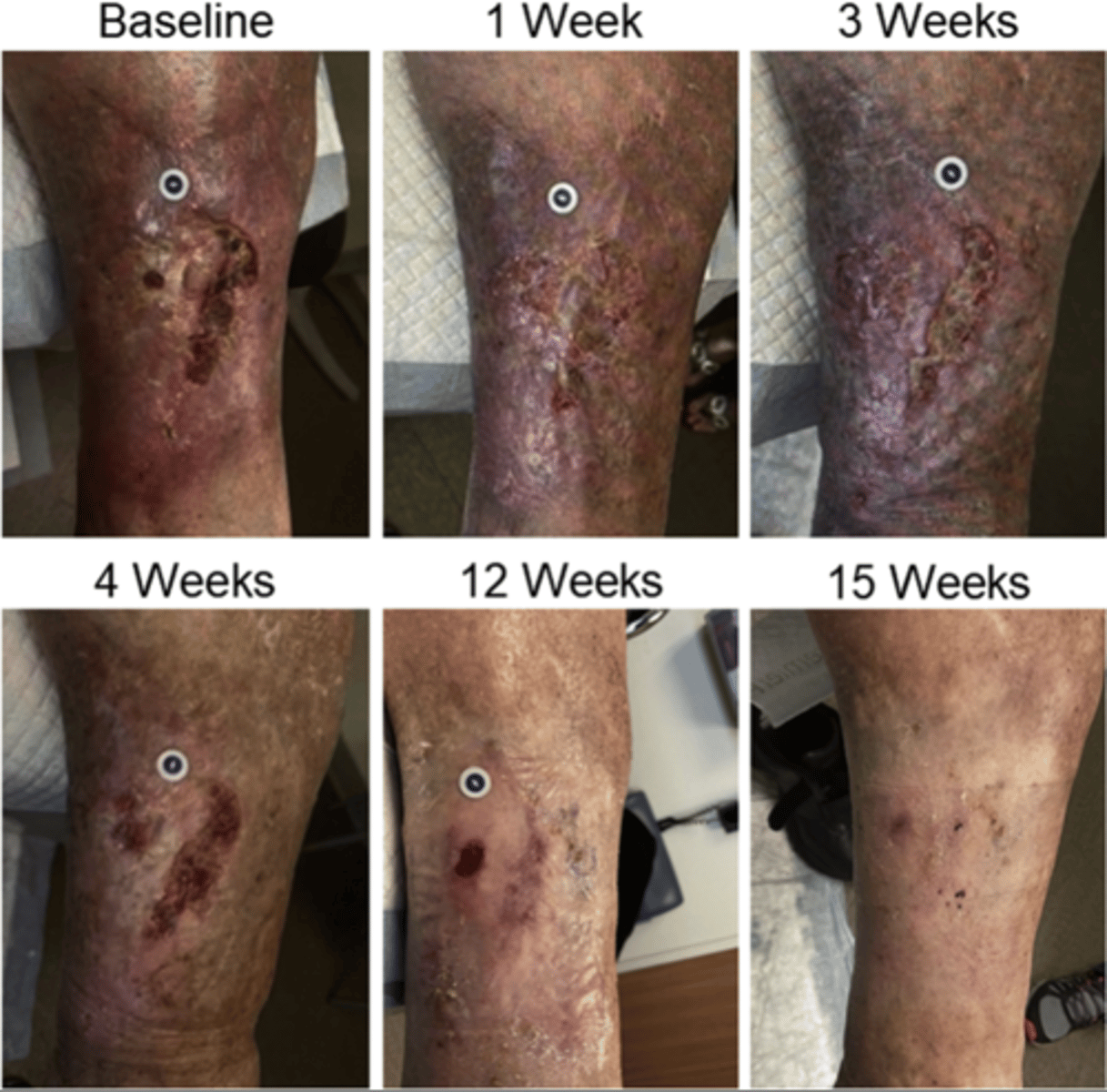
Venous Ulcers
Arterial Ulcers
Found in tibial area/bony areas
Severe pain
Deep, regular shape
Little bleeding
Shiny skin, hair loss, thickened toe nails
Venous Ulcers
Found near medial malleolus
Minimal pain
Shallow, irregular shape
Oozing/Odor
Stasis dermatitis/brown discoloration/varicosities
Venous Hypertension
Due to damaged valves, muscle atrophy, or poor ankle flexion
Increased venous pressure compromises capillary exchange
Low flow within capillaries leads to leukocyte trapping = damaged capillary membranes
Ambulatory Venous Hypertension
Pressure doesn't drop when walking
Intravenous pressure increases during exercise
Seen more in superficial and distal veins
Primary Varicose Veins
Tortuous superficial veins
Hereditary/congenital absence of valves
Secondary Varicose Veins
Obstruction of deep system - pregnancy, DVT, obesity
Reticular Veins
Smaller than varicose veins, larger than spider veins
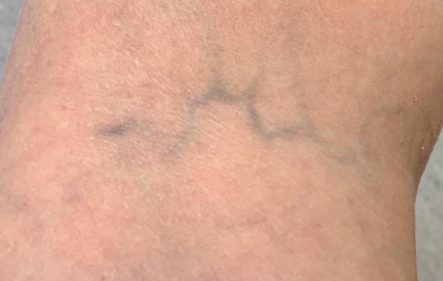
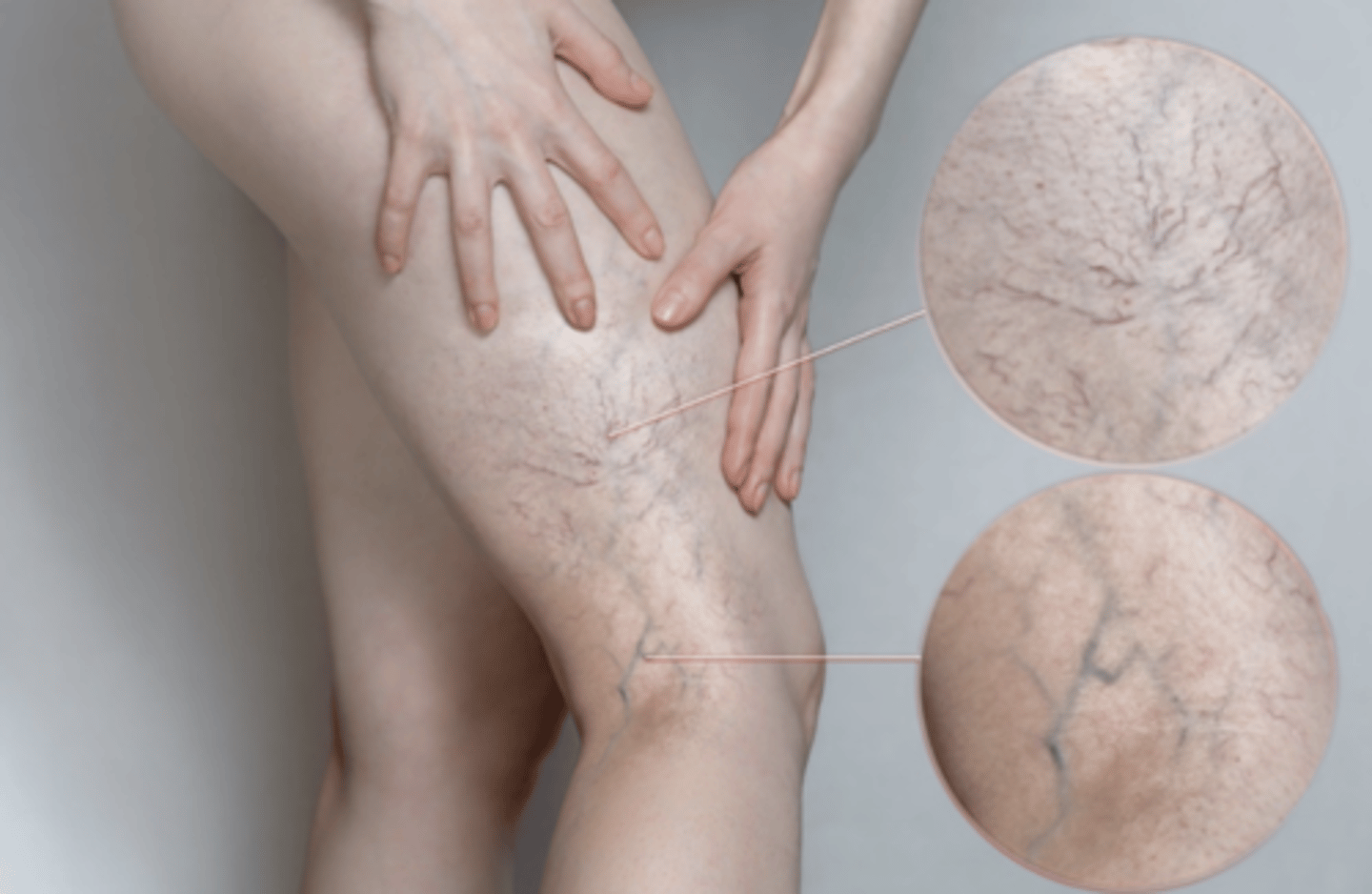
Spider veins/Telangiectasia
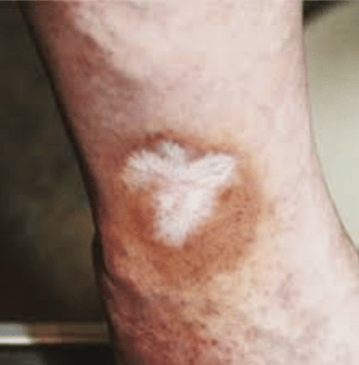
Atrophie Blanche
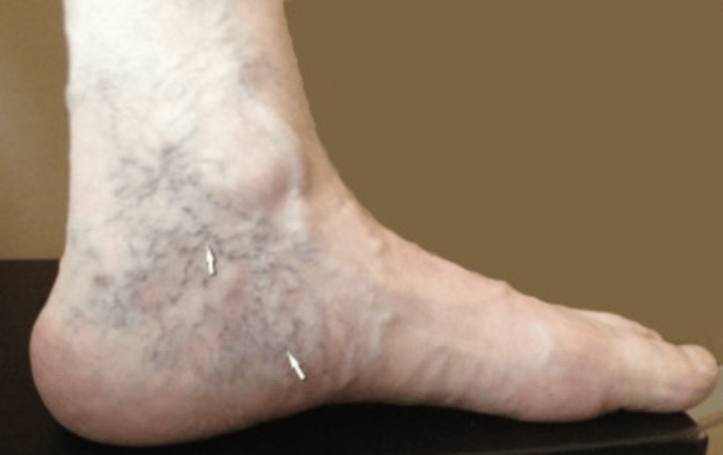
Corona Phlebetcatica
Discoloration from grouping of spider veins
Phlebitis
Inflammation of a vein
Thrombophlebitis
Inflammation of a vein with clot formation
Lipedema
Accumulation of fat and fluid in the tissues just under the skin
Lymphedema
Swelling due to an accumulation of lymph fluid around tissues
CEAP Classification System
C = Clinical Classification
C0: no venous insufficiency signs or symptoms
C1: telangiectasias and/or reticular veins (<3 mm in diameter)
C2: varicose veins (≥3 mm in diameter)
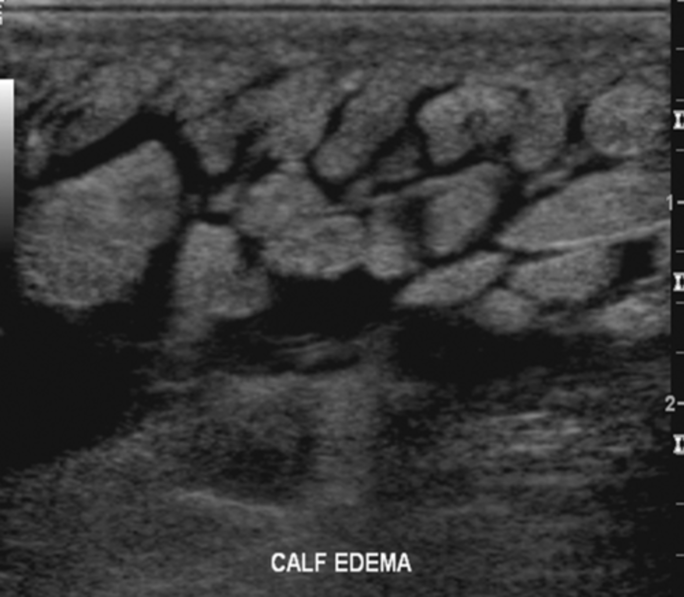
C3: edema
C4A: minor skin changes
C4B: major skin changes - lipodermatosclerosis or atrophie blanche
C4c: major skin changes - corona phlebetcatica
C5: healed skin ulcers
C6: open skin ulcers
E = Etiological Classification
Ep: CVVI is the major cause
Es: CVI or CVVI is secondary to deep venous thrombosis or other pathology
Ec: CVI or CVVI has a congenital origin
En: unknown etiology
A = Anatomic Classification
Ad: affects deep veins
As: affects superficial veins
Ap: affects perforating veins
An: no venous anatomy identified
P = Pathophysiologic Classification
Pr: reflux or reverse venous flow
Po: chronic venous obstruction
Pro: a pathologic combination
Pn: no venous pathophysiology identified
Clinical Severity Score
Attempts to determine a numerical, quantifiable index for research comparisons
Each attribute is given a score from 0-3
Pain
Varicose veins
Edema
Skin pigmentation
Inflammation
Induration
Number of active ulcers
Duration of active ulcers
Size of active ulcers
Compressive therapy
Disability Score
0: asymptomatic
1: able to carry out usual activities without compressive therapy
2: able to carry out usual activities only with compressive therapy and/or leg elevation
3: unable to carry out usual activities even with compressive therapy and/or leg elevation
Contraindications to CCVI Exam
Advanced CVI with obvious varicose veins and/or severe reflux
Patients who are susceptible to fainting, motion sickness, dizziness, nausea
Patients who are handicapped or have extreme discomfort while standing
Patient Positioning for CCVI Exam
Standing/gravity dependent but NOT weight bearing
Reverse Trendelenburg if cannot stand
Complete symptomatic thigh or calf first
GSV - knee rotated outward
SSV - back facing sonographer
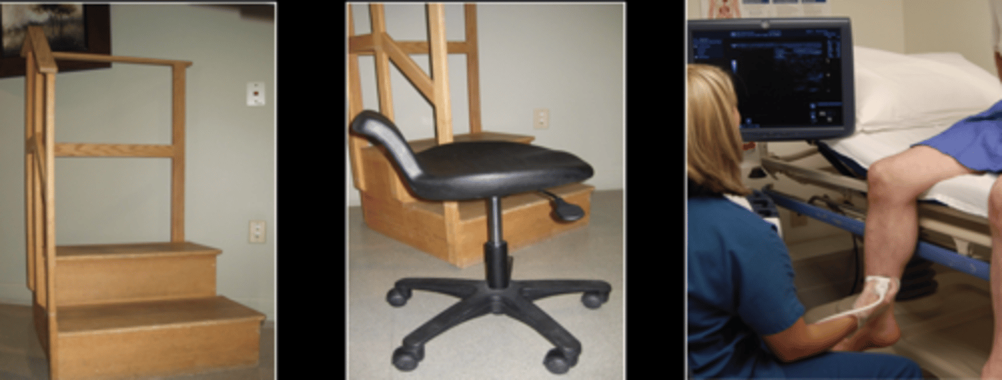
CCVI Techniques
Color, PW, CW each segment
Use auto cuff inflator for augmentation if available
Valsalva
Note any flow reversal & length of time
Augmentation
Promotes forward flow by squeezing calf
Valsalva
Increases abdominal pressure
Not useful in calf
Do not use in patients with arrhythmias
Proximal Compression
At groin
Mimics valsalva
Digital Release
Creates negative pressure distally
Normal Physiologic Reflux
Small amount of flow in the opposite channel is normal
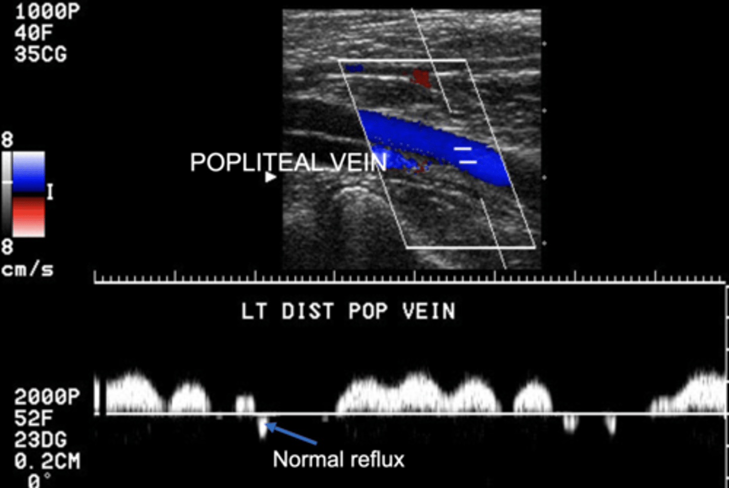
Deep System Reflux
≥ 1 sec
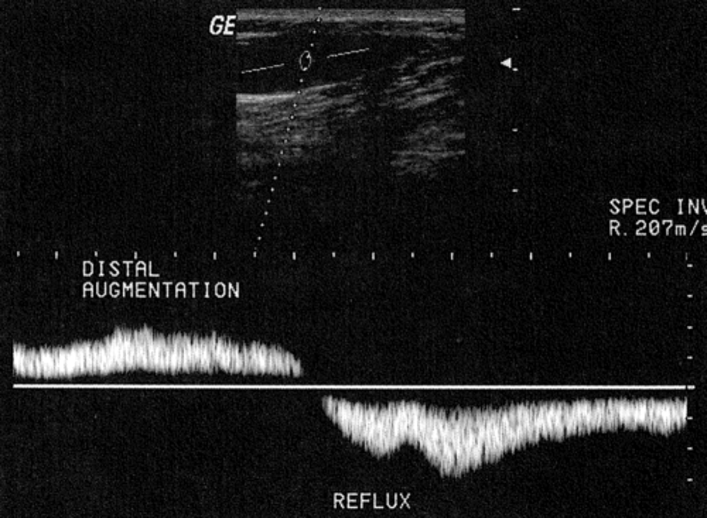
Superficial System Reflux
≥ 0.5 sec
GSV Reflux
If terminal valve is incompetent, usually all of it is
Reflux Photoplethysmography (PPG)
Evaluates venous valve competency
Easy & reliable results
Positive findings must be confirmed with other studies
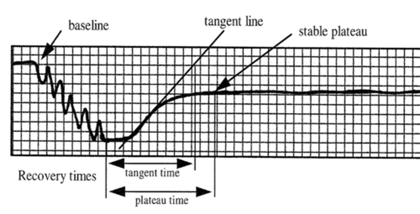
PPG Testing Protocol
Relaxed limb - normal baseline
Dorsiflex foot 5-6x - to empty calf veins
Venous refilling time - time from when exercise stops to stabilization period of ≥ 5 seconds
Normal PPG
Refill time/time to baseline > 20 seconds
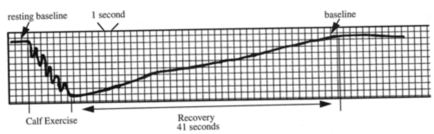
Abnormal PPG
Does not return to baseline
PPG - Tourniquet Test
Differentiates superficial from deep system
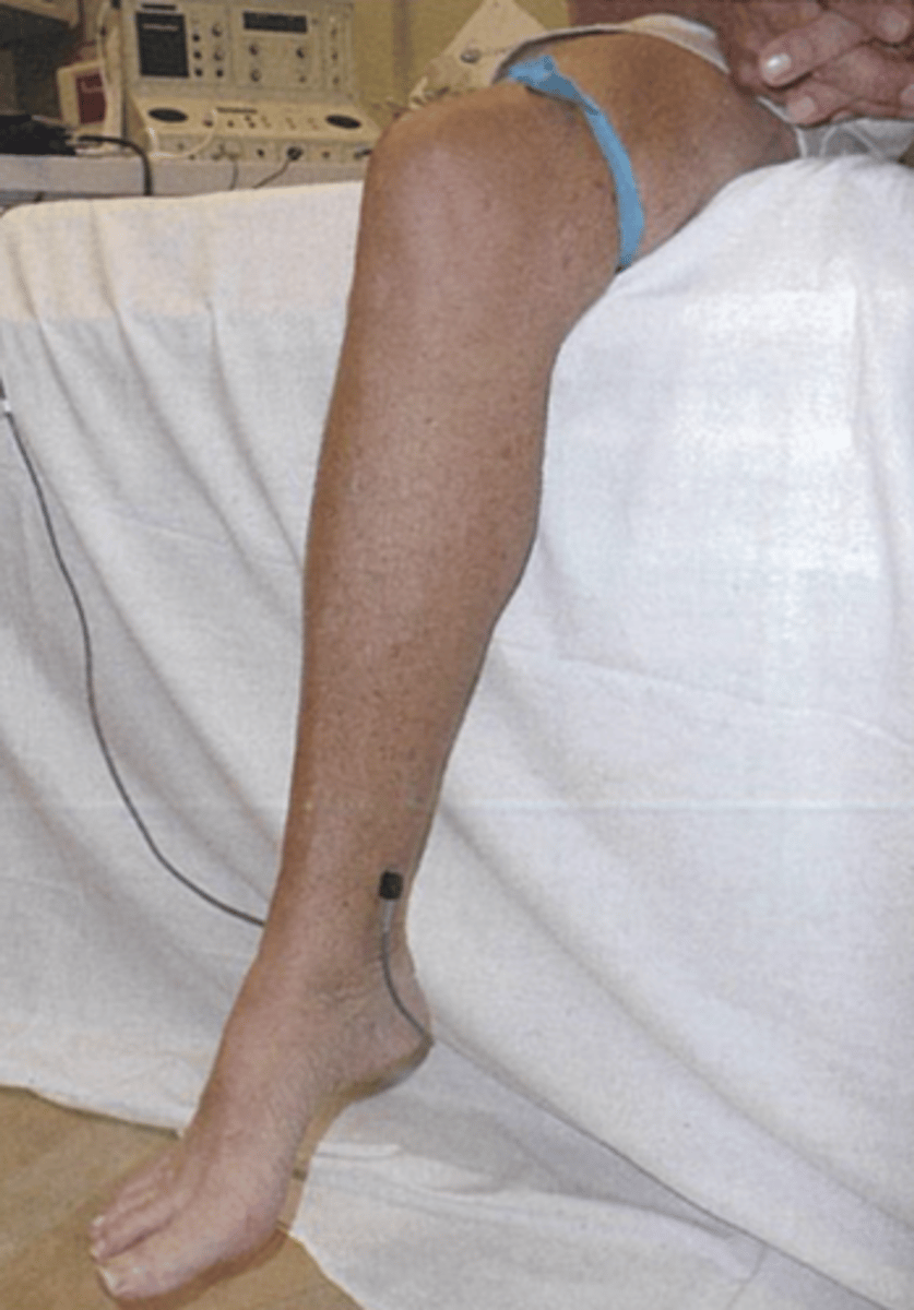
GSV Reflux PPG
Refill time < 20 sec
Normalizes with tourniquet above knee
SSV Reflux PPG
Refill time < 20 sec
Normalizes with tourniquet below knee
Incompetent Deep Valves PPG
Consistent reflux < 20 sec
CW CCVI Testing
Evaluates for obstruction & incompetence
Cannot use with extrinsic or intrinsic obstructions, bifid systems, or collateral flow
Air Plethysmography (APG)
Recommended for chronic venous insufficiency
Cuff inflated to 10 mmHg
Leg is elevated
Patient quickly stands - bearing weight on non-tested leg
Distribute weight evenly
1 toe raise
10 toe raises
Return to supine and elevate tested leg
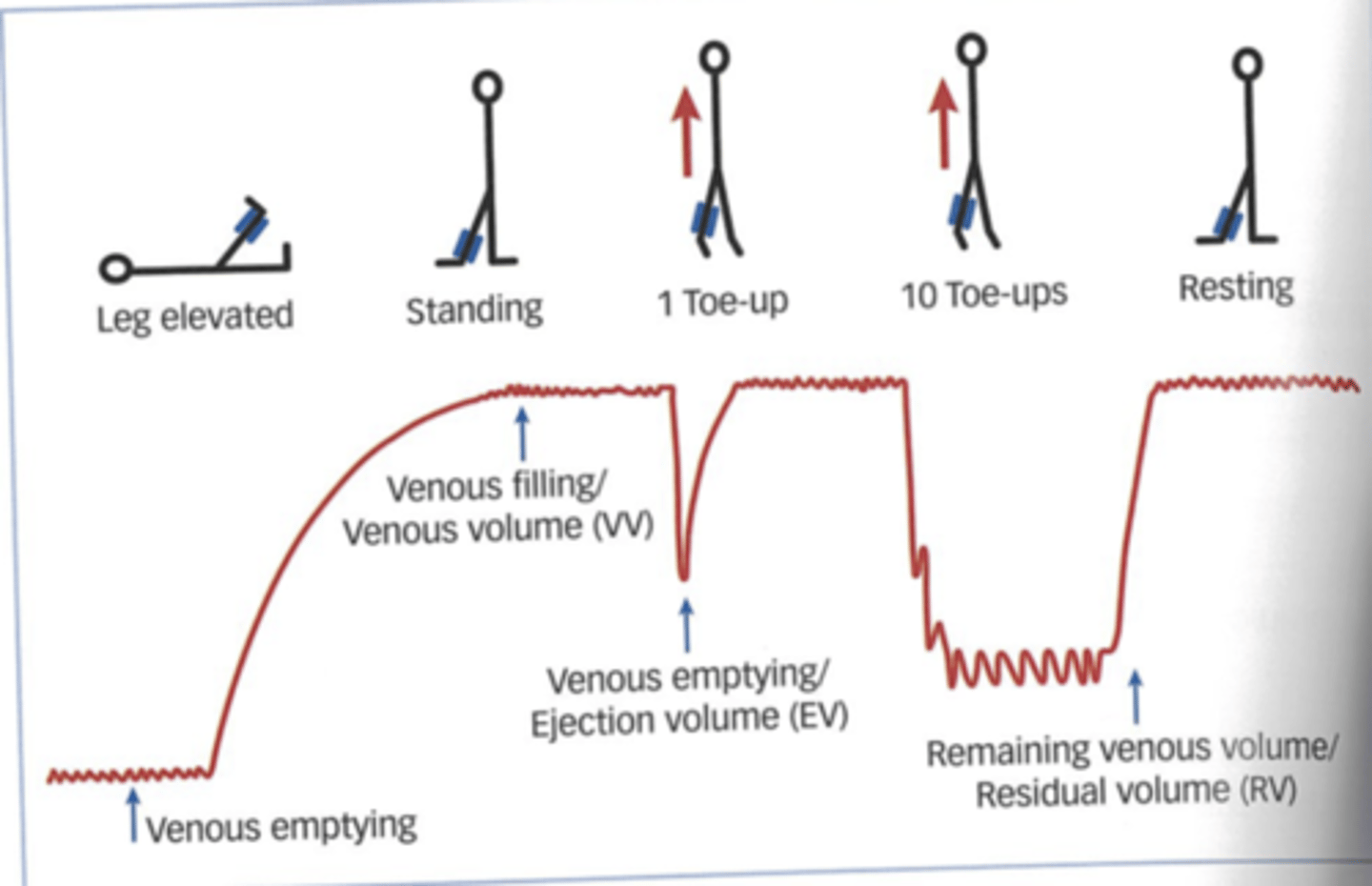
Venous Volume
Accumulated when patient moves from supine to standing
Filling Time
How long to accumulate blood in calf to 90% venous volume
NL = > 25 seconds
Ejection Fraction
After one toe
> 60% = normal
Volume Filling Rate
Blood accumulated per unit time
NL = < 2 mL/s
Residual Volume
Percentage of venous volume
How much volume is pumped from calf after 10 toe raises
NL = < 20 - 35%
Volumetric Emptying Rate
After thigh cuff deflation
Purpose of Vein Mapping
Bypass procedures use superficial veins
Ablation procedures
Dialysis fistulas/grafts
Reduces need for surgical exploration
Identify suitability for use as a graft
Vessel Mapping Techniques
Mark leg if surgery is planned
Place transducer over vein and center in view
Mark the skin directly over the vein
Move 1.5-2 inches and repeat down leg
Connect markings with a marking pen
Mark accessory (smaller) veins with dotted line
Use light pressure
High frequency transducer
Vein Diameter for Graft
≥ 2.5 mm
2.0 mm = smallest possible usable diameter
*Expand 2x when arterialized
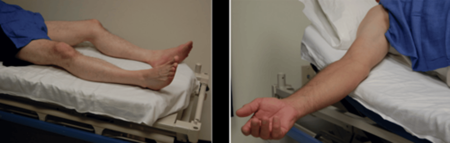
Patient Prep & Positioning for Venous Mapping
No lotion/powers - interferes with skin marking
Keep patient warm
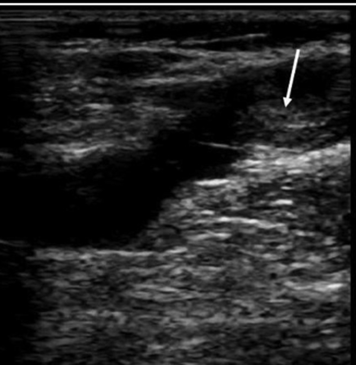
Thrombus
May be remnants of previous clot
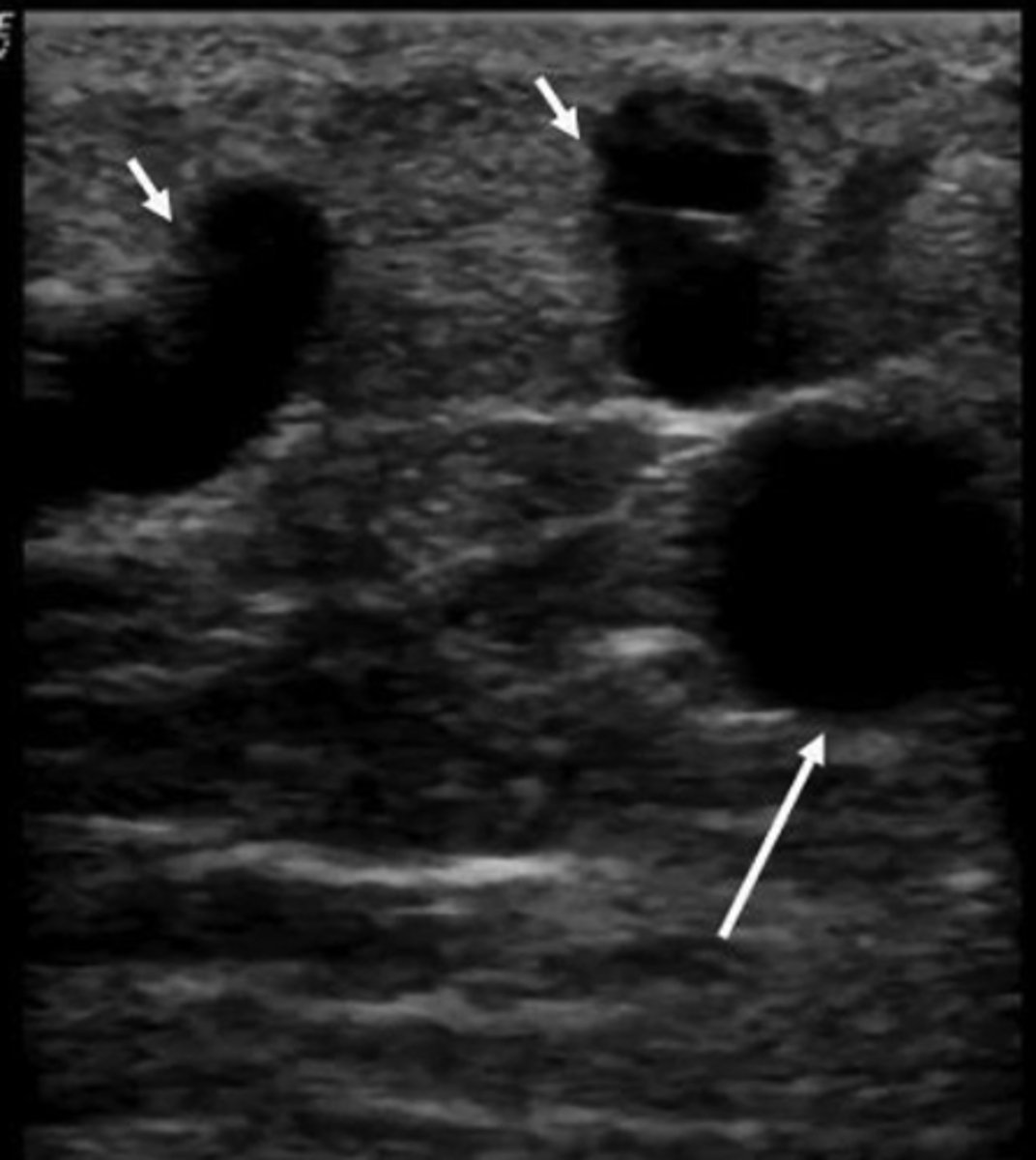
Varicosities
Dilated tortuous veins
Impact ability to use vessel as bypass
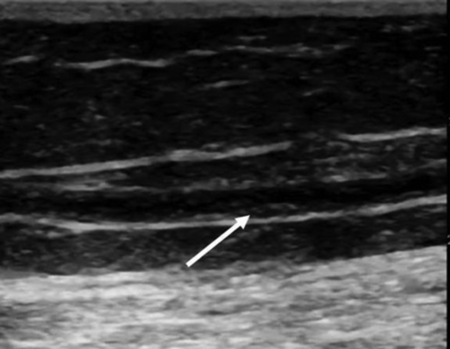
Recanalization
Irregular intimal surface
Cannot be used for arterial bypass
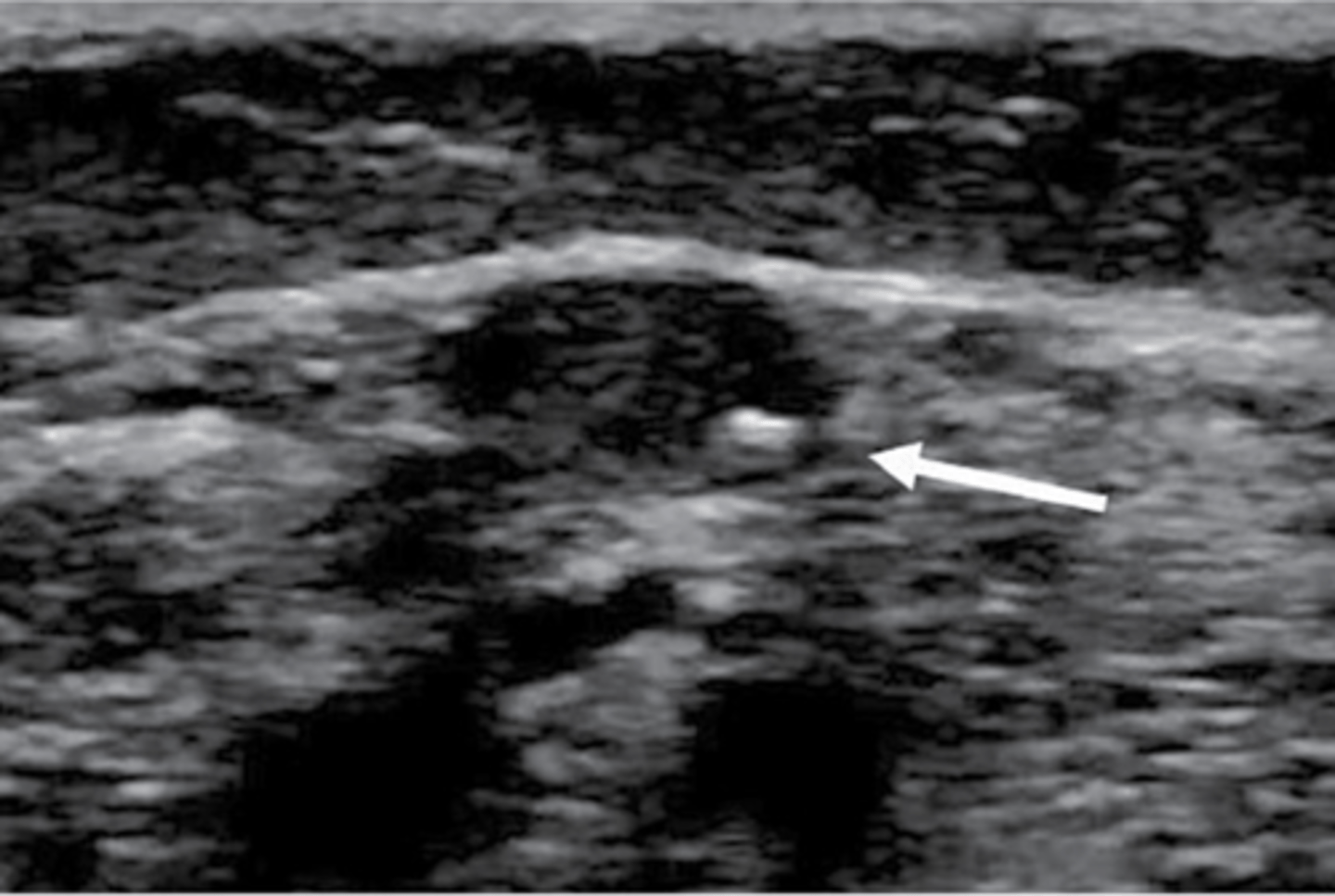
Vein Wall Calcifications
Less common than arterial
Vein could still be usable
Valve Abnormalities
Frozen leaflets
Vein could still be usable
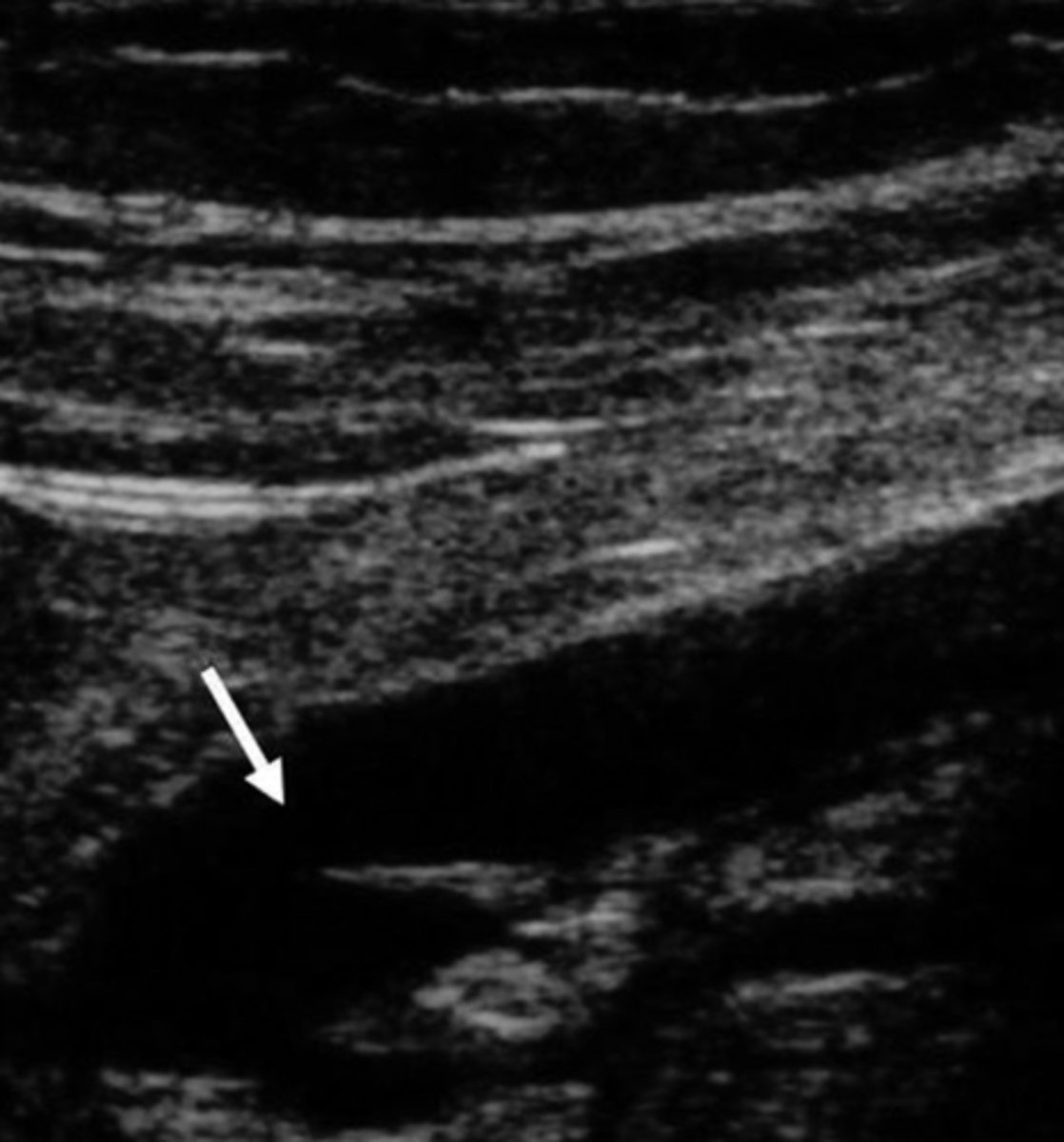
Vein Stripping
Surgical removal of varicose veins
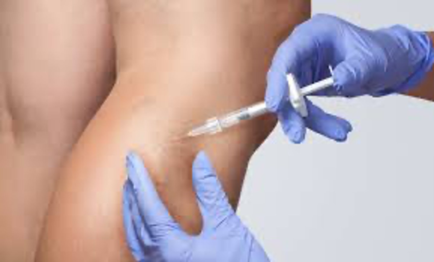
Chemical Ablation
Chemical injected directly into vein = obliteration
Thermal Ablation
Vein closed from within itself via laser/catheter
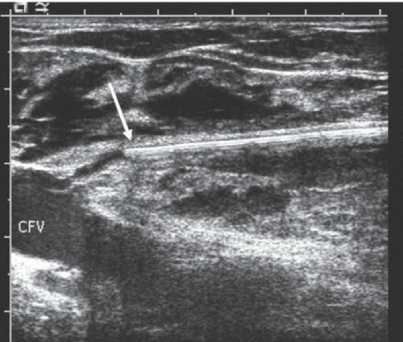
Tumescent Anesthesia
Anesthetizes tissues
Prevents tissue/heat damage
Compresses vein - empties
Injected via ultrasound guidance