5 Conjunctiva + Cornea Pt 1
 Call Kai
 Learn
 Practice Test
 Spaced Repetition
 Match
 Flashcards
 Knowt Play
1/25

There's no tags or description
Looks like no tags are added yet.
26 Terms
What is Hyperemia / Injection? Mention the cause.
→ engorgement of blood vessels
without accompanying exudation or infiltration
anterior ciliary & palpebral arteries
.
Cause
Trauma / Irritation
Mechanical manipulation
Dryness
Infection
Environmental
Smoke / smog / chemical fumes
Wind
UV radiation
Prolonged topical instillation of vasoconstrictors

What is Hemorrhages? Mention the causes.
→ escape of blood from vessels
can be flat, elevated, whispy - its apperance is variable
.
Cause
Trauma
with brusing, it breaks down and resorbs it back making it look
but it can look similar to the colours turning red → brownish/yellow
Infection (Viral, Bacterial – infrequent)
What is Subconjunctival Hemorrhage? Mention the sx.
→ Bleeding between conjunctiva & sclera
often in one sector of eye ⭐
May spread
.
Symptoms
Asymptomatic
Cosmesis
FBS (elevated if there was damage to conj, or if it looks elevated)
.
Causes
Valsalva maneuvers
Anticoagulation medications
Infection
Trauma / surgery
Systemic vascular disease
Bleeding disorders
.
Treatment
Spontaneous resolution 1-3 weeks
Education & reassurance (want to know how long it takes them to heal)
Assessment for ocular trauma
Rule out penetrating injury
ATs (cool artificial tears)
Consideration of blood work
Avoidance of anticoagulants (ASA, NSAIDs)


What is Petechial Hemorrhages? Mention the causes.
→ pinpoint hemorrhaging from capillaries ⭐
can be result of asphyxiation of vasculat ⭐
crushing the vessels and creating these pinpoint hemorrhaging
👥 pt allergic to things, neck of strangled pt
.
Causes
Prolonged straining
Medical conditions
Meds
Trauma
.

What is Chemosis? Mention the causes.
→ Conjunctival swelling (esp on bulbar conj)
Freq with accumulation of fluid within or beneath
may protrude through closed lids if severe
.
Causes
Hypersensitivity rxn
Manipulation

What are True Membranes? Mention what it can occur in association with.
→ superficial conjunctival layers
can’t be peeled → conjunctival epithelium tearing ⭐
.
Associations:
SJS
adenoviral infection
HSV conjunctivitis

What are Pseudomembrane? Mention what it can occur in association with.
→ Coagulated exudate adherent to inflamed tissues
can be peeled off leaving underlying epithelium intact (but doesn’t bleed)
.
Associations:
adenoviral infection
HSV conjunctivitis

What is Infiltration?
→ Cellular recruitment to the chronic inflammation site
Freq accompanies papillary reaction
Loss of detail of normal tarsal conjunctival vessels (looks pale)
More pronounced upper lid ⭐

What is Cicatrization /Subconjunctival Scarring? Mention the complications + causes.
→ Destruction of stromal tissue + scarring of conj tissue
Associated with loss of goblet cells & accessory lacrimal glands → poor tear film
damage to goblet cells → overproduction/underproduction of mucus
.
Complications
Cicatrizing entropion
DES
Trichiasis
Foreshortening of fornix
Keratinization
Acquired Ankyloblepharon (lids will scar to one another⭐)
.
Causes
Trachoma (chylmydia subset)
Cicatrizing conjunctivitis

What are Follicles? Mention the causes.
→ Multiple, discreet, slightly elevated lesions on bulbar conj
collections of lymphocytes
resembles grains of rice ⭐
small, dome-shaped nodules without a vascular core
.
Causes
Viral, chlamydial (b/c follicles are viral)
Carinaud oculoglandular syndrome
Medication hypersensitivity

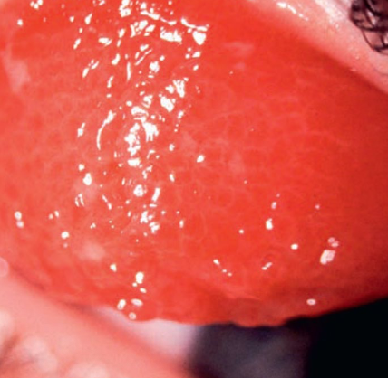
What are Papillae? Mention the causes.
→ Nodule with fibrovascular core that occur palpebral conj & limbal bulbar conj
due to inflammation
Appearance depends on location
Tarsal = flattened
Limbal = dome-shaped


What are the characteristics Conjunctival Degenerations?
→ Decomposition & deterioration of the tissue elements & functions
Age-related
Disease-specific
Chronic environmental exposures
Unilateral or bilateral
Asymmetric
What is a Pinguecula? Mention the causes, signs, sx, treatment.
→ Elastotic degeneration of the collagen fibers of conjunctival stroma
Extremely common
Actinic degeneration
bilateral, asymmetric
.
Cause
UV exposure
Conjunctival injury
dry eyes, dust, wind (irritant of the eye), OUTDOORS⭐
.
Signs
yellow/white elevation on bulbar conjunctiva
adjacent to limbus
DOESN’T extend onto cornea
within palpebral fissure
Nasal > temporal
may develop calcification or pigmentation
.
Symptoms
Cosmesis
FBS
Tearing
DES
.
Treatment
None
Lubrication – ATs, gels
UV protection
Surgical excision

What is Pingueculitis? Mention the treatment.
→ Acute inflammation of pinguecula
may lead to dellen (due to drying) ⭐
.
Treatment
None if asymptomatic
Lubrication
Short course of weak topical steroid
FML, Loteprednol
Excision if repeatedly inflamed or large causing irritation, cosmesis

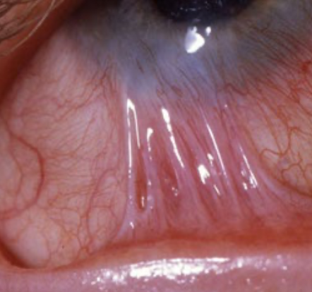
What is Pterygium?
→ Triangular, fibrovascular subepithelial ingrowth (“Surfers Eye”)
invades superficial corneal layers 👁
common in warm climates ⭐
👥 long h/o UV exposure or chronic surface dryness; Men > women; 20-30 yo onset
forms in interpalpebral zone
Nasal > temporal
may be double (nasal & temporal)

List the parts of a Pterygium.
Cap
Head
Body

What are the sx Pterygium?
Small lesions asymptomatic
Irritation & grittiness
Dellen formation at advancing edge
CL intolerance
↓ vision
Induced irregular astig
Interference with visual axis
Intermittent inflammation
Cosmesis

What is a Pseudopterygium?
→ Band of conj sticking to compromised cornea (looks like a pterygium)
response to acute inflammation or inciting event
How can you treat Pterygium?
None, most are asymptomatic
UV protection
Medical:
Tear substitutes
Topical steroids
Surgical:
Excision (but will come back 50% of the time)
Autograft
Mitomycin C
Amniotic graft
What are Concretions?
→ Multiple, tiny cysts with yellow/white deposits (epithelial debris) due to chronic inflammation of palpebral conj
can become calcified
can erode overlying epithelium if it’s big
most common inferiorly
Extremely common; Age-related
.
Symptoms
None
FBS
.
Treatment
none
Excision with needle with topical anesthesia if it’s eroding ocular surface

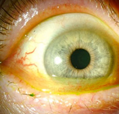
What is Conjunctivochalasis?
→ Loose, redundant, non-edematous conjunctival folds (“dermatochalasis of the conj”)
blocks normal tear flow
degenerative condition
.
Epidemiology
👥 elderly pt
bilateral, asymmetric
List the risk factors of Conjunctivochalasis.

Age
Female
CL wear
Rigid lens wearers tend to have worse severity than soft
Hyperopia / short axial length
Pinguecula
Dry eye / ocular surface disease
How does Conjunctivochalasis clinically present?
Dry eye sx (stinging, FB sensation)
Redundant conjunctival tissue *
Extend beyond lid margin
May obstruct puncta
Injection
Inferior conjunctival & corneal staining with Rose Bengal
Epiphora / pseudoepiphora with sx of dryness
Mucous discharge

How can you evaluate for Conjunctivochalasis?
Look for redundant conj folds over lid margin that move with blink
physically move conjunctiva with lid
Vital dye staining
NaFL to observe conjunctival folds
Lissamine or Rose Bengal staining
Evaluation of tear film
Evaluate tear meniscus
Delayed tear clearance
How can you treat Conjunctivochalasis?
→ Based on sx & severity
.
Order of therapy
Asymptomatic
Education & observation
Lubricants
Symptomatic
Medical therapy
Objectives: improve tear function & decrease inflammation to reduce pt discomfort
Lubricants
Topical steroids – usually a short course
Management of other ocular surface disease
Surgical therapy
Reserved for symptomatic pts who fail with medical therapy
Directed at removing/anchoring redundant conj to prevent disruption of tear film
What is Retention / Epithelial Inclusion Cyst?
→ Fluid-filled cyst of lymphatic tissue
Fluid = clear to turbid
No discomfort
.
Treatment
None if asymptomatic
Puncture with needle to drain if pt bothered
