topic 1B: mammalian transport system
 Call Kai
 Learn
 Practice Test
 Spaced Repetition
 Match
 Flashcards
 Knowt Play
1/31

There's no tags or description
Looks like no tags are added yet.
32 Terms
transport of substances in single cellular vs multicellular organisms
transport of substances in & out of cells by diffusion
relies on concentration gradient
SINGLE CELLULAR
only need diffusion of nutrients and wastes between cell and external environment
short diffusion distance even in innermost parts + large surface area to volume ratio ⇒ sufficient to supply all needs
MULTICELLULAR
metabolically active ⇒ needs constant supply of O2 and glucose for respiration + removal of waste
small surface area to vol. ratio
importance of mass transport system
allow bulk movement of gas and liquid in one direction along vessels
allow diffusion / osmosis / active transport over short distances
carry substance (e.g. oxygen / glucose)
transport chemicals (e.g. hormones)
feature of mass transport systems
more surfaces for the exchange of materials into and out of the transport system
widespread and branching system of vessels which are tubes for providing specific route to carry substances to different sites
ensure substance in / out in the right direction
transport materials rapidly (e.g. blood by pumping action of heart) to maintain concentration gradient for exchange
include a suitable transport medium for substances to dissolve in it
adapt rate of transport to different needs of organisms (e.g. by controlling blood pressure)
circulation system
closed system with blood transported in tubes
except insects: blood circulate in large, open spaces
single circulation
IN FISH
gills for gas exchange
only pass through heart once
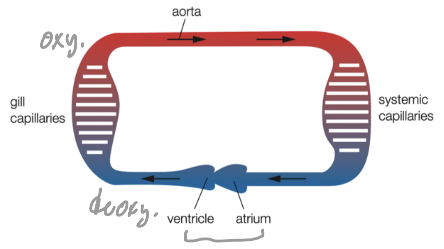
double circulation
IN BIRDS AND MAMMALS
high energy demand for body temp. regulation, movement and support
air sacs in lungs for gas exchange
pass through heart twice for a complete circulation
pressure can be increased to make blood flow more quickly
flow can be directed more precisely to organs that need most oxygen and nutrients

single vs double circulation

benefits of double circulation
support high metabolic rate for movement and body temperature regulation
systemic circulation: provide oxygenated blood from heart to rest of body cells with high blood pressure
pulmonary circulation to transport deoxygenated blood to lungs for gas exchange under low blood pressure
won’t damage vessels
blood functions
transport O2 and CO2
body defence
distribute heat
blood clotting
contents of blood
plasma
red blood cells (erythrocytes)
white blood cells (leucocytes)
blood platelets
plasma
fluid (50% of blood)
transport
respiratory gases
digested food products (e.g. glucose)
nutrients from storage to site needed
excretory products from cells to organs
chemical messages to target sites (e.g. hormones)
plasma proteins (e.g. fibrinogen)
maintain steady body temprapture
transfer heat around from organs to skin
as buffer to pH change
red blood cells
produced in bone marrow
mature: 120 days lifespan
no nucleus: ↑ space to pack haemoglobin
biconcave disc shape: ↑vol.S.A. for O2 diffusion
white blood cells
produced in bone marrow (some mature at thymus gland)
with nucleus and colourless cytoplasm, some with granules
change shape to squeeze through capillary
for body defense against pathogens + inflammation
blood platelets
tiny fragments of megakaryocytes (large bone marrow cells)
for blood clotting
transport of oxygen
by haemoglobin to form oxyhaemoglobin reversibly
each haem group carry 1 oxygen molecule max.
explanation for shape of oxygen dissociation curve
at low partial pressure of oxygen, haemoglobin has low affinity for oxygen, so first oxygen molecule binds to haemoglobin slowly
first oxygen binding changes conformation of haemoglobin molecule ⇒ makes it easier for subsequent oxygen molecules to bind ⇒ increasing haemoglobin affinity for oxygen
at this point, a small increase in pO2 causes a large increase in haemoglobin saturation
at high partial pressure of oxygen, most haem groups are occupied ⇒ harder for final oxygen molecules to bind
at this point, increasing pO2 by a large amount only has a small effect on the % saturation of Hb

explain how haemoglobin is adapted for oxygen transport (Hb O2 saturation against pO2)
in lungs where partial pressure of oxygen (pO2) is high, haemoglobin has high affinity for oxygen and becomes 100% saturated
In respiring tissues where pO2 is low, haemoglobin has lower affinity for oxygen and oxygen readily dissociates
because (oxygen dissociation) curve is steep at tissue pO2 levels, a small decrease in pO2 results in large release of oxygen for aerobic respiration
at low pO2, dissociation slows again as there are few oxygen molecules left on binding sites, release of final oxygen molecule is more difficult

high & low affinity for oxygen
high affinity:
Hb binds O2 easily and dissociates slowly
low affinity:
Hb binds O2 slowly and dissociates easily
transportation of carbon dioxide
tissue to blood
by plasma
CO2 dissolves in water to form H2CO3 carbonic acid by carbonic anhydrase
carbonic acid further separate to form H+ and HCO3− in RBC / plasma
by haemoglobin
carbaminohaemoglobin
hydrogencarbonate ions pass out and chloride ions move in (chloride shift)
Hb act as buffer and accept H+ to form haemoglobinic acid
avoid changing pH of blood

bohr effect
Respiring tissues produce high amounts of CO₂ as a waste product
CO₂ diffuses into the blood and combines with water to form carbonic acid (H₂CO₃), catalysed by carbonic anhydrase
Carbonic acid dissociates into hydrogen carbonate ions (HCO₃⁻) and hydrogen ions (H⁺), which lowers the blood pH, making it more acidic
H⁺ ions bind to haemoglobin, causing a conformational change that reduces haemoglobin's affinity for oxygen
Haemoglobin release oxygen more readily to the active, respiring cells that need it
curve shifts to the right
At any given pO₂, haemoglobin is less saturated with oxygen when pCO₂ is high, meaning more oxygen is released to the tissues

fetal vs maternal haemoglobin
fetal Hb higher affinity
fetal haemoglobin can remove oxygen from the maternal blood even when proportion of oxygen is relatively low
maternal and fetal blood run in opposite directions (countercurrent)
makes concentration gradient between mother’s blood and that of her fetus as steep as possible ⇒ maximising oxygen transfer to the blood of the fetus
oxygen dissociation curve shifts to the left
at any pO2, fetal haemoglobin has higher % saturationț

oxygen dissociation curve in high altitudes
less air ⇒ less oxygen ⇒ breathe faster and deeper to compensate (more air more oxygen)
genetic adaptations
↑ RBC ⇒ ↑ Hb ⇒ ↑ oxygen carrying capacity
↑ affinity for oxygen (curve shifts to left) ⇒ release oxygen more readily

importance of blood clot
prevent loss of blood volume which may be fatal
prevent entry of pathogens through the wound
formation of blood clot
flow of plasma, blood cells and platelets in the vessel
platelets in contact with components of tissues (e.g. collagen)
platelets release serotonin and thromboplastin
serotonin
cause contraction of smooth muscle on wall of blood vessels
↓ lumen ⇒ narrower blood vessels ⇒ ↓ blood flow to damaged area ⇒ ↓ bleeding
thromboplastin
enzyme to catalyse the reactions involved in blood clotting
process of blood clotting
thromboplastin catalyses the conversion of prothrombin to thrombin in plasma (with right conc. of calcium at the site)
prothrombin: large soluble protein, biologically inactive
thrombin: more active
thrombin converts soluble fibrinogen (plasma proteins) into insoluble fibrin to form fibres at the wound
more platelets and red blood cells at the wound will by trapped by fibre to form, eventually a clot
some proteins cause internal contraction of blood platelets to tighten the blood clot as to form a scab to skin protection and wound healing
blood circulation route
superior / inferior vena cava ⇒ right atrium ⇒ tricuspid valve ⇒ right ventricle ⇒ semilunar valve ⇒ pulmonary artery ⇒ lungs ⇒ pulmonary vein ⇒ left atrium ⇒ bicuspid valve ⇒ left ventricle ⇒ semilunar valve ⇒ aorta ⇒ arteries ⇒ arterioles ⇒ capillary of body tissues ⇒ venules ⇒ veins ⇒ vena cavae
artery
carries blood away from heart
highest blood pressure but decreasing when move further away from heart to peripheral arteries
carries oxygenated blood (usually high oxygen content) except pulmonary / umbilical artery
branches into arterioles with smaller lumen size
STRUCTURAL FEATURES
external layer
thick wall
tough tissue
lots of collagen
withstand high blood pressure in artery (by pumping action of heart)
middle layer
elastic fibres: stretch walls of arteries to accommodate the surge in blood volume without being damaged ; recoil to return to original length and diameter ⇒ maintain blood flow by squeezing blood inside (pulse)
smooth muscle fibres: contract and relax to regulate the diameter of lumen of vessel ⇒ regulate amount of blood flow
lumen
small

artery wall content ratio

vein
carries blood towards heart
lowest blood pressure
carries deoxygenated blood (usually lowest oxygen content) except pulmonary and umbilical vein
venules ⇒ veins ⇒inferior (from lower parts of body) / superior (from upper parts of body) vena cava
STRUCTURAL FEATURES
surrounded by skeletal muscles ⇒ contraction squeezes blood
semilunar valves at frequent intervals ⇒ only allow blood flow towards one direction / prevent backflow

capillaries
carries blood from artery to organs for material exchange between blood and body cells by diffusion
greatest change in blood content
greatest drop in blood pressure
slowest blood flow rate
STRUCTURAL FEATURES
highly branched
↑ S.A. for material exchange by diffusion
closer to cells ⇒ ↓ diffusion distance with body cells
small individual lumen size
slow blood velocity ⇒ more time for material exchange by diffusion
one-cell thick
no elastic fibres / smooth muscles / collagen
↓ diffusion distance ⇒ rapid diffusion of substances

blood property changes graph

heart basic info
left and right heart (separated by septum) work in sync (atrium & ventricle contract / relax at the same time)
left side: receives blood from lungs ; pump to other parts of body
right side: receives blood from body ; pump to lungs
made up of cardiac muscles that contract regularly without getting fatigued
receives continuous blood supply of oxygenated blood from coronary arteries
contains a lot of myoglobin, which has stronger affinity for oxygen that haemoglobin ⇒ store oxygen for rsp to keep heart contracting