Found 3.12 Liver
 Call Kai
 Learn
 Practice Test
 Spaced Repetition
 Match
 Flashcards
 Knowt Play
1/63

There's no tags or description
Looks like no tags are added yet.
64 Terms
Liver Anatomy
%BW, where?
lobes?
segments? I?
Largest internal organ
~1.5 kg, ~2% of body weight
RUQ; spans ribs 7–11 at the right midaxillary line
Anatomic lobes
Right and left lobes, divided by the falciform ligament
Caudate and quadrate lobes inferiorly
8 Functional segments
8 Couinaud segments (I-VIII), defined by hepatic and portal veins
Each has its own vascular inflow and biliary drainage
Caudate lobe (segment I) drains directly into the IVC, so it is spared and hypertrophies in hepatic venous outflow obstruction.

Liver Vascular
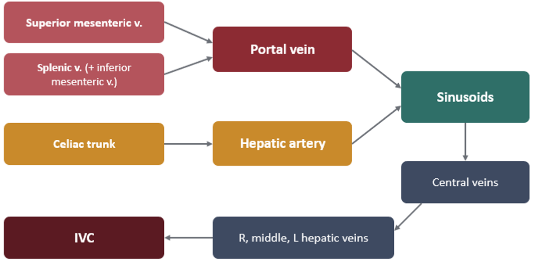
blood flow chart
what organs drain into portal? 6
oral vs IV meds
unique characteristics about the portal system
So the liver is perfused by the portal vein and the hepatic artery. dual blood supply.
You have right, middle, and left hepatic veins, and all of those drain directly into the inferior vena cava.
So the portal vein drains, because it's made-up of the superior mesenteric vein and the splenic vein, it drains the stomach and the small and large intestine, spleen, pancreas, and the gallbladder
So this is why you have something called first pass metabolism because things that are taken orally or absorbed through the intestines first go through the liver before they go to the rest of the body.
And which is why IV medications tend to be a lower dose than PO medications because they're not subject to first pass metabolism like PO medications are.
The portal system is valveless. That's unique for the portal system. And because it's valveless, it's a low pressure venous inflow between two capillary beds.
Hepatic artery branches from the abdominal aorta via the celiac trunk
Liver Innervation
SNS from? aka? fx?
PSNS from? fx?
receptors on arteries/veins? control?
Sympathetic fibers T3–T11 (via celiac plexus)
Constrict hepatic arterioles, sinusoids, and venules
Parasympathetic (vagal): ACh and VIP relax arterioles and sinusoids
Hepatic artery: α and β receptors
Portal circulation: α only
Venule tone sets hepatic blood volume (reservoir)
The Lobule: Structural Unit - makeup
portal triads vs central vein?
directional flow?
Shape: hexagonal cylinder, 0.8–2 mm in diameter; 50,000–100,000 lobules
Central vein drains into the hepatic veins, then the IVC
Portal triads at the corners: portal venule, hepatic arteriole, bile ductule, plus lymphatics and nerves
Hepatocyte plates radiate like spokes, usually 1–2 cells thick
Sinusoids carry mixed portal and arterial blood to the central vein
Bile canaliculi run between hepatocytes to bile ducts in the septa
Blood flows inward toward the central vein.
Bile flows outward toward the portal triad.
The Acinus & Metabolic Zonation
Zone 1: 2, aka?
Zone 2: 1, aka?
Zone 3: 3, aka?
hepatocytes are the cells; the acinus is an arrangement of those cells based on their blood supply.
P. Triad → Zone 1 → Zone 2 → Zone 3 → Central Vein
Falling O₂ and nutrient content along the sinusoid
Zone 1 · Periportal
Most O₂ and nutrients
Oxidative metabolism: gluconeogenesis, β-oxidation, urea synthesis
Zone 2 · Midzonal
Intermediate O₂ and function
Zone 3 · Centrilobular
Least O₂; anaerobic metabolism
most vulnerable to hypoxia ("shock liver")
Highest CYP450 content
Reactive intermediates concentrate here
most vulnerable to toxic metabolites: acetaminophen (NAPQI), halothane.

Hepatic Cell Types
Hepatocytes 4
Kupffer cells 1
endothelium 3
Stellate cells 3
Hepatocytes
80% of total cellular volume
Metabolism, synthesis, bile; smooth ER holds CYP450
Kupffer cells
Resident macrophages from monocytes
Phagocytize ≥99% of portal bacteria
Sinusoidal endothelium
Pores up to ~1 µm
no basement membrane
Plasma proteins pass into the space of Disse
Space of Disse: perisinusoidal space between endothelium and hepatocytes; drains to lymphatics in the interlobular septa.
Stellate (Ito) cells
Quiescent in the space of Disse; store vitamin A
With prolonged injury, secrete collagen: fibrosis, cirrhosis
So usually they're not very active.
Dual Blood Supply Numbers
total CO%? ml/min? %O2 consumption?
portal vs hepatic art
ml/min vs %
%O2 consumption?
↑ %O2 requirements solution?
Total hepatic blood flow
~1350 mL/min
~25% of cardiac output
liver uses ~20% of body O₂
Portal vein - low pressure
~1050 mL/min, ~75% of flow
~50% of O₂
partly deoxygenated
Hepatic artery - high pressure
~300 mL/min, ~25% of flow
~50% of O₂
even though the hepatic artery is a third of the amount of flow that the portal vein is, it provides just as much oxygen content as the portal vein
Increased hepatic O₂ demand is met mainly by increased O₂ extraction, not by further increases in flow.
oxyhemoglobin dissociation curve, when it pushes more to the right
It favors offloading that oxygen.
Hepatic venous pressure gradient (HVPG)
portal vs central pressure?
gradient in L/min?
normal vs abnormal?
HVPG = wedged hepatic venous pressure − free hepatic venous pressure
Portal vein ~7–10 mmHg
↓
Sinusoids very low resistance
↓
Hepatic vein ~0–5 mmHg
A gradient of only a few mmHg drives >1 L/min because sinusoidal resistance is so low.
Normal 3–5 mmHg.
A rise in hepatic venous pressure to >5 mmHg defines portal hypertension causes plasma-like fluid to transude from the liver surface: the basis of ascites.
≥10–12 mmHg: clinically significant; varices expected
Blood Reservoir
holds ml? % of blood volume?
can store extra ml?
SNS activation?
Holds ~450–500 mL (~10% of blood volume)
And so when it meets resistance and can't push the blood forward efficiently, that backflow, the liver catches a lot of it up to an additional 500 milliliters than it normally has.
Stores up to ~1L when right atrial pressure is high
Ex: during a central line, if someone's really dry, you can actually press on their liver and you'll see that the internal jugular vein dilate whenever you press on the liver.
Sympathetic activation = venoconstriction → expels up to ~500 mL into IVC
Hepatic lymph
how much lymph goes through here? why?
Protein ~6 g/dL (near plasma)
1/3 to 1/2 of all body lymph
The hepatic lymph or the lymph flow, usually Whenever there's arterial perfusion to a part of the body, the veins pick up about 90% of the fluid that left the vascular space and brings it back into the venous space.
And that last remaining 5 to 10% is picked up by the lymph.
But in the liver, because of how high the protein concentration is in the cells themselves.
About one third to half of all the body lymph is actually in the liver as opposed to all the other tissues.
So even though it, you know, even though it gets roughly 25% of the blood flow in the body, it can have almost half of the body lymph compared to the rest of the body.
Regulation of Hepatic Blood Flow
Portal 3
Hepatic artery 3
SNS tone 3
Portal vein
Passive, low-pressure system
Flow set by splanchnic arteriolar tone and intrahepatic resistance
Not autoregulated: follows gut and spleen outflow
Hepatic artery
High-pressure, high-resistance system
Depends on cardiac output and autonomic tone
Local regulation via the HABR
Sympathetic tone
Constricts arterioles, sinusoids, and venules: total flow falls
Venule constriction shifts blood into the systemic circulation
Key compensation in hemorrhage
Hepatic arterial buffer response (HABR)
flow r/ts for portal & artery
↑ SNS does what? 2
If Portal flow falls → Less adenosine washed out → Hepatic arterioles dilate → arterial flow rises to compensate and it may not fully replace the lost portal flow. (Doesn’t fix underlying issue)
arterial flow can rise by as much as 100%
HABR is semireciprocal: changes in portal flow inversely affect arterial flow.
When you have an ↑ in sympathetic tone, this constricts arterials and sinusoids and venules and ultimately decreases flow.
And the venual constriction will shift blood into systemic circulation.
lowers O₂ delivery to zone 3 and slows clearance of high-extraction drugs.
What Reduces Hepatic Blood Flow?
Volatile anesthetics
sevo
iso
des
halo
Dose-dependent ↑↑; desflurane moderate, isoflurane and sevoflurane minimal; halothane greatest. Isoflurane is a direct hepatic vasodilator, so safer.
meaning the higher the dose, the more hepatic blood flow is decreased.
Mostly vasodilate, but they can still reduce total hepatic blood flow. The main reason is that volatile anesthetics can lower cardiac output and blood pressure, which reduces portal-vein flow. The hepatic artery may dilate to compensate, but that compensation may not fully make up the difference
For liver perfusion, isoflurane is generally considered safer than halothane. Isoflurane tends to preserve hepatic arterial flow; halothane can reduce both hepatic arterial and portal flow, with larger decreases reported in some studies
What Reduces Hepatic Blood Flow?
Induction of anesthesia - why?
↓ HBF as much as %?
HBF may fall 30–50%, mainly via ↓ MAP and ↓ cardiac output with reflex splanchnic vasoconstriction
Induction of anesthesia can decrease hepatic blood flow and really flow of everything, but it's primarily because of its effects on map and cardiac output.
What Reduces Hepatic Blood Flow?
Neuraxial block - causes? outcome?
Sympathectomy-induced HPT = ↓ splanchnic flow
A spinal (subarachnoid) anesthetic causes a rapid sympathectomy, so blood pressure may drop before compensatory responses can catch up
Ex: used in C-section or joint replacement
An epidural can also cause sympathectomy, but its onset is generally slower and less abrupt.
What Reduces Hepatic Blood Flow?
Positive pressure ventilation, PEEP
↑ Intrathoracic and hepatic venous pressure, ↓ venous return and cardiac output, reflex sympathetic tone
Positive pressure ventilation and PEEP, this increase in angiothoracic pressure and hepatic venous pressure will ultimately decrease venous return and cardiac output.
So if you have less blood returning to the left side of the heart, then you have less to actually pump out of the heart. Therefore, cardiac output decreases.
What Reduces Hepatic Blood Flow?
PaCO₂ - ph?
Hypercapnia and acidosis vasodilate (↑ flow); hypocapnia and alkalosis vasoconstrict (↓ flow)
PACO2, hypercapnia and acidosis, vasodilate vessels to the liver so that it increases flow
hypocapnia and alkalosis vasoconstrict and ultimately decrease flow.
What Reduces Hepatic Blood Flow?
Upper abdominal surgery - 3
Largest decreases: traction, compression, catecholamine release
This is the largest decreases whenever there's traction or compression or catecholamine release, especially if the procedure involves the liver itself.
If they put some traction on the liver and pinch off the IVC or something, you can have a very, very large abrupt acute drop in blood pressure.
Liver Fx:
Carbohydrate - buffer system?
Gluconeogenesis
Glycogenolysis
clears % insulin on first pass
Glucose buffer function: when BGL ↓, Glycogen stored after meals is releases into blood stream triggering glycogenolysis resulting in ↑ BGL
Poor liver function: postprandial glucose can rise 2–3× normal
Gluconeogenesis from lactate, pyruvate, amino acids, and glycerol
Glycogenolysis triggered by epinephrine and glucagon
Clears ~50% of insulin on first pass
Liver Fx:
Fat
Beta Oxi?
Synthesizes 3
% of what? become what?
β-oxidation produces acetyl-CoA beyond the liver's needs; exported as acetoacetate
Synthesizes most cholesterol, lipoproteins and phospholipids
Nearly all fat synthesis comes from carbohydrate and protein
~80% of hepatic cholesterol becomes bile salts
Liver Fx:
Protein
synthesis of which type of AA? albumin?
Deamination
Urea cycle?
synthesis of nonessential amino acids
makes ~90% of plasma proteins (not γ-globulins); 15–50 g/day
Albumin: main determinant of oncotic pressure; t½ ~3 weeks
Deamination of amino acids for energy or conversion
Urea cycle removes ammonia from gut and tissues
Loss of urea formation, or portal flow that bypasses the liver, lets ammonia accumulate: the basis of hepatic encephalopathy.
Liver Fx:
Hemostasis
Makes all clotting factors except? 3
Vitamin K-dependent proteins 6
Thrombopoietin
All clotting factors except III, IV, VIII
Vitamin K-dependent proteins which helps activate the following: II, VII, IX, X, C, S
Thrombopoietin: which signals the bone marrow to make platelets
Liver Fx:
Storage
Vit A
Vit D
Vit B
Fe
Glycogen
Blood
Fx reserve
Vitamin A: up to ~10 months' supply
Vitamin D: ~3–4 months
Vitamin B12: ≥1 year
Iron: apoferritin binds iron as ferritin, a blood iron buffer
Glycogen: ready glucose state meant to be broken down asap for ↑ BGL
Blood (reservoir 500-1000 ml)
Large functional reserve: significant disease can exist before laboratory tests become abnormal.
Liver Fx:
Bile Formation & the Biliary Tree
production ml/day?
how and where is it released? relax it? 2
Fx of bile? 3
Production
~500 mL/day
Bile salts, bilirubin, cholesterol, phospholipids
Route for drug and metabolite excretion
Release
Between meals: high Oddi tone diverts bile to gallbladder
Duodenal fat in small intestine → release of cholecystokinin (CCK) → GB contracts, Oddi relaxes → content comes out
All opioids can cause sphincter of Oddi spasm; the more potent the opioid, the greater the rise in biliary pressure.
But one of the ways is reversing the opioids with Narcan (not recommended)
Glucagon is another one that can cause the sphincter to relax
Function
Micelles absorb fat and vitamins A, D, E, K
Enterohepatic recycling of bile salts
No bile = steatorrhea, vitamin K deficiency in days
Now, just because someone's had a cholecystectomy or having their gallbladder removed doesn't mean that they can't excrete drugs through the bile.
They still can. They just won't be in the gallbladder. It'll go from the liver straight to the duodenum.

Liver Fx:
Detoxification & excretion
drugs leave via? 2
hormone inactivation 3 (3)
Kupffer kill rate?
Drugs excreted into bile or made renally excretable
Hormones inactivated: thyroxine, catecholamines, steroids (estrogen, cortisol, aldosterone)
Liver damage → hormone excess from buildup
Kupffer cells filter 99% of gut bacteria
Liver Regeneration
restore up to %?
growth factor? 3
halted by? impaired by?
Restores mass after loss of up to ⅔–70%
Driven by hepatocyte growth factor, EGF, TNF, IL-6
TGF-β halts proliferation
Impaired by fibrosis, inflammation, viral infection
a fibrotic liver that essentially can't regenerate.
When Function Fails: Physiologic Consequences
Albumin synthesis 2
Clotting factors 2
Plasma cholinesterase 2
Albumin synthesis
Falls slowly (t½ ~3 weeks)
↓ Oncotic pressure (edema, ascites); ↑ free fraction of protein-bound drugs (drugs bound to protein are inactive)
If someone's emaciated, or someone has low albumin, then they're more sensitive to the propofol because there's not as much protein for it to be bound to
Clotting factors
↓ Synthesis; short half-lives (factor VII)
PT/INR prolongs quickly with acute injury
Plasma cholinesterase
↓ Synthesis
Helps in succs metabolism = prolonged action; ↑ ester local anesthetic exposure
When Function Fails: Physiologic Consequences
Glucose homeostasis
Urea cycle
Fluid compartments
Glucose homeostasis
↓ Glycogen stores, gluconeogenesis, insulin clearance
Hypoglycemia (severe disease) or hyperinsulinemia
Urea cycle
ammonia can't be converted into urea to be removed by the kidneys = ammonia accumulation
Encephalopathy; ↑ sensitivity to sedatives
Fluid compartments
↓ Oncotic pressure, Na retention
↑ Volume of distribution of many drugs
more drugs outside the bloodstream—into tissues or body fluids—so less remains in the plasma.
Bilirubin Formation & Conjugation
rbc age? goes where? to make? 2
how much unconjugated bili is made?
what happens in hepatocytes?
After ~120 days, macrophages in spleen (RES: reticuloendothelial system), split hemoglobin into globin and heme
Heme oxygenase → biliverdin → unconjugated (free) bilirubin ~200–300 mg bilirubin/day
Unconjugated bilirubin is lipid-soluble and travels bound to albumin to liver
In hepatocytes: ~80% glucuronide (UGT), ~10% sulfate, ~10% other; now water-soluble
Actively secreted into canaliculi → bile → intestine
Bacteria form urobilinogen: some → stercobilin (stool); some reabsorbed,
~5% reaches urine as urobilin, even w/ liver failure % doesn’t change

Jaundice
normal? abnormal?
is there a specific lab test?
Normal plasma bilirubin ~0.5 mg/dL, almost all unconjugated
Skin yellows >1.5 mg/dL (≈3× normal)
The Van den Bergh reaction is a lab test: It distinguishes bilirubin in a blood sample by how it reacts: conjugated bilirubin reacts directly, while unconjugated bilirubin needs an added accelerator.
Jaundice: Classification
Prehepatic (hemolytic) -
Unconjugated rises
Production exceeds hepatic excretory capacity
↑ Urobilinogen in gut and urine
Normal stool color
Jaundice: Classification
Hepatocellular -
Mixed picture
Impaired uptake, conjugation, or secretion
Hepatitis, cirrhosis, drugs
Gilbert syndrome; immature neonatal conjugation
Jaundice: Classification
Obstructive - aka
s/s 3
how does renal help?
sx?
aka cholestatic jaundice
No bile in gut → fat-soluble vitamin K malabsorption
Delayed excretion of drugs and metabolites cleared in bile
↓ Factors II, VII, IX, X and proteins C, S
Conjugated rises; refluxes into blood
Total obstruction: no urinary urobilinogen, clay-colored stools
Rarely >35 mg/dL (renal excretion)
because the kidneys can remove some of the water-soluble, conjugated bilirubin into the urine (dark)
PT corrects with parenteral vitamin K if hepatocytes can still synthesize factors
blockage: need cholecystectomy
Unconjugated bilirubin effects on body - 3
Vitamin K administration? 2
Lipid-soluble; when unbound it crosses the blood-brain barrier and is neurotoxic
Albumin binding is protective; drugs competing for albumin raise free bilirubin
Conversely, bilirubin and bile acids displace drugs from binding sites, raising free drug fraction
Neonates: immature glucuronidation raises risk of bilirubin encephalopathy (kernicterus)
Vitamin K response separates malabsorption (obstruction) from synthetic failure (hepatocellular disease).
Bile duct obstruction: Little bile reaches the intestine, so vitamin K isn’t absorbed well. The liver may still be able to make clotting factors, but it lacks vitamin K. Giving vitamin K by injection bypasses the gut; PT/INR improves.
Hepatocellular synthetic failure: The liver cells can’t make enough clotting factors. Adding vitamin K doesn’t fix that, so PT/INR stays prolonged or improves little.
Drug Metabolism
Phase I: functionalization - 3
Oxidation (most common): CYP450 'mixed function oxidase' needs NADPH and O₂
Reduction: occurs when O₂ is low (e.g., halothane)
Hydrolysis: ester bonds, often outside the liver: remifentanil, succinylcholine, esmolol, ester local anesthetics
Products may be inactive, active, or toxic (reactive epoxides detoxified by glutathione)
Fx Most affected by liver disease and aging
Both phases convert lipophilic → hydrophilic.
Some drugs go through Phase I, then Phase II. vice versa.
Some only does one or the other
Drug Metabolism
Phase II: conjugation - 2
“attaching a water-friendly tag” so the drug or its metabolite can leave in urine or bile.
Glucuronidation (UGT), sulfation, acetylation, glutathione conjugation
Adds a large polar group: water-soluble for bile or urine
Propofol (glucuronide), morphine (M3G inactive, M6G active), midazolam's active α₁-hydroxy metabolite
N-acetylation: isoniazid
Relatively preserved until late liver disease: not as affected during early liver disease
Both phases convert lipophilic → hydrophilic.
Lorazepam, oxazepam, temazepam need only phase II.
Drug Metabolism
The Cytochrome P450 System - fx, where? 2
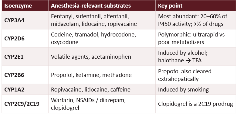
So cytochrome P450 is really important for metabolism in the liver and upregulation and downregulation of cytochrome P450 enzymes can increase or decrease metabolism of a lot of medications that you'll see.
Heme proteins on hepatocyte smooth ER ("microsomal") and these are riches in zone 3 of the liver. Also in gut, kidney, lung, adrenal, but it's primarily in the liver.
So these cytochrome P450 enzymes have different names and different ones metabolize different medications.
The Cytochrome P450 System Table -
CYP2D6 in Pediatrics
which drug?
Codeine is a prodrug that CYP2D6 converts to morphine.
Poor metabolizers may get little pain relief; ultrarapid metabolizers can produce too much morphine, causing dangerous respiratory depression
Codeine is contraindicated for patients under 18 after tonsil or adenoid surgery
If you have to give it, make sure its in PACU and they don’t go home after
Enzyme Induction vs. Inhibition
↑ Enzyme expression; develops over days to weeks
Parent drug levels fall → tolerance, ↑ dose requirement
More reactive intermediates possible (glutathione depletion)
Inducers: ethanol, barbiturates (phenobarbital), phenytoin, carbamazepine, rifampin, smoking
Example: ↑ requirement for sedatives, opioids, vecuronium, rocuronium
This is why alcoholics require more anesthesia is because of their cytochrome P450 induction.
Prodrugs reverse the rule: induction raises active metabolite; inhibition lowers it (e.g., codeine via 2D6).
Enzyme Induction vs. Inhibition
Drugs compete for the enzyme's active site; onset within hours
Parent drug levels rise → prolonged effect or toxicity
Inhibitors: cimetidine, azole antifungals, macrolides, ritonavir, amiodarone, diltiazem, verapamil, grapefruit juice (3A4)
Example: erythromycin + midazolam → prolonged sedation
So is there the timing of reversing their paralytic will be longer if you know the patient takes something like cimetidine or another medication that inhibits subchrome P-450.
Prodrugs reverse the rule: induction raises active metabolite; inhibition lowers it (e.g., codeine via 2D6).
Liver Disease & Drug Disposition Table -

Volatile Agents & the Liver
Sevo
Iso
Des
Halo
Sevo: no TFA; forms HFIP and inorganic fluoride
2% metabolized
Iso and des form trace TFA: rare cross-sensitivity
0.2% and 0.02% metabolized
Halothane
CYP2E1 oxidation → trifluoroacetyl (TFA) adducts on liver proteins → neoantigens
20% metabolized
Mild ALT rise in ~10–30%; fulminant hepatitis ~1 in 10,000–30,000
Risks: female, middle age, obesity, repeat exposure; zone 3 necrosis
Cirrhosis & Portal Hypertension
backward vs forward?
outcomes? 5
Backward: fibrosis and regenerative nodules ↑ intrahepatic resistance = hard going out
Stellate cells drive fibrosis; portal pressure can reach 20–30 mmHg.
Forward: splanchnic vasodilation (nitric oxide) ↑ portal inflow = alot coming in
↓
PORTAL HYPERTENSION - HVPG >5 mmHg (≥10–12 clinically significant)
Esophageal and gastric varices; portosystemic shunting
Splenomegaly: Platelet sequestration → thrombocytopenia
Ascites: Sinusoidal hypertension + ↓ albumin + Na retention
Encephalopathy: Gut toxins bypass the liver
Hyperdynamic state: ↓ SVR, ↑ CO; RAAS/ADH → HRS
Hyperdynamic Circulation
of cirrhosis/portal hypertension - 4 steps
This card is describing the hyperdynamic circulation of cirrhosis/portal hypertension. The confusing part is that total blood volume can be high while the arteries are still underfilled.
1. Splanchnic vessels dilate, largely due to nitric oxide (NO).
2. Blood pools in the expanded abdominal vascular bed, so effective arterial volume falls.
3. The heart compensates with ↑ heart rate and ↑ cardiac output. But because vessel resistance falls (↓ SVR), blood pressure may still be low or low-normal.
4. Vasodilation also makes vessels less responsive to sympathetic signals and vasopressors.
Here, NO means nitric oxide—not nitrous oxide (N₂O), the anesthetic gas.
Cirrhotic Cardiomyopathy
r/ts of CO vs peripheral vascular resistance
response to stress? 2
Autonomic neuropathy %
1. Even with ↑ CO there is ↓ peripheral vascular resistance
2. Systolic and diastolic dysfunction; blunted response to stress
β-adrenergic hyporesponsiveness
Electrophysiologic changes: prolonged QTc in 30% (Child A) to 60% (Child C)
Autonomic neuropathy in ~43%
So you have remodeling and decreased responsiveness to medications with chronic liver failure.
the heart may have a normal ejection fraction at rest but still have poor reserves and a blunted response when stress requires it to increase cardiac output.
Ex: stress of induction of anesthesia = pt coding
Hemostasis in Liver Disease:
Coagulation factors
what ↓ and ↑?
VTE risk?
↓ fibrinogen in %?
lab accuracies?
↓ Procoagulants and ↓ anticoagulants (protein C, antithrombin)
↑ Factor III, VIII and vWF (made outside hepatocytes) - to compensate
Thrombin generation often normal; VTE risk not reduced
Dysfibrinogenemia in 70–80%
PT/INR measures only procoagulants, so it predicts bleeding poorly in cirrhosis. The balance is fragile and tips either way.
Hemostasis in Liver Disease:
Platelets
thrombocytopenia in %?
Thrombocytopenia in 30–64%
Splenic sequestration (up to 90% of platelets)
solution there is to do a splenectomy because cirrhosis raises pressure in the portal vein system. That pressure backs up into the splenic vein, making the spleen congested and enlarged.
↓ Thrombopoietin; marrow suppression
↑ vWF partly compensates adhesion
Hemostasis in Liver Disease:
Fibrinolysis - slow down or increase?
Hyperfibrinolysis in 30–46% of end-stage disease: break clots down too quickly
Liver normally clears tPA and makes plasminogen, PAI-1, TAFI
Net balance, not one value, determines bleeding between clot-building and clot-breaking proteins matters. Cirrhosis doesn’t automatically mean the blood can’t clot; the balance can shift toward either bleeding or clotting
Renal Dysfunction
what is activated for too long? what e-?
↓ Effective arterial volume → SNS, RAAS, vasopressin prolonged activation
Na and water retention; dilutional hyponatremia, hypokalemia
Renal prostaglandins maintain perfusion = ↑ NSAID sensitivity
HRS: functional prerenal failure; kidneys work after transplant
Type 1: creatinine doubles in <2 weeks; Type 2: indolent, refractory ascites
So functionally they have pre renal failure because of the restricted blood flow to the kidneys. But if you correct the liver problems, then the kidneys will correct also as long as they're not failing.
Ascites - 3 combo
SAAG?
threshold?
what organ is next?
Most common complication of cirrhosis
Sinusoidal hypertension + ↓ oncotic pressure + renal Na retention
Fluid is protein-rich; can cause relative intravascular hypovolemia
SAAG ≥1.1 g/dL indicates portal hypertension (~97% accuracy)
↓ FRC and atelectasis from diaphragmatic splinting
Onset of ascites marks a threshold: ~50% mortality within 3 years. HRS risk with ascites: 18% at 1 year, 39% at 5 years.
hepatorenal syndrome—kidney dysfunction that can develop in advanced liver disease, often when ascites is present.
Pulmonary Complications: HPS - IPVD
dead space vs shunt?
aa gradiet?
hallmark?
response to O2?
Hepatopulmonary syndrome (HPS)
Core problem
Intrapulmonary vascular dilation (IPVD)
Mechanism
Capillaries dilate from 8–15 µm to 50–500 µm: diffusion–perfusion defect, functional shunt
Criteria
Liver disease + PaO₂ <80 mmHg or A–a gradient ≥15 mmHg on room air + IPVD on bubble echo
Hallmark
Orthodeoxia: hypoxemia worsens upright, improves supine
Also common: ascites and hepatic hydrothorax restrict lung volumes; 50–70% of patients report dyspnea.
Response to O₂
Type I improves with 100% O₂; failure suggests true shunt
Prevalence
Up to 20% of transplant candidates
Pulmonary Complications: PoPH - patho
Hallmark 2
Portopulmonary hypertension (PoPH)
Core problem
Portal hypertension/liver disease → pulmonary artery narrowing and remodeling → ↑ pulmonary vascular resistance (PVR) → ↑ pressure the right ventricle must pump against → right ventricular strain.
Mechanism
Endothelin, shear stress from high CO, hormonal and immune factors
Criteria
Portal HTN + mPAP ≥25 mmHg, PAOP <15 mmHg, PVR >240 dyn·s·cm⁻⁵ (3 WU)
Hallmark
Right ventricular strain; severity by mPAP (<35, 35–50, >50)
Response to O₂
Hypoxemia is not the main feature
Prevalence
2% of portal HTN; 4–6% of transplant candidates
Hepatic Encephalopathy
ammonia from?
liver dmg?
astrocytes
glutamine
GABA tone (reversal?)
Main pathway:
Gut bacteria produce ammonia
Other toxins: mercaptans, short-chain fatty acids, manganese
A damaged liver or blood bypassing the liver (shunting) lets more of them reach the bloodstream and brain.
Brain support cells called astrocytes convert ammonia into glutamine as a detox step.
Glutamine can build up inside astrocytes, drawing in water and causing swelling, edema.
“↑ GABA tone” means the brain’s inhibitory, calming signal is stronger—like turning up the brain’s “brakes.” That can contribute to sleepiness and slowed thinking in hepatic encephalopathy.
“Endogenous benzodiazepine-like substances” means naturally occurring substances in the body that may enhance signaling at the same receptor as benzodiazepines. It doesn’t mean the person took a benzodiazepine.
Flumazenil helps transiently
Hepatic Encephalopathy: Risk Factors - 4
ammonia r/ts severity
The precipitants lists things that can push someone with liver disease into an episode:
GI bleeding (protein load)
Infection, sepsis
Sedatives, especially benzodiazepines
Hypokalemia, alkalosis, hypoxia, hypovolemia
Ammonia levels correlate poorly with severity
West Haven grade - 4
Grades 1-4
Trivial lack of awareness, euphoria or anxiety, short attention span
Lethargy, minimal disorientation, personality change, inappropriate behavior
Somnolence to semistupor but responsive; confusion, gross disorientation
Coma, unresponsive to verbal or noxious stimuli
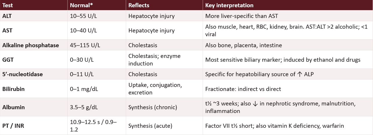
The liver test, you can read through these. I'm not really worried about super memorizing like normal levels. I do think it's important to know like the interpretation if it's high or low. -
No single test measures liver function. Use the pattern and the trend.
Patterns of Injury:
Hepatocellular
what labs are affected?
disease?
AST and ALT markedly ↑
ALP mildly ↑
Hepatitis (viral, toxic, ischemic)
Patterns of Injury:
Cholestatic
what labs are affected?
ALP, GGT, 5′-NT markedly ↑
Direct bilirubin ↑
AST and ALT mildly ↑
Patterns of Injury:
Synthetic failure
what labs are affected?
Albumin ↓ (chronic)
PT/INR ↑, not vitamin K-responsive
Platelets often ↓
Beware Misdirection of:
Passing a CBD stone
Isolated mild ↑ unconjugated bilirubin or GGT
early what labs are affected? looks like?
later what labs are affected? looks like?
Passing a common bile duct stone:
Passed CBD stone; AST/ALT rise early and can mimic hepatitis → Hepatocellular pattern.
ALP and GGT rise later → Cholestatic pattern.
Isolated mild ↑ unconjugated bilirubin or GGT often needs no workup.
Severity Scoring: Child-Pugh
Class A: good prognosis. -
Class C: limited life expectancy and high perioperative risk.

Severity Scoring: MELD
4 labs tracked
Bilirubin, INR, creatinine (± sodium)
Predicts 3-month mortality
Used for transplant allocation
Renal function is one of only three core variables: reflects HRS physiology
High-Yield Takeaways - MK
portal vs hepatic artery (flow & O2 req)
zone 3: 4
HABR 3
HVPG 4
↑ vs ↓ ER drugs
cirrhosis 5
PT/INR and albumin
Portal vein: ~75% of flow, ~50% of O₂. Hepatic artery: ~25% of flow, ~50% of O₂
Zone 3: least O₂, most CYP450 → first hit by hypoxia, acetaminophen, halothane
HABR: ↓ portal flow → adenosine accumulates → hepatic artery dilates
HVPG >5 mmHg = portal hypertension; ≥10–12 = varices and systemic disease
High-ER drugs are flow-limited; low-ER drugs are capacity- and binding-limited
Cirrhosis: hyperdynamic (↑CO, ↓SVR), rebalanced hemostasis, HRS, HPS/PoPH, HE
PT/INR reflects acute synthesis (factor VII); albumin reflects chronic (t½ ~3 wk)