A&P Exam 3 Muscles
 Call Kai
 Learn
 Practice Test
 Spaced Repetition
 Match
 Flashcards
 Knowt Play
1/44

There's no tags or description
Looks like no tags are added yet.
45 Terms
3 types of muscles
skeletal cardiac and smooth
skeletal and smooth muscle
elongated and called muscle fibers
myo mys and sarc
prefixes for muscle
skeletal muscle
• Organs attached to and cover bony skeleton
• Elongated cells called muscle fibers
• Striated (striped)
• Voluntary (i.e., conscious control)
• Responsible for overall motility
• Contract rapidly; tire easily; powerful
• Require nervous system stimulation
cardiac muscle
• Only in heart; bulk of heart walls
• Striated
• Can contract without nervous system
stimulation
• Involuntary
Smooth Muscle
• In walls of hollow organs, e.g., stomach,
urinary bladder, and airways
• Not striated
• Can contract without nervous system
stimulation
• Involuntary
Special Characteristics of Muscle
Tissue
• Excitability (responsiveness): ability to receive
and respond to stimuli
• Contractility: ability to shorten forcibly when
stimulated
• Extensibility: ability to be stretched
• Elasticity: ability to recoil to resting length
Muscle Functions
– Movement of bones or fluids (e.g., blood)
– Maintaining posture and body position
– Stabilizing joints
– Heat generation (especially skeletal muscle
Additional muscle functions
– Protects organs, forms valves, controls pupil size,
causes "goosebumps"
Gross Anatomy of skeletal muscle
Predominantly muscle fibers however blood
vessels, nerve fibers, and connective tissue
are present
Each skeletal muscle served by one artery, one nerve,
and one or more veins
– Enter/exit near central part and branch through
connective tissue sheaths
– Every skeletal muscle fiber supplied by nerve
ending that controls its activity
– Huge nutrient and oxygen need; generates large
amount of waste
Epimysium
dense irregular connective tissue
surrounding entire muscle; may blend with fascia
Perimysium
fibrous connective tissue surrounding
fascicles (groups of muscle fibers)
Endomysium
fine areolar connective tissue
surrounding each muscle fiber

Direct skeletal muscle attachment
epimysium fused to periosteum of bone
or perichondrium of cartilage
Indirect skeletal muscle attachment
connective tissue wrappings extend
beyond muscle as ropelike tendon or sheetlike
aponeurosis
Microscopic Anatomy of skeletal muscle
• Long, cylindrical cell
– 10 to 100 μm in diameter; up to 30 cm long
• Multiple peripheral nuclei
• Sarcolemma = plasma membrane
• Sarcoplasm = cytoplasm
– Contains large amounts of Glycosomes for
glycogen storage, myoglobin for O2 storage
• Modified structures: myofibrils, sarcoplasmic
reticulum, and T tubules
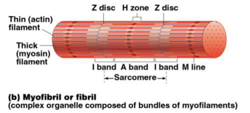
Myofibrils
• Densely packed, rodlike elements
• Each cell contains hundreds of these
• ~80% of cell volume
• Contain sarcomeres - contractile units
– Sarcomeres contain myofilaments
• Exhibit striations - perfectly aligned repeating
series of dark A bands and light I bands
H zone:
lighter region in midsection of dark A band
where filaments do not overlap
M line
line of protein myomesin bisects H zone
Z disc (line)
coin-shaped sheet of proteins on
midline of light I band that anchors thin filament and connects myofibrils to one another
Thick filaments
run entire length of an A band
Thin filaments
run length of I band and partway into
A band
Sarcomere
region between two successive Z discs. Composed of thick and thin myofilamentsmade of contractile proteins
Excitability (responsiveness
ability to receive
and respond to stimuli
Contractility
ability to shorten forcibly when
stimulated
Extensibility
ability to be stretched
Elasticity
ability to recoil to resting length
T tubules
send signals conduct impulses deep into the muscle fiber
Elastic filament
– Composed of protein titin
– Holds thick filaments in place; helps recoil after
stretch; resists excessive stretching
Dystrophin
Links thin filaments to proteins of sarcolemma
Sarcoplasmic Reticulum (SR)
• Network of smooth endoplasmic reticulum
surrounding each myofibril
– Most run longitudinally
• Pairs of terminal cisterns form perpendicular
cross channels
• Functions in regulation of intracellular Ca2+
levels
– Stores and releases Ca2+
cross
bridges
Occurs when myosin heads bind to actin
Activation (at neuromuscular junction)
• Must be nervous system stimulation
• Must generate action potential in sarcolemma
Excitation-contraction coupling
• Action potential propagated along sarcolemma
• Intracellular Ca2+ levels must rise briefly
The phases leading to muscle fiber contraction

contraction
ap in a neuron → ap axon terminal → release of ACH→ ACH binds to sarcolemma ap → AP travels along sarcolemma down the t tubles → Ca+ dumped into cell by SR → Ca+ binds to troponin→ Tropomyosin moved out of the way → Myosin heads bind to actin and pull if ATP is present (cross bridging)
generation of a motor neuron action potential, neurotransmitter release, generation of a muscle cell action potential, release of calcium ions from SR, ATP-driven power stroke, sliding of myofilaments