NBEO Part II Things to Remember
 Call Kai
 Learn
Learn Practice Test
 Spaced Repetition
 Match
Match Flashcards
 Knowt Play
1/140

There's no tags or description
Looks like no tags are added yet.
Name | Mastery | Learn | Test | Matching | Spaced | Call with Kai | Chat |
|---|
No analytics yet
Send a link to your students to track their progress
141 Terms
What are the formulations of cyclosporin?
- Restasis (cyclosporin 0.05%)
- Cequa (cyclosporin 0.09%)
- Verkazia (cyclosporin 0.1%)
- Vevye (cyclosporin 0.1%)
How many times is Oxervate (cenegermin-bkbj 0.002%) dosed for neurotrophic keratopathy?
6x/day for 8 weeks
What are the formulations of loteprenol etabonate?
- Alrex (loteprednol etabonate 0.2%)
- Lotemax (loteprednol etabonate 0.5%)
- Zylet (tobramycin / loteprednol etabonate )
What is the treatment for fungal keratitis?
- Natacyn (natamycin 5%) Q1hr
- amphotericin B 0.5% Q1hr
- both with loading doses
- voriconazole 400mg PO BID
What is the treatment for acanthamoeba keratitis?
chlorhexidine 0.02% and propamidine isethionate 0.1%
- Q1hr around the clock for 1 day
- Q2hr when awake/Q4hr while asleep for days 2-7
- TID for the remaining 4 months
olopatadine brand name and dosage?
- Pataday Extra Strength (olopatadine 0.7%) QD
-- There's also a 0.2% QD form
- Patanol (olopatadine 0.1%) BID
***OTC
ketotifen brand names and dosage?
- Zaditor or Alaway (ketotifen 0.025%) BID
**OTC
azelastine brand name and dosage?
- Optivar (azelastine 0.05%) BID
bepotastine brand name and dosage?
- Bepreve (bepotastine 1.5%)
**BID
Alcaftadine brand name and dosage?
- Lastacaft (alcaftadine 0.25%) QD
Go-to treatment(s) for horeola and preseptal cellulitis
#1 500mg Keflex QID (cephalexin) PO x 10 days
#2 500mg Augmentin QID (amoxicillin/clavulonic acid) x10 days
If penicillin allergy:
- Z-pak (Azithromycin 250mg/2 tablets on day 1, then 1 tablet on days 2-5)
If MRSA suspected (nursing home/hospital acquired)
- Bactrim BID (sulfamethoxazole 400mg/trimethoprim 80mg)
valacyclovir dosing for HSV (active infection)
Valtrex (valacyclovir) 500mg TID PO
valacyclovir dosing for HSV prophylaxis
Valtrex (valacyclovir) 500 mg QD
valacyclovir dosing for HZO
Valtrex (valacyclovir) 1000mg TID
nepafenac: use and dosage
- Nevanac (nepafenac 0.1%) TID
- Ilevro (nepafenac 0.3%) QD
**Best for CME prevention and tx post CE/PCIOL
**Best macular penetration because its a prodrug converted to amfenac inside ocular tissues
bromfenac: use and dosage
- Bromsite (bromfenac 0.09%) QD
- Prolensa (bromfenac 0.075%) QD
**Good macular penetration/CME prevention
ketorolac: use and dosage
- Acular (ketorolac 0.5%) QID
- Acuvail (ketorolac 0.45%) QID
**fair macular penetration/CME prevention but best used for pain/ant seg inflammation
diclofenac: use and dosage
- Voltaren (diclofenac 0.1%) QID
**poor macular penetration/CME prevention so should not be used for post seg conditions
what is the most likely dx for a pupil that constricts to 0.125% pilocarpine?
Constricts to 0.125% pilo → Adie's
**Parasympathetic supersensnviity
What drops should you use to aid in the diagnosis of a Horner's pupil?
Cocaine: NE reuptake inhibitor; will not dilate Horner's eye d/t lack of NE in synaptic cleft
1% hydroxyamphetamine: potentiates NE release from healthy post-ganglionic neurons, so will dilate central/pre-ganglionic Horner's lesions
1% aparaclondine: weak alpha 1 agonist, will dilate any Horner's lesion d/t super sensitivity
latanoprost formulations
- Xalatan (latanoprost 0.005%)
- Xelpros (latanoprost 0.005%): BAK free
- Iyuzeh (latanoprost 0.005%): PF
Non-latanoprost PGAs (5)
- Travatan Z (travaprost 0.004%): SofZia
- Lumigan (bimatoprost 0.1%)
- Latisse (bimatoprost 0.03%)
- Zioptan PF (tafluprost 0.0015%)
- Vyzulta (latanoprostene bound 0.024%): nitric oxide
Beta blocker formulations (5)
- Timoptic (timolol maleate 0.5% and 0.25%)
- Timoptic XE PF (timolol maleate 0.5% and 0.25%): gel
- Timoptic Ocudose PF (timolol 0.5% and 0.25%)
- Istalol (timolol maleate 0.5%)
- Betimol (timolol 0.5% and 0.25%): hemihydrate instead of maleate
Alpha agonist formulations (3)
- Alphagan P (brimondine 0.15% or 0.2%)
- Lumify (brimondine 0.025%)
- Iopodine (apraclondine 0.5%)
CAI formulations
- Truspot (dorzolamide 2%)
- Azopt (brinzolamide 1%)
Combo IOP lowering formulations
- Cosopt (dorzolamide 2%/timolol 0.5%)
- Combigain (brimondine 0.2%/timolol 0.5%)
- Simbrinza (brinzolamide 1%/ brimonidine 0.2%)
- Rocklatan (netarsudil 0.2%/latanoprost 0.005%)
How to translate A1c to average blood glucose?
Ex. An HBA1c of 7.5% is equal to was PG level?
(30*A1c)-50
Ex. An HBA1c of 7.5% is equal to was PG level?
(30*7.5)-50= 175 mg/dL
Lens subluxation vs. dislocation
Subluxation = displacement
Dislocation = detached from zonules, lying outside of the hyaloid fossa (AKA ectopia lentis)
In which direction does subluxation occur most commonly in Marfan's vs. homocystinuria?
Marfan's: down and out/temporally, with zonules and therefore accommodation still intact
Homocystinuria: down and in/nasally, with zonules ruptured d/t lack of support from homocysteine; iridodesis and/or phacodonesis (shaking of iris/lens) may occur
Both are at higher risk of an RD
What is the class of drug and MOA of Warfin (Coumadin)?
Anticoagulant
Prevents the formation of vitamin K dependent clotting factors
What is the class of drug and MOA of Heparin?
Anticoagulant
Prevents conversion of prothrombin to thrombin (via activation of antithrombin III to inhibit Factor Xa; indirect)
Eliquis is a direct anticoagulant because it directly blocks Factor Xa
What type of conjunctival lesion is being described?
Adjacent to the limbal region or caruncular area.
Can appear flat or elevated.
Well-defined borders, can be moved freely over the sclera.
Cystic spaces within are commonly observed — key diagnostic sign.
Pigmentation varies.
May grow and change pigmentation during puberty.
Conjunctival nevus
Signs of malignancy include:
Vascularization
Atypical location (fornix, palpebral conjunctiva)
Growth onto cornea
Rapid growth or pigmentation change, especially after puberty
What type of conjunctival lesion is being described?
Found in middle-aged Caucasians.
Typically appears after age 45.
Observed as flat brown lesions that can be moved over the sclera.
May change over time; size and pigmentation can increase or decrease.
Primary acquired melanosis
PAM without atypia is benign, limited to the basal layer of the conjunctiva.
PAM with atypia has 50% chance of becoming malignant, extends to all conjunctival layers.
Elevated or vascularized areas should raise suspicion for malignancy.
What conjunctival lesion is being described?
Congenital, evenly pigmented lesion.
Appears black/dark brown.
Has well-demarcated borders.
Does not move freely over the scleral surface.
Slow-growing, but can increase in size over time.
melanocytoma
What type of conjunctival lesion is being described?
Seen in middle-aged to elderly patients.
Lesions are usually elevated and vascularized.
May have large feeder vessel at lesion site.
Frequently occurs at limbus, typically darkly pigmented. Can also be amelanotic.
conjunctival melanoma
Can develop from:
PAM
Pre-existing nevi
Spontaneously
May metastasize, especially to:
Preauricular nodes
Anterior cervical lymph nodes
Palpation of these areas is crucial if melanoma is suspected.
What is the upper limit of normal for Caucasian and African American patients for exophthalmos measured via a Hertel exophtalmometer?
22mm for Caucasians
24mm for African Americans
What conditions are most commonly associated with angioid streaks?
PEPSI
Pseudoxanthoma elasticum (strongest association)
Ehler Danlos
Piaget's
Sickle Cell
Idiopathic
What is Pseudoxanthoma elasticum?
Caused by mutations responsible for transporting pyrophosphate leading to an accumulation of calcium and phosphate in the elastic fibers, causing them to become stiff and brittle.
Patients with PXE typically have characteristic signs of very loose skin folds and yellow skin papules that are commonly observed in the neck region, axillae, and on flexor aspects of joints.
These patients also frequently suffer from cardiovascular disease caused by accelerated atherosclerosis, and have an increased risk of developing gastrointestinal bleeds, which can be life threatening.
The combination of PXE and angioid streaks is referred to as "Gronblad-Strandberg syndrome."
What retinal findings are often associated with angioid streaks?
Optic disc drusen
Peau d'orange: yellow, speckled, molted fundus appearance mid peripherally (AKA lepord-skin spotting)
CNVM
What is the most common causative organism of acute postoperative endophthalmitis?
Staph epidermis, followed by staph areus, then gram negative organisms
The most common cause of delayed onset post operative endophthalmitis is P. acnes
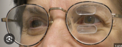
How are Peli prisms placed on lenses for field expansion?
For a right hemianopsia, two 40 diopter fresnel prism segments are placed BO over the right lens
For a left hemianopsia, two 40 diopter Fresnel prism segments are placed BO over the left lens. Esch segment is placed 7mm above and 7mm bellow the midline of the pupil. This allows for about 20 degrees of filed expansion (40*0.57).
This is different from placing base right prism over both eyes for a right hemianopsia, which is not field expansion at all. This causes a shift in the visual field for patients with a midline shift or hemi filed neglect.
What is Sheard's criteria?
Compensating relative fusional vergence (blur point) should be 2x the patient's deviation.
Adapted to be used for eso or exo;
Prism needed = 2/3 phoria - 1/3 compensating fusional vergence
Eso has poor BI/divergence/NFV and needs BO/PFV to fix it
Exo has poor BO/convergence/PFV and needs BI/NFV to fix it
What does SILO mean in VT?
These are vergence cues used when training variable tranoglyohs or vectograms
When converging, the image should become closer to the patient and smaller (small in)
When diverging, the image should get further away and larger (large out)
Retinitis pigmentosa
- Inheritance pattern:
- Mutation:
- Age of onset:
- Key findings:
- Prognosis:
- Inheritance pattern: AR most common
- Mutation: RPE65
- Age of onset: teens-20s (later if AD)
- Key findings: bone spicules, attenuated arterioles. waxy pallor, CME, PSC, night blindness, VF constriction
- Prognosis: progressive VF loss with lat loss of central vision
*ERG: reduced scotopic early, then reduced photopic

Stargardt disease
- Inheritance pattern:
- Mutation:
- Age of onset:
- Key findings:
- Prognosis:
- Inheritance pattern: AR
- Mutation: ABCA4
- Age of onset: early childhood-teens
- Key findings: yellow pisiform (fish tail) flecks in posterior pole, beaten bronze macula
- Prognosis: central VA reduced, periphery spared, blindness possible mid-life

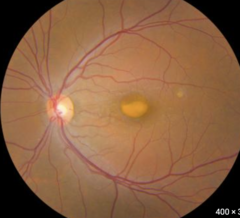
Best's disease (Viteliform macular dystrophy)
- Inheritance pattern:
- Mutation:
- Age of onset:
- Key findings:
- Prognosis:
- Inheritance pattern: AD
- Mutation: BEST1
- Age of onset: childhood
- Key findings: egg-yolk lesion at macula
- Prognosis: good VA early, central vision declines as egg breaks up
*Normal ERG, abnormal EOG (
Gyrate atrophy
- Inheritance pattern:
- Mutation:
- Age of onset:
- Key findings:
- Prognosis:
- Inheritance pattern: AR
- Mutation: ornithine aminotransferase deficiency
- Age of onset: childhood-teen
- Key findings: scalloped and well demarcated areas of chorioretinal atrophy starting peripherally
- Prognosis: progressive VF loss, blindness by mid-life if untreated, some respond to B6
*Reduced ERG and EOG

Choroideremia
- Inheritance pattern:
- Mutation:
- Age of onset:
- Key findings:
- Prognosis:
- Inheritance pattern: X-linked
- Mutation: CMH
- Age of onset: childhood-teens
- Key findings: patchy RPE and chorioretinal atrophy beginning in midperiphery
- Prognosis: progressive blindness by mid-life
*Reduced ERG and EOG

Cone dystrophy
- Inheritance pattern:
- Mutation:
- Age of onset:
- Key findings:
- Prognosis:
- Inheritance pattern: AD most common
- Mutation:
- Age of onset: Childhood-20s
- Key findings: photophobia, CV loss, central scotoma, bull's eye maculopathy
- Prognosis: progressive central vision loss, rods affected later
*Redcued photopic ERG, normal EOG

Ocular albanism
- Inheritance pattern:
- Mutation:
- Age of onset:
- Key findings:
- Prognosis:
- Inheritance pattern: AR or X-linked
- Mutation:
- Age of onset: birth/early childhood
- Key findings: ITD, foveal hypoplasia, nystagmus
- Prognosis: Life long reduced VA
*Normal ERG and EOG, abnormal VEP

Leber's hereditary optic neuropathy
- Inheritance pattern:
- Age of onset:
- Key findings:
- Prognosis:
- Inheritance pattern: mitochondrial
- Age of onset: young adult males
- Key findings: subacute central vision loss, optic disc hyperemia, RNFL selling early with later optic atrophy
- Prognosis: central vision loss
*Normal ERG and EOG, abnormal VEP
How to calculate CL power for apical alignment and spherical over refraction of plano using K's and SRx?
- TLP corrects some of the SRx
- Steeper fit = creates plus TLP
- Flatter fit = creates minus TLP
*If this diagnostic lens is not available in the fitting set, select the next flattest base curve, which creates apical touch equal to the difference between BCs (flatter so contributes minus to TLP)

What is dimple veiling?
- Bubbles of CO2 trapped under a lens, leaves tiny indents in epi that pool with FL
- Can be asymptomatic or caused irritation and blur
- Much more common in GPs
- Due to poor fit (too steep), must flatten the lens by decreasing OAD or OZD
A patient with a VA of 20/100 has a JND of?
JND = 1.00
For sphere refraction show +/- 0.50
For cyl refraction, show 1.00
How to select spherical vs. front toric, back toric and bitoric?
Spherical: less than 2.50 Kcyl and less than 0.75D RA
Front troic: less than 2.50 Kcyl and greater than 0.75D RA
Back toric: greater than 2.50 Kcyl and SRx cyl is 1.5x Kcyl
Bitoric: greater than 2.50 Kcyl and SRx cyl is not 1.5x Kcyl
Describe a spherical power effect (SPE) bitoric
These lenses compensate for their toric back surface with a toric front surface and thus have a combined spherical power
- Used to improve fitting/comfort on patient with a lot of corneal astig but do not need correction for astig in their CLs (
Describe a cylindrical power effect (CPE) bitoric
These lenses have toric back surface for proper fitting on a cornea with a lot of toricity and a front toric surface to correct for residual astigmatism. They have a combined toric power.
- Used to improve fitting/comfort on patients with a lot of corneal astigmatism who also need correction for astigmatism in their CLs (>0.75D RA)
LASIK complications: Epithelial ingrowth
- Occurrence:
- Cause:
- RF:
- Tx:
- Occurrence: days-weeks
- Cause: proliferation of surface epi cells into flap interface, caused by poor flap contact (epi cells aren't surrounded by other epi cells)
- RF: enhancement surgeries, older pt, BM dystrophy, hx/o RCE, eye rubbing, diabetics, belphrospasm
- Tx: If limited to the periphery of flap interface = monitor in 2 weeks; if reduced VA and progression; lift flap and remove cells

LASIK complications: diffuse lamalar keratitis (sands of Sahara)
- Occurrence:
- Cause:
- RF:
- Tx:
- Occurrence: 2-5 days
- Cause: diffuse inflammatory infiltrates across periphery of interface, not penetrating the stroma or extending into the flap which will increase to cover the flap inter face and a central haze develops (looks like staining but epi intact)
- RF:
- Tx: topical steroids, possible lifting of flap with debridement of interface
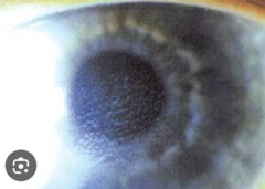
ANSI standards for sphere tolerance

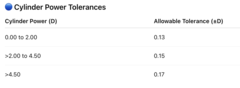
ANSI standards for cylinder tolerance
ANSI standards for axis tolerance

ANSI standards for horizontal and vertical prism tolerance

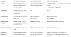
Minimum FDA requirement for impact resistant labeling
- FDA enforces that all ophthalmic lenses must be impact-resistant unless a specific medical exemption is documented by the prescriber.
All finished ophthalmic lenses (including Rx) must pass the drop ball test:
- ⅝ inch (16 mm) steel ball dropped from 50 inches (127 cm) onto the center of the lens.
- The lens must not fracture.
- Every glass lens must be individually tested.
- Plastic (polycarbonate, Trivex, CR-39, etc.) lenses may be batch tested, provided manufacturing controls are documented.
- The FDA considers this test the minimum requirement for "impact-resistant" labeling. It does not guarantee industrial-level eye protection.

FDA requirements for basic impact vs. high impact safety eyewear
Safety eyewear designed for occupational use must meet ANSI Z87.1 standards, which define two protection levels:
*Basic Impact*
- 1-inch (25.4 mm) diameter steel ball dropped from 50 inches.
- Lens must not fracture, chip, or dislodge.
- Lenses are marked "Z87" (no "+" sign).
- Frames must also bear "Z87" marking.
- Usage: Suitable for low-risk environments with minimal projectile or mechanical hazards.(Example: laboratory eyewear, general clinical use)
*High Impact*
- Lenses must pass both the Basic Impact test and an additional High-Velocity Impact Test:
- ¼ inch (6.35 mm) steel ball is fired at 150 ft/sec (45.7 m/s).
- Lens must not fracture, dislodge, or allow the projectile to pass through.
- Frame and lens combination are tested together
- Lenses and frames must both be permanently marked:
"Z87+"
- Frames marked "Z87-2+" for prescription frames; "Z87+" for plano.
- Side shields (if present) must also bear the Z87 marking. Side shields must be able to withstand a projectile at 150ft/sec at 3 points.
- Usage: Required for industrial, manufacturing, construction, and healthcare settings with projectile or high-velocity debris risk
FTC: The Eyeglass Rule (16 CFR Part 456)
- Must automatically release the spectacle Rx immediately after refraction, even if not requested.
- No conditions: cannot require purchase of glasses or extra payment before release.
- No waivers: patients cannot sign away their right to a copy.
- Must include: patient name, exam date, prescriber name, sphere/cylinder/axis, prism (if applicable), and expiration date.
- Verification: must confirm Rx accuracy when another seller requests it.
FTC: The Contact Lens Rule (16 CFR Part 315)
- Must automatically release the CL prescription upon completion of fitting (after trial lenses & follow-ups).
- Cannot require the patient to purchase lenses from your office.
- Verification: Third-party sellers must verify the Rx.
If no response from the prescriber within 8 business hours, it's passively verified.
- Documentation (2020 update): Must obtain patient signature confirming receipt of CL Rx, or document electronic release.
- Maintain record for 3 years.
- Expiration: Must list an expiration date (usually 1 year unless medically justified otherwise).
- No alteration: Sellers cannot substitute brand or parameters without approval.
FTC: Fairness to Contact Lens Consumers Act (2003)
Federal law that authorizes and defines the FTC Contact Lens Rule.
The FCLCA is the law, and the Contact Lens Rule is the implementation of that law.
FTC: Truth in Advertising (FTC Act, 15 U.S.C. § 41-58)
- Advertising claims: must be truthful, supported by evidence.
- Pricing: advertised exam or eyewear prices must represent the actual total cost and clearly disclose limitations.
- "Free exam" or "discount" ads: cannot mislead or include hidden conditions.
- Endorsements/testimonials: must represent honest experiences; any paid relationship must be disclosed.
- Prohibits: deceptive statements about frame quality, warranties, lens type, or insurance acceptance.
OSHA
OSHA enforces use of protective eyewear meeting ANSI Z87.1 standards.
Required for: ddging/grinding, using hazardous chemicals, splash or projectile risk.
- Contact lenses ≠ PPE → must wear safety eyewear over
- Employer provides eyewear labeled Z87 or Z87+.
What are atoric lenses?
Atoric = Aspheric + Toric
Free-form aspheric lens design in which each meridian has its own unique aspheric curvature.
In contrast to aspheric lenses (which have the same aspheric curve in all meridians), atoric lenses have different aspheric curves in the two principal meridians.
This allows for more precise correction of off-axis aberrations, like marginal and oblique astigmatism.
Standard frame alignment order of adjustments
1️⃣ Horizontal alignment (bridge skew)
- Ensure both lenses are level when the frame sits on a flat surface.
- Horizontal misalignments arise from bridge skew, not lens tilt. Always correct bridge first.
2️⃣ Vertical alignment (4-point touch)
- Ensure both lenses and both endpieces touch a flat surface
- Xing: Top and bottom of frame not parallel (looks like an "X" from side view) --> heat bridge; rotate hands in opposite directions until eye wires are parallel.
- Variant planes: One lens closer to face than the other (but lenses still parallel) --> heat bridge; place thumbs near pads and push eye wire away on the side that sits closer.
- *Vertical misalignment = skew along z-axis; correct at bridge, not temples*
3️⃣ Open temple alignment
- Ensure both temples open symmetrically with correct temple spread.
- Check: Open temples and view from above — both should angle evenly outward.
- Temple spread = distance between temples when open; correct by adjusting endpieces, not bending temples.
4️⃣ Temple parallelism
- Ensure both temples are parallel and have even pantoscopic tilt.
- Check: Place frame upside down on a flat surface; if it wobbles, temples aren't parallel
- Temple parallelism relates to pantoscopic tilt — correct hinge angle before final temple bends.
5️⃣ Temple bend alignment
- Ensure both temples curve downward and inward equally behind the ears.
- Check: Set frame right-side up on table — should rest evenly without wobble.
6️⃣ Temple fold angle
- When folded, both temples should cross at the center of the frame front.
- Check: Close temples — they should overlap symmetrically in the middle.
Frame adjustment: the left lens sits higher on the patient's face than the right lens
Lower the left lens by adjusting the left temple upward (or raising the opposite temple).
Spherical abberation
- Peripheral rays are bent more strongly than paraxial (central) rays when passing through a spherical surface, so they focus at different points along the optical axis.
- Causes blurred image even on-axis.
- Most significant for large pupil sizes (wide apertures).
Correction / Minimization:
- Use aspheric lens surfaces (flatten periphery).
- Reduce aperture (smaller pupil).

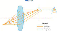
Coma
- Off-axis point objects produce a comet-shaped blur — asymmetric distortion due to different magnification for rays entering at different heights.
- Problematic for off-axis light and wide pupils.
Correction / Minimization:
- Use aspheric surfaces
Oblique/marginal astigmatism
- When light from an off-axis point passes through the lens, the tangential and sagittal rays focus at different points, forming two line foci instead of one.
- The eye naturally experiences this in peripheral gaze.
Correction / Minimization:
- Controlled using correct base curve selection (Tscherning ellipse).
- Minimized in corrected curve lenses and free-form designs.

Curvature of filed
- The image of a flat object forms on a curved surface (Petzval surface) instead of a plane, so the image cannot be in focus across the entire field simultaneously.
- Center may focus while edges blur, or vice versa.
- The retina is curved, which partially compensates for this aberration.
Correction / Minimization:
- Combine with oblique astigmatism correction to flatten the field.
- Corrected curve lenses balance COF and oblique astigmatism
- Use aspheric surfaces
Distortion
- Magnification changes progressively across the field, so straight lines appear bent or curved.
- Barrel distortion: magnification decreases toward edges (minus lenses).
- Pincushion distortion: magnification increases toward edges (plus lenses).
Correction / Minimization:
- Lens design balance between magnification and curvature.
- Corrected with aspheric optics or digital surfacing.
Longitudinal chromatic abberation
- Different wavelengths (colors) of light are focused at different distances along the optical axis.
- Blue (short λ) focuses anteriorly; red (long λ) focuses posteriorly.
- Produces blurred focus and colored fringes around images.
Correction / Minimization:
- Use materials with high Abbe value (low dispersion).
- Combine lenses of different materials (achromatic doublet).
Longitudinal/transverse chromatic abberation
- Different wavelengths focus at different image heights
- Causes colored fringes at lens periphery, especially in high-power or off-axis viewing.
- Worse in progressives and prism lenses.
Correction / Minimization:
- Use high Abbe value materials.
- Ensure optical centers align with visual axis to reduce off-axis effects.
Toxocariasis...
What organism causes this infection?
What type of organism is it?
What is the treatment?
Toxocara canis
Nematode (multicellular parasite/helminth)
Labs:
Tx: prednisone/corticosteroids, because the ocular inflammation is a result of eye's reaction to the dead/decaying organism

Toxoplasmosis
What organism causes this infection?
What type of organism is it?
What is the treatment?
Toxoplasma gondii
Unicellular parasite/protozoan
Labs: Serum IgG/IgM, PCR of aqueous/vitreous
Tx: self-limiting in immunocompetent individuals; Pyrimethamine, sulfadiazine, and corticosteroids
Histoplasmosis
What organism causes this infection?
What type of organism is it?
What is the treatment?
Histoplasma capsulatum
Fungus
Tx: none/Anti-VEGF for CMVN
Lyme disease
What organism causes this infection?
What type of organism is it?
What is the treatment?
Borrelia burgdorferi
Spirochete bacterium
Lab: ELISA and Western blot
Tx: Doxycycline PO or IV ceftriaxone for neuro involvement
Tuberculosis
What organism causes this infection?
What type of organism is it?
What is the treatment?
Mycobacterium tuberculosis
Acid-fast bacillus (bacterium)
Labs: Quantiferon-TB Gold, PPD skin test; chest imaging
Tx: RIPE: Rifampin, Isoniazid, Pyrazinamide, Ethambutol
Syphilis
What organism causes this infection?
What type of organism is it?
What is the treatment?
Treponema pallidum
Spirochete bacterium
Labs: Non-treponemal (VDRL/RPR) + confirmatory treponemal test (FTA-ABS or TPPA); CSF VDRL if neuro involvement
Tx: IV Penicillin G
Cat scratch disease
What organism causes this infection?
What type of organism is it?
What is the treatment?
Bartonella henselae
Gram-negative bacillus
Labs: IgG/IgM serology; PCR of ocular fluid if available
Tx: Doxycycline or azithromycin (2-6 weeks); add rifampin for severe neuroretinitis
Mucin balls
- White pearl-like debris on the posterior surface of the CL
- More common in SiHi lenses that are too flat and used for extended wear
- May cause dimpling of the corneal surface
- Fixed by steepening the BC, decreasing amount of wear time, adding ATs, diferent MPS, or adding a surfactant cleaner

Dimple veiling
- CO2 bubbles trapped under a GP lens, leaving small indentations on the surface of the cornea
- Fixed by flattenting the BC or decreasing the OAD

Protein deposits
- Superfical, white, sligtly translucent depositions on the CL surface
- More common in hydrogels
- Can irritate upper lid and cause GPC
- Fixed by swiching to daily replacment, different MPS, or adding enzymatic cleaner
Jelly bumps
- White/grey deposits on the front surface of soft CLs
- Composed of calcium-protein complexes
- Increased incidence in lenses with scratches, ridges, polishing errors, or patinets with metabolic disorders
- Fixed by proper digital cleaning or adding surfactant clearner

Choroidal nevi that express what characteristics are suspicious for malignancy?
- patient is sympotmatic; metamorphopsia, or flashes
- greater than 5mm in diameter
- thickness greater than 1mm (B-scan)
- lipofuscin present on the surface; accumulation of metabolic debris from inabiltiy to phagocytose PR outter segments indicating growth and metabolic activity
- lesion is within 3mm of ONH
- there is an assosicated serous RD present
What is the most common site of choroidal melanoma metastasis?
What are the most common sites of melanoma metastasis from other sites for men vs. women?
Choroid to liver
Men: lung to choroid
Women: breat to choroid
What is Foster Kennedy syndrome and what posterior segment findings does it present with?
Space occupying lesion of the frontal lobe and olfactory groove meningioma. Leading to vision loss and olfactory defects (anosmia).
Causes compression of the ipsilateral optic nerve leading to optic atrophy and a central scotoma. As the tumor grows, it increases intracranial pressure, causing optic neuritis/swelling of the contralateral nerve. Swollen nerves do not cause VF defects (unless chronic).

Identify this developmental/congenital cataract
Zonular cataract (can be nuclear, lamellar or capsular)
d/t drop in Ca levels during development. Looks larger in a child and smaller in an adult as it gets push toward the center of the lens.

Identify this developmental/congenital cataract
Coronary/supranuclear cataract
Mostly genetic. Opacification deep in the cortex. No visual impact.

Identify this developmental/congenital cataract
Sutural opacities
Formed when lens fibers do not meet properly such that extra space between the suture is filled with albuminoid substance

Identify this developmental/congenital cataract
Cerulean/blue dot cataract
Blue opacities in the cortex, nonprogressive and not visual significant.

Identify this developmental/congenital cataract
Polar cataract (can be anterior or posterior)
Anterior polar cataract: associated with PPM, aniridia, Peter's anomaly, and anterior lenticonus
Posterior polar cataract: associated with Mittendorf dot (remnant or hyaloid artery)

Identify this developmental/congenital cataract
Galactosemia / oil droplet cataract
bilateral, d/t lack of galactosemic enzyme in GI tract
