Human Brain Quiz 4
 Call Kai
 Learn
 Practice Test
 Spaced Repetition
 Match
 Flashcards
 Knowt Play
1/152

There's no tags or description
Looks like no tags are added yet.
153 Terms
How is hearing a distance sense?
• Touch often reports something already at the body
• Sound can reveal an event before contact and outside the current line of sight
• Distance becomes lead time for orienting, avoidance, communication, and preparation
How does sound reach the ear and what reaches it?
• Sound is a mechanical pressure disturbance transmitted through a medium
• Frequency, amplitude, and phase describe different properties of a waveform
• Natural sounds contain many frequencies whose amplitudes change over time
• The auditory system must infer the event that produced the waveform
Why are real sounds not sine waves?
Real sources (voices, instruments, footsteps) produce complex spectra with multiple frequencies, whereas sine waves are simple, single-frequency periodic waves.
Different acoustic patterns can support the same perceptual pitch or source identity
What are the three components that can vary partly independently in a complex sound?
Harmonic structure
Temporal envelope
Temporal fine timing/structure
What is the path from sound to hearing within the ear?
Air pressure waves travel down the ear canal
The tympanic membrane moves in and out
Ossicles transmit and amplify the motion
The stapes moves at the oval window, creating fluid displacement in the cochlea

How does the outer and middle ear solve a transfer problem?
Air and cochlear fluid have very different acoustic impedances
Sound travels through air, but the inner ear (cochlea) is filled with fluid
(They overcome the impedance mismatch)
Tympanic membrane and ossicles convert air-pressure variations into mechanical motion
The ossicular system increases the pressure delivered to the oval window
overcomes the impedance mismatch between air and fluid so sound energy can actually enter the inner ear instead of bouncing off
Why is the cochlea not a microphone?
The cochlea actively filters, breaks down, and processes sound.
A pressure change launches a traveling wave along the cochlear partition
Mechanical properties vary continuously from base to apex
The cochlea begins separating frequencies before the signal reaches the brain
This is sensory computation in the receptor apparatus itself
How does frequency differ in place within the year
• The basilar membrane is relatively stiff and narrow near the base
• It becomes wider and more compliant toward the apex
• High frequencies peak nearer the base; lower frequencies peak farther apically
• The resulting frequency map is tonotopy
What are the two hair-cell classes and their jobs?
• Outer hair cells primarily regulate cochlear mechanics
• Inner hair cells provide the principal afferent output
• Damage to outer hair cells reduces sensitivity and sharpens less; damage to inner hair cells removes neural input more directly
How does the cochlea convert sound vibrations into electrical signals?
Mechanotransduction: converting physical movement (sound vibrations) into an electrical signal the nervous system can understand.
Bending toward the tallest stereocilium tightens tip links, opening channels so K⁺ enters → depolarization.
Bending away from the tallest stereocilium loosens tip links, closing channels → hyperpolarization.
This creates a graded receptor potential that converts sound vibrations into electrical signals.
Why is the receptor not the first-order sensory neuron in audition?
• Inner hair-cell depolarization opens voltage-gated Ca2+ channels
• Ca2+ triggers glutamate release at ribbon synapses
• Spiral-ganglion neurons generate the action potentials carried in cranial nerve VIII
• Hair cell = receptor cell; spiral-ganglion neuron = first-order sensory neuron
Inner hair cell → spiral ganglion neuron (1st order) → cochlear nerve / CN VIII→ cochlear nuclei (2nd-order neurons)
How does auditory coding keeps place and time?
place coding (tonotopic mapping) and temporal coding (phase locking)
Cochlear position preserves a place code for frequency
At lower frequencies, auditory-nerve spikes can synchronize to the waveform phase
For low-frequency sounds, individual neurons do not just fire randomly; they fire in strict synchronization with a specific phase (like the peak) of the sound wave.
The timing of the nerve impulses directly mirrors the timing of the sound wave itself.
Populations can preserve timing even when no single neuron fires on every cycle
Individual nerve cells have a refractory period and cannot fire on every single cycle
so a population of neurons works together Neuron A fires on cycle 1, Neuron B on cycle 2 to capture the exact timing of every wave cycle
Hearing therefore uses where activity occurs and when it occurs
Why does the ear preserve timing so precisely?
• Tiny timing differences between the ears carry information about source direction
• Temporal fine structure also contributes to pitch and sound segregation
• The auditory pathway preserves sub-millisecond information because behavior can use it
• The code is built for the problems the organism must solve
Does the cochlea have a map? How does the brain figure out location?
No, it does not.
The cochlea is organized purely by frequency (tonotopy), sorting sounds from high to low pitch along its length
A 500-Hz source on the left and a 500-Hz source on the right activate similar cochlear regions
Identical sound frequencies hit the exact same spot on the basilar membrane regardless of whether the sound comes from the left, right, front, or back.
External location is therefore not laid out directly on the receptor sheet
Auditory space must be reconstructed from transformations imposed before the sound reaches the two ears
The brain figures out location by calculating physical changes that happen to sound waves before they hit the receptors—such as arrival time delays, head shadow volume differences, and pinna frequency filtering.
This is the central spatial problem of audition
Where does the auditory pathway become bilateral?
When it goes from the cochlear nuclei to the inferior colliculus

Why does the auditory pathway becomes bilateral so early?
Spiral-ganglion axons first reach the ipsilateral cochlear nuclei
From there, crossed and uncrossed pathways distribute information to both sides
Secondary neurons split: some cross over to the contralateral side while others stay ipsilateral, sending auditory information from both ears to both halves of the brainstem.
Binaural comparison begins in the brainstem before thalamus and cortex
Because computing spatial differences (like time and level delays) requires comparing inputs from both ears instantly, the brainstem sets up cross-talk early rather than waiting for higher-level processing centers.
The anatomy makes sense when viewed as a computation rather than a tract list
Its primary design constraint is building dedicated neural circuits to perform rapid computational comparisons for sound localization
What is Interaural Time Difference (ITD)?
A lateral sound usually reaches the nearer ear first
The useful delay is measured in microseconds
The maximum time difference for a sound to travel around the head is tiny—roughly 700 microseconds (0.7 milliseconds)—requiring extreme physiological precision from neurons
Low-frequency temporal structure allows especially precise timing comparisons
Low-frequency sounds (< 1,500 Hz) permit "phase-locking," where auditory nerve fibers fire in precise synchronization with the peaks of the sound wave, giving the brain an accurate time-stamp for comparison
Direction can be inferred from a difference that is far smaller than conscious reaction time
Sub-millisecond temporal calculations happen entirely automatically and unconsciously in the lower brainstem (Medial Superior Olive)

What are Interaural Level Differences (ILD)?
At many higher frequencies, the head creates an acoustic shadow
Explanation: High-frequency sounds have short wavelengths that cannot easily bend around the human head, meaning the solid mass of the head blocks and reflects the sound wave, creating an acoustic "shadow" of lower volume on the far side.
The nearer ear receives a stronger signal than the farther ear because of the head shadow effect
The nervous system compares these level differences across the two sides to compute where the sound is located horizontally.
ITD and ILD are overlapping cues, not two perfectly separated frequency channels
What frequencies do ITD and ILD dominate?
ITD dominates at low frequencies (where wavelengths are too large to be blocked by the head)
ILD dominates at high frequencies (where the head casts a strong shadow)
They are complementary, overlapping mechanisms rather than strict, isolated systems.
How can two ears still leave ambiguity?
Different locations can produce similar binaural timing and level differences
Because ITD and ILD are based purely on horizontal time and volume differences, a sound coming from directly in front, directly behind, or overhead can sometimes create the exact same interaural values.
A cone of confusion contains positions that are difficult to distinguish from binaural cues alone
This refers to a 3D conical surface extending out from the ears where every point shares identical ITD and ILD values, making it impossible to tell if a sound is in front, behind, or above you using only two ears.
The shape of the pinnae, head, and torso adds direction-dependent spectral filtering
The complex, asymmetrical folds of your outer ear (pinna), along with your shoulders and head, bounce and reflect sound waves, creating a unique pattern of frequency peaks and notches (the Head-Related Transfer Function).
Elevation and front–back judgments depend strongly on these spectral cues
Because binaural cues fail along the cone of confusion, the brain relies on monaural spectral filtering—reading how the pinna alters the frequency spectrum based on the sound's vertical angle—to determine height and distinguish front from back.

How does the body solve the ambiguity that two ears leave?
The complex, asymmetrical folds of your outer ear (pinna), along with your shoulders and head, bounce and reflect sound waves, creating a unique pattern of frequency peaks and notches (the Head-Related Transfer Function - HRTF).
Pinna filtering depends on the listener's own anatomy
Because everyone has unique ear shapes, head sizes, and torso geometries, incoming sound waves are filtered in a completely personalized way (via individual HRTFs).
The nervous system must learn how that body transforms sounds from different directions
The brain is not born with a universal map of auditory space; it must actively learn and adapt to the specific acoustic distortions created by your unique physical body as you grow and change.
Spatial hearing is therefore calibrated to the organism that receives the signal
Auditory localization is an embodied process—your brain interprets sound cues relative to the exact physical dimensions and acoustic properties of your body
How does moving the head change the ambiguity of having two ears?
• A small head turn changes ITDs, ILDs, and pinna cues in a lawful sequence
• Motor and somatosensory information can be compared with those acoustic changes
• Active sampling converts one ambiguous measurement into a more informative series
• Localization is a sensorimotor process even when the percept feels immediate

What happens if we change the ears, aka alter the shape of the pinna with molds?
Hofman and colleagues altered adult listeners' pinna shapes with molds
Elevation and front–back localization initially deteriorated.
With their familiar pinna geometry disrupted, participants immediately lost their ability to judge elevation and distinguish whether sounds were coming from in front or behind them.
The acoustics reaching the ears had changed, while the external world had not
The acoustic "code" generated by the ear's spectral filtering no longer matched the actual spatial coordinates of sounds in the environment, creating a mismatch between sensory input and reality.
This creates a direct test of whether spatial calibration can be relearned

Can adult spatial hearing be recalibrated?
Yes!
• Localization improved with continued experience using the altered pinnae
• When the molds were removed, the original calibration remained available
• The nervous system can learn a new mapping without erasing the old one
• Plasticity can mean recalibration of cue use rather than wholesale cortical takeover
Where does orienting auditory information begin?
Orientation begins before the auditory cortex
Auditory information converges in the inferior colliculus (midbrain) before reaching the thalamus (Medial Geniculate Nucleus) and higher cortical areas.
Midbrain auditory circuits interact with systems for orienting and target selection
The inferior colliculus projects directly to the superior colliculus (tectum), which coordinates multisensory space (vision, sound, touch) to align eyes, head, and body toward environmental targets.
Hearing is linked to action before a cortical percept is complete
Automated behavioral responses—like flinching or turning your head toward a sudden loud noise—occur via rapid midbrain circuits milliseconds before the auditory cortex fully perceives or identifies what the sound was.
This returns to the tectal sensory plan from Lecture 10
It highlights an evolutionary blueprint where midbrain structures (the tectum/colliculi) act as an ancestral command center, turning sensory inputs into rapid orienting movements without needing higher-level cortical intervention.

What does the auditory cortex preserve?
Auditory cortex preserves maps—but not a piano keyboard: it is not a simplistic, rigid layout where each neuron represents just one musical note
The primary auditory cortex lies on the superior temporal plane in and around Heschl's gyrus of the temporal lobe
Human auditory cortex contains multiple tonotopic gradients
The cortex features multiple mirrored, parallel frequency maps that help process different sound dimensions.
Cortical populations also respond to timing, bandwidth, intensity, spatial cues, and context
Tonotopy is real, but pitch and auditory objects are not one-note-per-neuron maps
Recognizing complex auditory objects (like words, animal calls, or musical chords) relies on distributed population codes across many neurons rather than isolated "single-note" detectors
What do our ears actually hear?
• We do not ordinarily experience frequencies, ITDs, or cochlear locations
• We hear a voice, a footstep, rain, a melody, or a car approaching
• The system must group acoustic fragments as likely consequences of the same event
• The resulting perceptual unit is an auditory object
What does the eardrum receive when sound occurs?
Unlike the eye (which can receive separate photons from distinct points in space), each eardrum receives only a single, combined air-pressure waveform that compresses all simultaneous sounds into one mixed signal.
Common onset, harmonic relations, shared modulation, continuity, and location provide grouping evidence
The brain uses acoustic Gestalt principles—such as sounds starting at the same time, sharing harmonic frequencies, fluctuating together, or coming from the same spatial coordinate—to determine which elements belong together.
Auditory scene analysis (ASA) separates probable sources from one combined pressure waveform
the computational process by which the brain unmixes the composite sound wave
The problem is inference about common cause, not detection of isolated features
it is an act of perceptual inference where the brain figures out which physical events in the outside world likely caused the acoustic patterns hitting the ear
How can hearing and recognition come apart?
A person can detect sound without reliably knowing what produced it
Environmental auditory agnosia can impair recognition of sounds such as water, barking, or a closing door
The deficit cannot be explained simply by peripheral deafness
A coherent auditory world requires more than successful transduction

What does clinical dissociation of auditory stimulus teach us?
• Sensory transmission, object formation, and semantic recognition are distinguishable operations
• Ordinary hearing hides these distinctions because they normally occur together
• A lesion can reveal the architecture by disrupting one operation more than another
• This parallels the tactile agnosia discussed in Lecture 10
How are vision and audition two distance senses that solve space differently?
• Auditory space must be reconstructed from timing, level, spectral filtering, and movement
• Vision begins with a two-dimensional receptor sheet on which neighboring locations are already laid out
• That geometric advantage does not make vision a direct picture of the world
• The retina immediately begins transforming the image
How does the camera analogy work briefly with vision?
Cornea and lens focus light
The iris regulates the aperture
An inverted image forms on a light-sensitive surface
Up to this point, comparing the eye with a camera is useful
What is the retina?
The retina is the neural tissue - it is an extension of the central nervous system containing multiple layers of interconnected neurons that begin processing visual information right inside the eye.
The simplest vertical pathway is photoreceptor → bipolar cell → ganglion cell
Horizontal cells provide lateral interactions in the outer retina
They create sideways connections across photoreceptors and bipolar cells, allowing for early computations like lateral inhibition to enhance edge detection and contrast.
Amacrine cells provide diverse lateral and temporal interactions in the inner retina
operate between bipolar and ganglion cells, introducing complex sideways and time-based computations (such as motion sensitivity and adaptation).
Ganglion-cell axons form the optic nerve
the final output layer of the retina
the axons bundled together to form the optic nerve that carries all visual signals out of the eye toward the brain
What does it mean when it is said that the signals move opposite to the incoming light in vision?
The retina has a counterintuitive "backwards" wiring layout where incoming light travels all the way through the transparent neural layers first, while the resulting electrical signals travel back in the opposite direction toward the front surface.
Light passes through the inner retina before reaching rods and cones
To reach these photoreceptors, incoming light must first physically pass through all the other transparent layers of the retina, including the ganglion cell layer and the bipolar cell layer before finally being captured by rods and cones at the back of the eye.
Neural signals then travel back toward bipolar and ganglion cells
After photoreceptors absorb light the computational flow reverses direction, moving forward from the back of the retina toward the inner surface where the optic nerve exits.
Photoreceptors and bipolar cells mainly use graded voltage changes
Ganglion cells convert retinal output into action potentials
Why does the retina have to trade acuity for sensitivity?
The eye cannot maximize both fine detail and low-light vision simultaneously; instead, different retinal circuits specialize either in sharp resolution (acuity) or dim-light detection (sensitivity).
Rod pathways favor sensitivity and extensive pooling
Pool their signals together into shared bipolar and ganglion cells,
Multiplying weak light signals so we can see in the dark, though at the cost of blurring fine spatial details
Cone pathways support faster responses, high spatial resolution, and color vision
Central Fovea circuitry preserves fine spatial detail
Minimal neural pooling ensures that each cone maintains a dedicated line to the brain, maximizing visual acuity.
Peripheral retina circuitry pools more broadly and improves weak-signal sensitivity
Numerous photoreceptors converge onto single ganglion cells, drastically boosting light capture at night while sacrificing sharp detail
What does high resolution require?
High resolution requires low convergence: To see fine details sharply, individual photoreceptors must maintain separate, unmixed pathways to the brain rather than bundling their signals together.
Near the fovea, cone signals can remain separated through small receptive-field pathways
Small receptive fields preserve information about exactly where light fell so the brain can accurately map the exact spatial coordinates of incoming points of light
Broader pooling increases sensitivity but loses some spatial precision
When multiple photoreceptors feed into a single ganglion cell, weak signals add up to catch faint light, but the exact origin of the light gets blurred.
The retina uses different solutions at different eccentricities
The fovea prioritizes high-acuity, low convergence for daylight, while the periphery prioritizes high-pooling sensitivity for dim conditions
Details about the retina (rods, cones, nerve fibers, and etc)
~100,000,000 rod photoreceptors: the vast majority of photoreceptors in the human retina, specialized for highly sensitive, monochromatic vision in dim light
~6,000,000 cone photoreceptors: packed tightly into the fovea to drive high-acuity daytime and color vision
~1,000,000 nerve fibers in the optic nerve: the eye experiences a massive "bottleneck" in transmission capacity,
so it must heavily process, filter, and compress the information locally—extracting edges, motion, and contrast—before sending the summarized data up to the brain
Convergence of information requires information processing and compression in the retina
The retina provides a good opportunity to discuss simple neural circuits
How does light hyperpolarize photoreceptors?
In darkness, rods and cones are relatively depolarized
Unlike standard neurons that sit quietly at rest with negative internal potentials, photoreceptors in the dark keep open sodium channels (called the "dark current"), maintaining a relatively depolarized state.
They continuously release glutamate (an excitatory neurotransmitter)
Because they are depolarized in the dark, photoreceptors constantly secrete glutamate onto downstream bipolar cells
A light increment makes the membrane potential more negative
When photons hit a photoreceptor, a biochemical cascade closes the sodium channels, causing the cell to hyperpolarize (its membrane potential becomes more negative) and reducing its glutamate output.
The first visual transformation therefore has the opposite sign many students expect
Light triggers an inhibitory response (hyperpolarization and less neurotransmitter) rather than excitation
How is darkness an active state in vision?
In darkness, cGMP keeps cyclic nucleotide-gated channels relatively open
Na+ and Ca2+ continue entering the outer segment while K+ leaves from the inner segment
This dark current keeps the photoreceptor relatively depolarized because positive ions are constantly entering the cell in the dark
Synaptic Ca2+ entry supports tonic glutamate release
The continuous depolarization keeps voltage-gated calcium channels open at the synaptic terminal, driving a steady, continuous ("tonic") stream of glutamate release onto downstream bipolar cells
How is the dark current closed in vision?
Photon → activated opsin → transducin → PDE6
PDE6 lowers cGMP, which previously kept ion channels open.
CNG channels close and inward current falls
The cyclic nucleotide-gated (CNG) channels open; their removal causes these channels to snap shut, halting the inward flow of positive sodium and calcium ions.
The photoreceptor hyperpolarizes and releases less glutamate
With positive ions no longer entering while potassium continues to leave, the cell's membrane potential drops (hyperpolarizes), which shuts down voltage-gated calcium channels and causes the cell to release less glutamate.
How can glutamate neurotransmitter mean opposite things within vision?
A cone releases glutamate onto both ON and OFF bipolar cells
A single cone photoreceptor synapses directly onto two distinct classes of bipolar cells simultaneously, releasing the same neurotransmitter (glutamate) onto both.
OFF bipolar cells use ionotropic glutamate receptors and preserve the sign (hyperpolarize)
when light decreases glutamate release, these channels close and hyperpolarize the cell (preserving the sign of the photoreceptor's hyperpolarization).
ON bipolar cells use mGluR6 signaling and invert the sign (depolarize)
ON bipolar cells express metabotropic glutamate receptors (mGluR6) that normally keep ion channels closed in the dark
opening channels and depolarizing (exciting) the cell.
The effect of a transmitter depends on the receptor and the receiving cell
Why does the Retina Splits Signals into ON and OFF Pathways?
ON pathways preferentially report local light increments
specifically tuned to signal when illumination increases in a given part of the visual field, helping us perceive bright objects and highlights.
OFF pathways preferentially report local light decrements
Dedicated to signaling when illumination decreases, ensuring we are just as sensitive to shadows, dark shapes, and outlines as we are to light.
Both changes can be represented by increased activity in different populations
Neurons have a baseline resting firing rate, making it hard to signal drops in light simply by slowing down
By using two separate populations that both increase their firing rate (ON cells fire more for light, OFF cells fire more for dark), the brain achieves high-fidelity signaling for both events.
The retina has already separated two behaviorally useful kinds of change
By dividing visual data into parallel bright-detecting and dark-detecting streams right at the first retinal synapse, the nervous system optimizes information processing for critical survival tasks
What state is visual stimulus in before the optic nerve leaves the eye?
An optical image has already become a neural code
The physical patterns of light and shadow are fully translated into electrical and chemical neural languages.
Photoreceptors change voltage and transmitter release rather than copying pixel values
The retina immediately transforms light photons into graded voltage shifts and modulated neurotransmitter levels
Parallel ON and OFF pathways assign opposite signs to local changes
The retina actively categorizes environmental changes rather than just passively recording them
Next: the retina begins comparing each location with its neighbors
Having established local values, subsequent retinal circuitry (such as horizontal and amacrine cells) starts performing lateral computations to compare points of light against their surrounding neighborhood, setting the stage for edge and contrast detection.
Macula and Fovea
The macula covers a broader region of central vision, while the fovea sits at its center to handle our sharpest, high-acuity focus
The pit at the very center of the fovea (foveola) contains exclusively cone photoreceptors tightly packed together to maximize spatial detail
In the central fovea, the overlying ganglion cells, nerve fibers, and bipolar cells are pushed laterally to the sides.
This allows light to strike the cones directly without passing through intermediate cell layers first, minimizing light scattering
Moving away from the foveal center toward the peripheral retina changes the composition from cones only to a mix of rods and cones, and eventually to mostly rods in the far periphery.
What is Macular Degeneration Progression?
Normal Fovea (Left Panel): Shows the healthy foveal pit with tightly packed, intact photoreceptors and properly aligned neural layers.
Wet AMD / Neovascular (Middle Panel): Characterized by the growth of abnormal, leaky blood vessels from the underlying choroid into the retina, causing fluid buildup, bleeding, and rapid damage to photoreceptors.
Dry AMD / Atrophic (Right Panel): Characterized by the accumulation of cellular debris (drusen) and the gradual breakdown and atrophy of photoreceptors and the retinal pigment epithelium at the fovea.

How does Large Receptive Fields Balance Acuity vs. Sensitivity? (Funnel Metaphor)
Large Receptive Fields / Wide Funnel (High Sensitivity / Scotopic):
Pools signals from a wide area of photoreceptors. While it is excellent for capturing faint signals in the dark (high sensitivity), the exact spatial origin of the light is lost because all inputs funnel into a single output channel.
Small Receptive Fields / Narrow Funnel (High Spatial Resolution / Photopic):
Restricts input to a tiny area (like foveal cone pathways). This preserves precise location information for sharp visual acuity, though it collects fewer total photons (lower sensitivity).

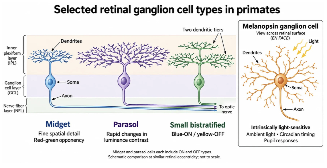
What are the different Retinal Ganglion Cell Types in Primates?
Midget Ganglion Cells (P-cells)
Explanation: Possess small dendritic arbors and narrow receptive fields specialized for high spatial resolution (fine detail) and red-green color opponency.
Parasol Ganglion Cells (M-cells)
Explanation: Feature large, widely branching dendritic arbors designed to detect low contrast and rapid changes in luminance (motion sensitivity).
Small Bistratified Ganglion Cells
Explanation: Display two distinct dendritic tiers within the inner plexiform layer (IPL) that carry Blue-ON / Yellow-OFF chromatic signals to the brain.
Melanopsin Ganglion Cells (ipRGCs)
Explanation: Intrinsically photosensitive retinal ganglion cells that express melanopsin to directly detect ambient light levels, driving non-image-forming functions like circadian rhythm regulation and pupil constriction.
ON/OFF Subtypes & Axonal Projections
Explanation: Both midget and parasol populations branch into separate ON and OFF subtypes, sending their axons through the nerve fiber layer (NFL) out into the optic nerve.
How are fovea and periphery a tradeoff?
• Densely packed foveal cones and small receptive fields preserve spatial detail
• With eccentricity, convergence generally increases, and receptive fields become larger
• Pooling improves weak-signal detection while sacrificing spatial precision
• Eye movements continually bring selected targets onto the high-resolution fovea because high acuity is restricted to a tiny central spot; rapid eye movements (saccades) constantly reposition the fovea onto interesting targets to inspect fine details in real time.
How is the optic nerve a bottleneck?
The retina contains far more photoreceptors than ganglion-cell axons
This creates a compression problem
Neighboring photoreceptors often receive highly redundant information
Retinal circuitry does not simply compress a photograph
It transforms the image before sending information to the brain
it must heavily process, filter, and compress the information locally—extracting edges, motion, and contrast—before sending the summarized data up to the brain
How does the surround changes an ON-center response with a stimulus vision map?
Center Illumination (Excitation):
A light stimulus falling directly on the ON-center of a retinal ganglion or bipolar cell causes depolarization and a high rate of action potentials (or increased excitation). [1, 2, 3]
Surround Illumination (Inhibition):
A light stimulus falling on the off-surround activates horizontal cells, which send inhibitory feedback that hyperpolarizes and suppresses the center response. [1, 2, 3]
Uniform / Full-Field Illumination (Cancellation):
When light covers both the ON-center and the inhibitory surround simultaneously, the opposing signals cancel each other out, yielding a low or baseline firing rate similar to complete darkness. [1, 2]
Contrast and Edge Detection: By subtracting the ambient luminance of the surround from the local luminance of the center, the neuron becomes insensitive to broad, uniform lighting changes and highly sensitive to boundaries, spots, and fine spatial edges. [1, 2, 3]
Core Circuit Takeaway
ON bipolar cells invert the cone signal, while bipolar cells excite ganglion cells. Horizontal cells provide lateral feedback at cone terminals to construct the antagonist surround.
How does retinal ganglion cells respond to edges?
Input Image (Cornea) vs. "Neural Image" (Retinal Ganglion Cells)
The cornea/lens projects a continuous, full-luminance picture onto the retina, but retinal ganglion cells transform it into an edge-filtered visual map where uniform areas are flattened to grey and boundaries are sharply outlined
Center-Surround Receptive Fields Emphasize Edges
Because center-surround antagonism cancels out uniform, widespread illumination, ganglion cells fire primarily where light levels change sharply, highlighting borders and visual contours.

What is the center-surround principle with vision?
Retinal ganglion cells compare light in the center of their receptive field with light in the surrounding area.
This comparison emphasizes local contrast (differences in brightness) rather than absolute light levels.
Lateral connections help neighboring regions provide the comparison signal.
This makes edges and changes in brightness stand out to the brain.
Key takeaway: Vision emphasizes differences between neighboring areas of light and dark rather than simply measuring overall brightness.
A retinal output can be expressed relative to a spatial reference
The reference is supplied by neighboring regions
Local contrast becomes more prominent than common illumination
The neuron is not simply a detector of a particular object or feature
What is cone opponency, and how does it support color vision?
Humans have three cone types: S (short), M (medium), and L (long) wavelengths.
Their sensitivities overlap, so no single cone can identify a specific color (univariance).
Retinal cells compare cone signals and organize them into opponent channels:
L vs. M: Red–green.
S vs. (L + M): Blue–yellow.
L + M: Light–dark (brightness).
These comparisons allow the brain to distinguish colors based on wavelength, not just brightness.
Key takeaway: Color vision relies on comparing cone activity through opponent channels rather than interpreting each cone independently.
How does wavelength affect photons and color coding?
Wavelength affects the probability that a photon will be absorbed by a particular pigment
Explanation: A photon's physical wavelength determines how likely it is to be captured by a specific visual pigment, but it does not guarantee absorption.
Once absorption occurs, the transduction event does not preserve the photon's wavelength
Explanation: The downstream biochemical cascade (closing cGMP channels and hyperpolarizing) is identical whether the photon had a long, medium, or short wavelength.
What remains is an absorption event
Explanation: The photoreceptor only registers that an event occurred, reducing wavelength data into a single graded voltage signal (univariance).
Information has already been discarded
Explanation: Because a single receptor loses wavelength specificity upon capturing a photon, the brain cannot tell color from a single photoreceptor alone, necessitating downstream cone-opponent comparisons.
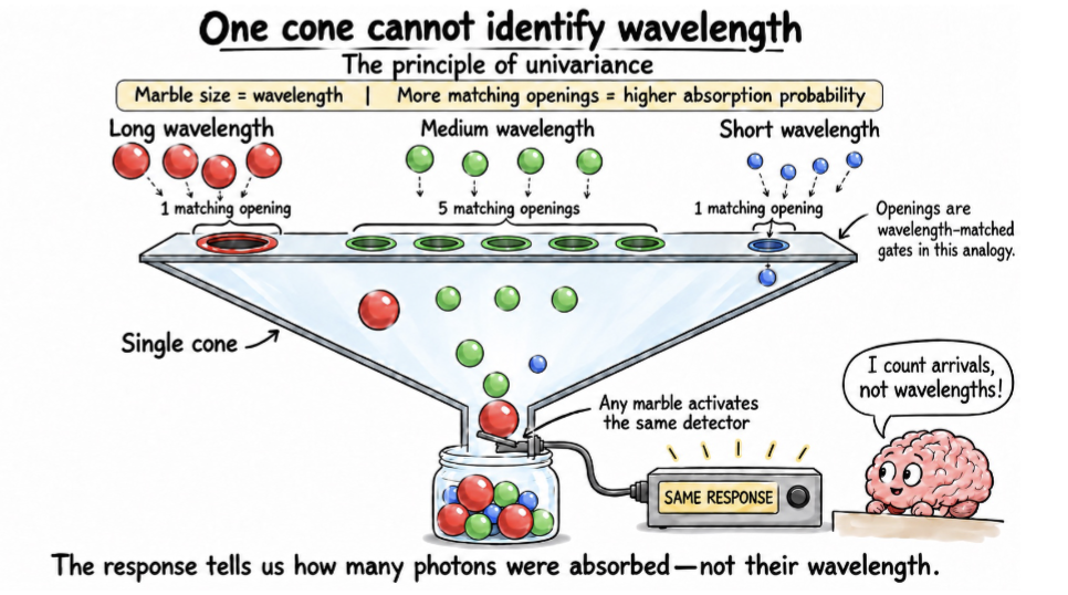
How does a cone have a fundamental ambiguity?
A cone reports how many photons its pigment absorbs
Different combinations of wavelength and intensity can produce the same response
No downstream neuron can recover information that never survived the receptor
This is the principle of univariance
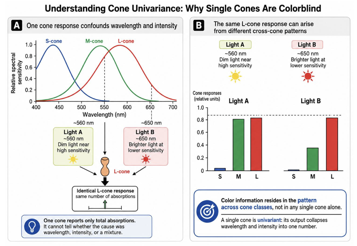
Why are single cones technically colorblind?
Panel A: One Cone Response Confounds Wavelength and Intensity
Explanation: Panel A shows that an L-cone produces an identical response (the exact same number of photon absorptions) to two completely different light sources
A single cone only reports total absorptions, making it impossible to tell whether the output was caused by wavelength, intensity, or a mixture
Panel B: The Same L-Cone Response Arises from Different Cross-Cone Patterns
Explanation: Panel B demonstrates that while the L-cone response is identical for both lights, the overall pattern across S-, M-, and L-cones differs significantly between Light A and Light B.
Takeaway: Color information resides in the pattern across multiple cone classes rather than any single cone alone, because a single cone is univariant and collapses wavelength and intensity into a single number.
How does early retinal output carry several comparisions?
L+M-related signals strongly represent broadband luminance changes
Explanation: Summing L and M cone inputs creates a high-sensitivity luminance channel that captures overall brightness variations, shadows, and contrast across the visual scene.
L-versus-M signals emphasize differences between strongly overlapping cone classes
Explanation: Subtracting M-cone input from L-cone input (or vice versa) isolates chromatic differences between these two closely neighboring spectral sensitivities, forming the basis of red-green opponency.
S-versus-(L+M) signals provide another chromatic comparison
Explanation: Comparing short-wavelength S-cones against the combined sum of L- and M-cones creates a second opponent axis, representing blue-yellow color differences.
These are physiological axes, not neural labels for experienced red, green, blue, or yellow
Explanation: The nervous system operates on raw mathematical ratios and electrical differences across cone populations at this stage; subjective color labels (like "red" or "yellow") are perceptual constructions built by later cortical processing in the brain.

What is the comparison between the Center-Surround Principle and Cone Opponency?
Center-surround circuitry compares neighboring regions of space
Explanation: Uses lateral feedback mechanisms to contrast a point in the visual field against its surrounding spatial neighborhood, extracting edges and relative illumination.
Cone-opponent circuitry compares receptor populations relative to adaptation
Explanation: Evaluates differences and ratios across S-, M-, and L-cone classes to overcome univariance and process spectral relationships.
The references and mechanisms are different
Explanation: Spatial center-surround processing relies on neighboring locations across the retinal sheet, whereas cone opponency relies on spectral comparisons between different cone types at the same retinal location.
Both create useful variables from relationships among inputs
Explanation: The core unifying principle of the retina is relational computation—transforming raw light captures into meaningful biological variables (spatial contrast and color opponency) before sending signals to the brain.
How does the visual output leave the retina after each stage of retinal processing?
Fine spatial detail favors small receptive fields
Weak-signal detection favors pooling
Rapid changes favor different temporal responses from sustained contrast
Chromatic signals require different combinations of cone input
The optic nerve therefore contains complementary descriptions of the same visual region
How does one visual hemifield reaches both eyes?
The right visual field falls on the left half of each retina
Explanation: Light originating from the right visual hemifield strikes the nasal retina of the right eye and the temporal retina of the left eye.
The left visual field falls on the right half of each retina
Explanation: Light from the left visual hemifield projects onto the nasal retina of the left eye and the temporal retina of the right eye.
Retinal anatomy and external visual location therefore cannot be treated as the same thing
Explanation: Because both eyes view the entire visual field and split inputs across their retinas, eye-centered retinal maps must be distinguished from external spatial coordinates.
Trace one example rather than memorizing four separate cases
Explanation: Rather than trying to memorize every permutation of left/right eyes and visual fields, following a single hemifield's trajectory through the optic chiasm makes the underlying projection logic intuitive.

How does the visual hemifield sort optic chiasm?
The nasal retina is the medial half of the retina (closest to the nose), while the temporal retina is the lateral half (closest to the temples)
Nasal-retinal ganglion-cell axons cross
Explanation: Axons originating from the nasal half of each retina decussate (cross over) to the contralateral side of the brain at the optic chiasm.
Temporal-retinal axons remain uncrossed
Explanation: Axons originating from the temporal half of each retina stay on the ipsilateral side (the same side of the brain) without crossing.
Signals from the same visual hemifield therefore enter the same optic tract
Explanation: By crossing nasal fibers while keeping temporal fibers uncrossed, the optic chiasm successfully sorts all information from a single external visual hemifield so that it travels together down the same optic tract to the primary visual processing centers.

How do lesions expose the coordinating visual system?
• Optic nerve damage produces monocular loss
• Chiasmal damage can produce bitemporal loss
• Postchiasmatic damage produces homonymous loss
• The pattern of failure reveals what the pathway represents at each location

How Does V1 magnify central vision?
A degree of visual angle near fixation occupies much more V1 than a degree in the far periphery
Explanation: The primary visual cortex devotes significantly more surface area to processing central degrees of visual space compared to identical eccentricities in the far periphery.
Cortical magnification continues the unequal sampling established in the retina
Explanation: The dense packing and high foveal cone counts seen at the retinal level are directly mirrored and amplified by disproportionate neural representation in V1.
High-resolution central vision consumes disproportionate neural territory
Explanation: Even though the central fovea covers a tiny fraction of the total visual field, it commands a massive percentage of processing space in the visual cortex.
Retinotopy (the orderly, spatial mapping of the visual field from the retina onto a structure in the brain) preserves neighborhood, not physical scale
Explanation: While adjacent points in the visual field project to adjacent points in V1 (topographic mapping), the actual physical distances and proportions of the visual world are heavily distorted.
What are the optic tract’s destinations?
• Detailed pattern vision depends strongly on the route through LGN to V1
• Other outputs influence pupil control, circadian timing, orienting, and gaze stabilization
• Retinal information is distributed according to behavioral use
• There is no requirement to construct one complete percept before visual information can
affect behavior

How is vision linked to action before conscious perception?
Vision is linked to action before the visual cortex
The superior colliculus participates in orienting and target selection
Explanation: Subcortical visual pathways project directly to structures like the superior colliculus to rapidly direct attention and orient gaze toward salient environmental targets before conscious cortical processing occurs.
Visual information can influence movements of the eyes and head
Explanation: Early visual reflexes bypass the visual cortex entirely, driving immediate motor responses to reposition the eyes and head toward sudden visual stimuli.
Other retinal outputs regulate physiology and stabilize gaze
Explanation: Specialized retinal outputs (such as ipRGCs and accessory optic pathways) project to non-cortical brain regions to manage non-image-forming functions like pupillary light reflexes, circadian rhythms, and gaze stabilization.
Sensory processing is already embedded in control system
Explanation: Vision is not merely a passive perception system that feeds a conscious brain; it is deeply integrated into motor and physiological control loops right from the earliest stages of neural architecture.
What is the path from the retina?
Retina->LGN->V1
• Retinal ganglion cells provide the principal driving visual input
• Cortical, inhibitory, and state-related inputs alter how that signal is transmitted
• The LGN regulates timing, gain, and reliability while preserving retinal relationships
• Relay describes the direction of information flow, not passive copying
WWhat organizational features of visual information does the LGN preserve from the retina?
• Neighboring visual locations remain neighbors: retinotopy (the orderly, spatial mapping of the visual field from the retina onto a structure in the brain)
• Major retinal-channel biases remain recognizable
• Dominant eye of origin remains strongly segregated
• Contralateral and Ipsilateral eyes are segregated by layers
• These organized inputs provide ingredients for cortical recombination
Why are parallel channels are not separate visual worlds?
Ganglion-cell populations differ in spatial, temporal, and chromatic weighting
Explanation: Different retinal ganglion cell subtypes process distinct features of the visual world, varying in their spatial receptive field sizes, temporal response speeds, and spectral color sensitivities.
No single population contains the complete scene
Explanation: An individual ganglion cell type only extracts specific properties (like edges, motion, or color contrasts), meaning no single parallel pathway holds a fully assembled picture of the visual environment on its own.
No retinal channel maps neatly onto one later perceptual faculty
Explanation: Retinal outputs do not represent isolated, standalone "perceptual building blocks" (such as a single channel dedicated solely to a specific object or final percept); instead, they provide foundational signals that get recombined across multiple higher brain areas.
Specialized outputs remain overlapping and complementary
Explanation: Rather than operating in total isolation, these parallel channels overlap across the retinal surface to provide rich, multi-dimensional descriptions of the exact same visual space.
Is the LGN a simple relay?
No, it is not a simple relay
• Retinal ganglion cells provide the principal driving visual input
• Cortical, inhibitory, and state-related inputs alter how that signal is transmitted
• The LGN regulates timing, gain, and reliability while preserving retinal relationships
• Relay describes the direction of information flow, not passive copying
V1 Orientation Selectivity & Receptive Field Construction
1. LGN neurons detect light, but not its direction
Retinal ganglion cells and LGN neurons have circular receptive fields.
They detect changes in light and dark in a small area.
However, they cannot tell whether a line is horizontal, vertical, or diagonal.
Think: Each LGN neuron detects one small circular spot, not the overall shape of a line.
2. V1 combines information from multiple LGN neurons
Multiple LGN neurons send signals to the same V1 neuron.
Their receptive fields are arranged next to each other in a straight line.
V1 combines (adds together) their signals.
Think: V1 puts several small pieces of information together to recognize a longer line.
3. What happens when a line matches the arrangement?
When a light bar matches the alignment of the LGN inputs, all the LGN neurons activate together.
Their excitatory signals add together.
This produces a strong response from the V1 neuron.
Result: The V1 neuron fires a lot because it recognizes its preferred orientation.
4. What happens when the line is rotated?
If the line is rotated, it no longer matches the arrangement of the LGN inputs.
Fewer LGN neurons activate together.
Their signals do not add together as effectively.
The V1 neuron responds much less.
Compare retina and V1
• Retina: compare one location with neighboring space
• Color circuitry: compare receptor populations relative to adaptation
• V1: combines local contrast signals according to their spatial arrangement
• The mechanisms differ, but each derives new information from relationships among inputs
How are the maps are preserved while the code changes in vision?
• Retina: photons become contrast, chromatic comparisons, and parallel outputs
• Chiasm: eye-based signals become hemifield-based signals
• LGN: mapped retinal relationships enter a recurrent thalamic circuit
• V1: mapped inputs are recombined into new spatial selectivities
What is Ocular Dominance Columns?
Stripe-shaped regions in the primary visual cortex (V1) where neurons respond preferentially to visual input from either the left eye or the right eye
Alternating Pattern: Input from the left and right eyes arrives from the lateral geniculate nucleus (LGN) and segregates into alternating, parallel stripes or bands across the cortex.
Layer Concentration: They are most sharply defined and densely clustered in layer 4 (specifically layer 4C in primates), though their influence extends vertically to other cortical layers.
Binocular Vision: Higher layers in the cortex combine these separate signals, allowing the brain to merge inputs from both eyes to create depth perception and binocular vision
Do the individual information received by both eyes differ?
• Each eye views the world from a slightly different position
• The two retinal images therefore differ slightly
• Those differences contain information about relative depth
• Depth begins with another comparison
What location leads to binocular disparity within the eye?
A fixated object falls on corresponding retinal locations in the two eyes
Explanation: When you stare directly at an object, its image lands on identical, matching points on the retinas of both the left and right eyes.
Objects at other depths fall at horizontally displaced positions
Explanation: Objects sitting closer to you or farther away than the fixation point do not land on matching retinal spots; instead, they are horizontally offset between the two eyes.
That interocular displacement is binocular disparity
Explanation: Binocular disparity is the exact spatial difference (shift) between where an object's image falls on the left retina versus the right retina.
The sign and amount of disparity provide information about depth relative to fixation
Explanation: The brain decodes whether the displacement is crossed (closer than fixation) or uncrossed (farther than fixation) to compute precise 3D depth.
What are disparity-selective neurons?
Effective left-eye and right-eye inputs can be offset in retinal position
Explanation: A binocular cortical neuron receives convergent inputs from both eyes, but its receptive fields in the left and right retinas are shifted slightly relative to one another.
The neuron responds most strongly when a feature occupies the preferred pair of locations
Explanation: Maximum firing occurs only when the binocular disparity of the visual stimulus matches the specific physical offset built into the neuron's left and right eye receptive fields.
Its firing varies systematically with interocular disparity
Explanation: As an object moves closer or farther, changing the degree of binocular disparity, the neuron's firing rate scales predictably based on how well the shift matches its tuning curve.
The preferred relationship can correspond to a particular relative depth
Explanation: Because every specific interocular disparity maps to a distinct 3D location relative to the fixation point, these neurons act as dedicated neural detectors for specific depths in space.
How are different retinal disparities produced?
An object nearer than fixation generally produces crossed disparity
Explanation: Because the object sits closer to the observer than the point of gaze, the lines of sight must cross in front of the fixation target, projecting the image to horizontally disparate positions (crossed disparity).
An object farther than fixation generally produces uncrossed disparity
Explanation: Because the object is beyond the fixation distance, the lines of sight do not cross before the object, resulting in an uncrossed retinal displacement.
The two eyes do not contain a labeled depth value
Explanation: Individual retinal images only capture flat 2D projections of light; neither eye possesses an inherent, pre-calculated "distance tag" or depth label.
Depth information must be extracted from the relationship between their images
Explanation: Depth perception is entirely relational—it is dynamically computed by the brain by comparing the horizontal offset (disparity) between the left and right retinal projections.
When does comparison of the visual cue between the two eyes start?
• Eye-specific signals remain strongly segregated through much of LGN and V1 input circuitry
• They converge extensively within V1
• Some neurons respond best to a particular displacement between the two eyes' images
• A binocular relationship has become a cortical response property
How does absolute change to relative disparity?
V1 strongly represents disparity relative to the current fixation geometry
Explanation: V1 neurons primarily encode absolute disparity—the raw distance offset of an object relative to where the eyes are currently pointing and converged.
Later cortex increasingly represents differences in disparity between neighboring objects or surfaces
Explanation: As visual processing moves up the cortical hierarchy into areas like V2 and V4, neurons shift to encoding relative disparity, comparing the depth of one surface directly against another nearby surface.
Relative disparity is more useful for describing which surface lies in front of another
Explanation: Relative disparity remains constant even when eye movements change fixation, providing a stable representation of 3D object relationships and spatial depth regardless of gaze direction.
The reference frame has changed again
Explanation: Visual coding transforms once more—moving from an eye-centered fixation reference frame in V1 to an object- or surface-centered relative frame in higher visual areas.
What is the computational pipeline of colore within vision?
Photoreceptor Activation (Cone Ratios)
Incoming light hits the S (blue), M (green), and L (red) cones simultaneously. Because each cone type has a different spectral sensitivity curve, any incoming wavelength generates a unique triplet ratio of excitation.
Retinal Opponent Arithmetic (Axis Calculations)
Retinal circuits calculate spatial and spectral differences instead of absolute light levels, transforming the cone outputs into two opponent chromatic axes and one luminance channel:
Cortical Coordinate Mapping (Hue Interpretation)
The visual system uses the two chromatic opponent values as a 2D coordinate pair:
Downstream cortical neurons in V1 and higher visual areas read these population coordinate vectors to build your conscious perception of distinct hues.
What happens beyond V1?
• V2 and later areas remain extensively interconnected with earlier cortex
• Receptive fields generally become larger
• Context extends across greater portions of visual space
• Local signals increasingly participate in contours, surfaces, depth, motion, and objects
What are the properties of V2
• V1 strongly represents local orientation, chromatic contrast, and absolute disparity
• V2 places greater weight on contour continuation, border relationships, and relative disparity
• Local signals are being recombined rather than merely passed onward
• Retinotopy remains even as the effective spatial context expands
What are the two broad cortical networks in V2?
Dorsal and Ventral pathways
Ventral visual networks place strong weight on form, surfaces, objects, and recognition (color-based cortex)
Dorsal visual networks place strong weight on motion, spatial transformations, orientation, and action (motion-based cortex)
The shorthand has been “what” versus “where” visual pathways or streams
A modern description emphasizes dominant computations rather than independent pathways
What does visual area specialization not imply?
Specialization does not imply isolation
• Different regions place different weights on different information
• Motion, form, color, depth, recognition, and action are partially differentiated
• The regions remain recurrently connected
What does V-4 circuitry emphasize?
emphasizes color and form
Responses reflect combinations of color, contour, curvature, texture, shape, and context
Color processing is prominent but not exclusive
Several human ventral regions show strong color-related responses
What is cerebral achromatopsia?
Color blindness
• Brain injury can disrupt color perception despite functioning retinal cone mechanisms
• Unilateral damage can produce color loss restricted to the contralateral visual field
• Ventral occipital and temporal territories contribute strongly to normal color experience
• The deficit is cortical, not a loss of photopigments
What is cerebral akinetopsia
Motion Blindness
• Patient LM had bilateral lateral occipitotemporal injury
• Motion could appear as a succession of positions rather than a continuous trajectory
• Pouring liquid and judging approaching traffic became difficult
• Static visual capacities were much less affected
Faces and Letterstrings (Puce et al., 1996):
Category-Specific Visual Processing Areas
Explanation: The human ventral visual stream contains anatomically distinct regions in the fusiform and inferior temporal gyri specialized for processing distinct high-level visual categories.
Right-Hemisphere Lateralization for Faces (Red/Yellow Clusters)
Explanation: Visual presentation of face stimuli drives robust neural activation localized primarily in the fusiform gyrus (Fusiform Face Area / FFA), showing strong right-hemisphere dominance.
Left-Hemisphere Lateralization for Letterstrings (Green Clusters)
Explanation: Visual presentation of text and letterstrings selectively recruits the left ventral occipitotemporal cortex (Visual Word Form Area / VWFA), reflecting left-lateralized language processing networks.
Double Dissociation of High-Level Form Perception
Explanation: Functional neuroimaging demonstrates an anatomical dissociation where spatial and structural processing for faces versus written language are segregated into specialized, hemisphere-biased cortical patches.
Prosopagnosia
Face blindness
Definition: Neurological inability to recognize familiar faces, often caused by damage to the ventral visual stream (fusiform face area).
Key Feature: Facial recognition fails, forcing reliance on superficial visual cues (e.g., hair, voice, clothing); a changed hairstyle removes a crucial identifying clue
Capgras Delusion
A familiar person seems an impostor
Definition: Neurological/psychiatric syndrome where an individual can visually identify a familiar person but believes they have been replaced by an identical-looking impostor.
Key Feature: Intact visual facial recognition paired with a disconnection from autonomic emotional responses (loss of the expected "emotional spark" upon seeing a loved one).
Frégoli Delusion
One person, many disguises
Definition: Delusion in which a person believes that different, unfamiliar people encountered in daily life are actually a single familiar person taking on various disguises.
Key Feature: Hyper-activation or misattribution of emotional familiarity to distinct, unrelated faces in the environment.
Topographic amnesia
Environmental Disorientation
The inability to recognize familiar buildings or landmarks, relying on street names and numbers for guidance

Filling in the Blind Spot (or Scotoma)
The Blind Spot (Optic Disc) Has No Photoreceptors
Explanation: Where the optic nerve axons exit the retina and blood vessels enter, there are no rod or cone photoreceptors, creating an absolute physiological blind spot in each eye's visual field.
Perceptual Completion (Active Cortical Infill)
Explanation: Rather than perceiving a gaping dark hole, the visual brain actively interpolates missing information by interpolating textures, colors, and continuous lines from the surrounding spatial context.
Cortical Mechanisms Beyond the Retina
Explanation: Filling-in is an active visual cortex operation (involving lateral and feedback connections in V1 and V2) that uses statistical predictions and border continuity to reconstruct the missing visual area.
Binasal/Binocular Masking and Pathological Scotomas
Explanation: In normal binocular vision, overlapping visual fields from the opposite eye cover the blind spot; however, even in monocular vision or pathological scotomas (damaged cortical/retinal patches), cortical completion seamlessly conceals the deficit.
Charles Bonnet Syndrome
When eye conditions (like age-related macular degeneration, cataracts, or glaucoma) reduce the amount of visual data sent to the brain, the brain's visual cortex can become hyperactive and start generating its own phantom images.
Alice in Wonderland Syndrome (Dysmetropsia)
a rare neurological disorder that causes temporary distortions in visual perception, body schema, and the sense of time

What happens when V1 is damaged?
Blindsight
Damage to V1 usually removes normal vision from the corresponding visual field
Large unilateral lesions can produce a homonymous field defect
Bilateral damage can produce profound cortical blindness
Yet visual information need not lose every influence on behavior
Residual abilities can include localization, motion discrimination, orientation judgments, orienting, or reaching
Performance is usually coarse and fragil
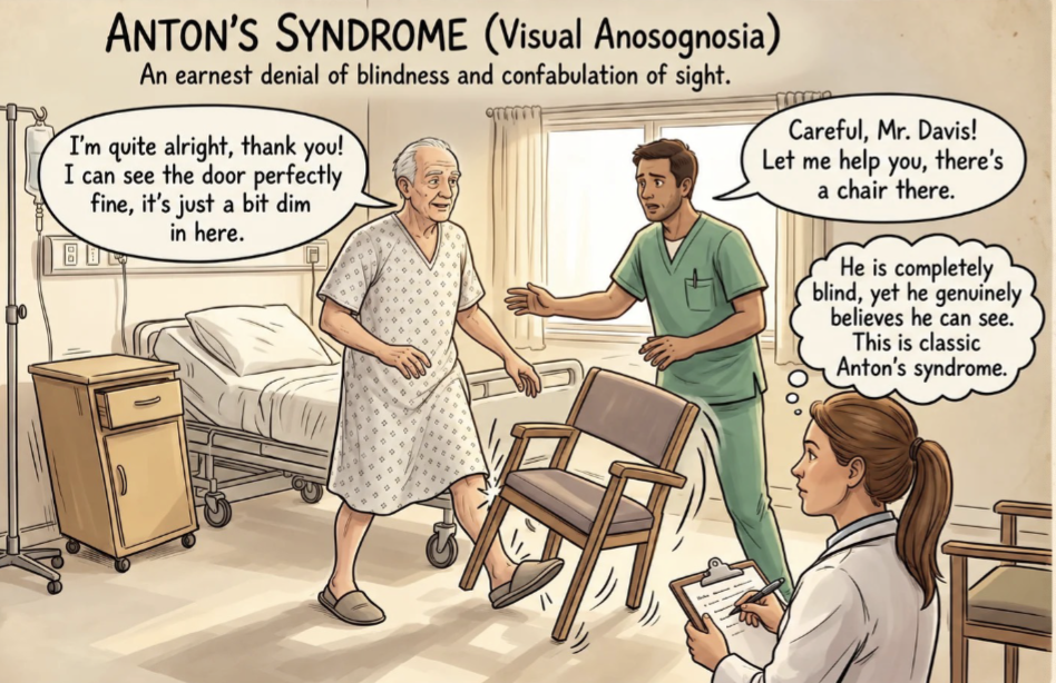
Anton’s Syndrome
Visual anosognosia
An earnest denial of blindness and confabulation of sight
Why is blindsight not one hidden pathway?
Alternative routes through the superior colliculus and pulvinar
Retinal information can reach extrastriate and motor-related systems through several routes
LGN–extrastriate, collicular–pulvinar, spared V1, and interhemispheric pathways may contribute
Different residual behaviors need not depend on the same route
Anatomy after injury may also differ from anatomy in the intact brain
What does blindsight establish?
• V1 is central to ordinary visual experience
• V1 is not the only route by which visual input can influence the nervous system
• Visual processing and normal visual awareness are therefore distinguishable
• A behavioral effect does not imply a complete conscious percept
How is recognizing an object another transformation
• Adequate acuity and visual fields do not guarantee recognition
• Local features must become coherent form
• Form must gain access to identity and meaning
• Meaning may then gain access to a name